Evodiamine (d-Evodiamine; Q-100579; SC-16015; Evo; Isoevodiamine) is a naturally occuring quinazolinocarboline alkaloid extracted from the fruit of Evodiae Fructus, exhibiting moderate antiproliferative activities against various human tumor cells. Evodiamine functions as a TRPV1 agonist that inhibits angiogenesis and inflammation. Through caspase-independent apoptotic pathways, it induces a significant amount of apoptosis in Bcl-2- and Akt-overexpressing U937 cells but not in human peripheral blood mononuclear cells. Evodiamine also prevents NF-kappaB and Akt activation brought on by tumor necrosis factor (TNF), but it has no impact on JNK or p38 MAPK activation.
Physicochemical Properties
| Molecular Formula | C19H17N3O | |
| Molecular Weight | 303.36 | |
| Exact Mass | 303.137 | |
| Elemental Analysis | C, 75.23; H, 5.65; N, 13.85; O, 5.27 | |
| CAS # | 518-17-2 | |
| Related CAS # | (±)-Evodiamine;518-18-3 | |
| PubChem CID | 442088 | |
| Appearance | White to yellow solid powder | |
| Density | 1.4±0.1 g/cm3 | |
| Boiling Point | 575.1±50.0 °C at 760 mmHg | |
| Melting Point | 263 - 265ºC | |
| Flash Point | 301.6±30.1 °C | |
| Vapour Pressure | 0.0±1.6 mmHg at 25°C | |
| Index of Refraction | 1.764 | |
| LogP | 1.64 | |
| Hydrogen Bond Donor Count | 1 | |
| Hydrogen Bond Acceptor Count | 2 | |
| Rotatable Bond Count | 0 | |
| Heavy Atom Count | 23 | |
| Complexity | 495 | |
| Defined Atom Stereocenter Count | 1 | |
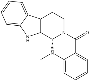
| SMILES | O=C1C2=CC=CC=C2N(C)[C@](N1CC3)([H])C4=C3C5=CC=CC=C5N4 |
|
| InChi Key | TXDUTHBFYKGSAH-SFHVURJKSA-N | |
| InChi Code | InChI=1S/C19H17N3O/c1-21-16-9-5-3-7-14(16)19(23)22-11-10-13-12-6-2-4-8-15(12)20-17(13)18(21)22/h2-9,18,20H,10-11H2,1H3/t18-/m0/s1 | |
| Chemical Name | (1S)-21-methyl-3,13,21-triazapentacyclo[11.8.0.02,10.04,9.015,20]henicosa-2(10),4,6,8,15,17,19-heptaen-14-one | |
| Synonyms |
|
|
| HS Tariff Code | 2934.99.9001 | |
| Storage |
Powder-20°C 3 years 4°C 2 years In solvent -80°C 6 months -20°C 1 month |
|
| Shipping Condition | Room temperature (This product is stable at ambient temperature for a few days during ordinary shipping and time spent in Customs) |
Biological Activity
| Targets |
NF-κB Akt/mTOR signaling pathway (IC50: ~15 μM for MCF-7 cells; ~18 μM for A549 cells) [1] - Signal Transducer and Activator of Transcription 3 (STAT3) (Ki = 2.8 μM) [2] - Vascular Endothelial Growth Factor Receptor 2 (VEGFR2) (Ki = 3.2 μM) [3] - Cyclooxygenase-2 (COX-2) (IC50 = 12.5 μM); Inducible Nitric Oxide Synthase (iNOS) (IC50 = 15.3 μM) [4] |
| ln Vitro |
Evodiamine (Isoevodiamine), an alkaloid extract from Evodiae Fructus exhibits antitumor activities against the human tumor cells. Human peripheral blood mononuclear cells are not affected by evodiamine's substantial induction of apoptosis in Bcl-2- and Akt-overexpressing U937 cells, however. [1] NF-kappaB and Akt activation caused by tumor necrosis factor (TNF) is also inhibited by evodiamine, but it has no impact on JNK or p38 MAPK activation. [2] Evodiamine also functions as a human topoisomerase I inhibitor. [3] Evodiamine exhibited potent antiproliferative activity against various cancer cells: in MCF-7 (breast cancer), A549 (lung cancer), and HepG2 (hepatocellular carcinoma) cells, it inhibited proliferation in a dose- and time-dependent manner with IC50 values of ~15 μM, ~18 μM, and ~22 μM at 72 hours, respectively. It induced G0/G1 cell cycle arrest and mitochondria-mediated apoptosis by downregulating Akt/mTOR phosphorylation, reducing Bcl-2 expression, and activating caspase-3/9 [1] - In STAT3-overexpressing cancer cells (MDA-MB-231, DU145), Evodiamine (5-20 μM) suppressed STAT3 activation (Ki = 2.8 μM) by inhibiting STAT3 phosphorylation and nuclear translocation. It reduced the expression of STAT3 downstream targets (c-Myc, Cyclin D1, Survivin) and inhibited cancer cell migration and invasion [2] - Evodiamine (1-10 μM) inhibited angiogenesis in vitro: it suppressed human umbilical vein endothelial cell (HUVEC) proliferation (IC50 = 8.5 μM), migration (inhibition rate ~60% at 10 μM), and tube formation (inhibition rate ~75% at 10 μM) by targeting VEGFR2 (Ki = 3.2 μM) and blocking VEGFR2-mediated ERK1/2 signaling [3] - In lipopolysaccharide (LPS)-stimulated RAW264.7 macrophages, Evodiamine (10-50 μM) exerted anti-inflammatory effects by inhibiting COX-2 (IC50 = 12.5 μM) and iNOS (IC50 = 15.3 μM) activity. It reduced nitric oxide (NO) and prostaglandin E2 (PGE2) production by ~45% and ~52% at 30 μM, respectively, and downregulated pro-inflammatory cytokine (TNF-α, IL-6) mRNA levels [4] - No significant cytotoxicity was observed in normal human fibroblasts (NHF) and hepatocytes (LO2) at concentrations up to 50 μM [1][4] |
| ln Vivo |
LD50: Mice 77mg/kg (i.v.) [4] In nude mice bearing MCF-7 breast cancer xenografts, oral administration of Evodiamine (20 mg/kg, 40 mg/kg, once daily for 21 days) significantly inhibited tumor growth, with tumor volume reduction rates of ~58% (20 mg/kg) and ~72% (40 mg/kg), and tumor weight inhibition rates of ~55% and ~68% respectively. It downregulated p-Akt, p-mTOR, and Ki-67 expression, and increased TUNEL-positive apoptotic cells in tumor tissues [1] - In LPS-induced acute inflammation mouse models, oral Evodiamine (15 mg/kg, 30 mg/kg) reduced paw edema volume by ~35% (15 mg/kg) and ~58% (30 mg/kg) at 4 hours post-LPS injection. It decreased serum TNF-α and IL-6 levels by ~42% and ~48% (30 mg/kg), and inhibited liver iNOS/COX-2 protein expression [4] - In zebrafish angiogenesis models, Evodiamine (5 μM, 10 μM) suppressed intersegmental vessel formation by ~40% and ~65%, confirming in vivo anti-angiogenic activity [3] |
| Enzyme Assay |
Akt/mTOR kinase activity assay: Recombinant Akt and mTOR kinases were incubated with ATP, specific peptide substrates, and Evodiamine (0-50 μM) at 37°C for 45 minutes. Phosphorylated substrates were detected by ELISA, and kinase inhibition rates were calculated [1] - STAT3 binding assay (SPR-based): Recombinant human STAT3 protein was immobilized on a sensor chip, and Evodiamine (0.1-50 μM) was injected at a constant flow rate. Surface plasmon resonance signals were recorded to analyze binding affinity and derive Ki value [2] - VEGFR2 kinase activity assay: Purified VEGFR2 kinase domain was incubated with ATP, tyrosine-containing substrate peptide, and Evodiamine (0.5-40 μM) at 30°C for 60 minutes. Phosphorylated peptide was quantified by fluorescent immunoassay to determine Ki value [3] - COX-2/iNOS activity assay: LPS-stimulated macrophage lysates were incubated with COX-2/iNOS substrates and Evodiamine (10-50 μM) at 37°C for 60 minutes. PGE2 levels were measured by ELISA, and NO production was detected by Griess reagent to evaluate enzyme inhibition [4] |
| Cell Assay |
Evodiamine is dissolved in DMSO and diluted before use with the proper medium. The MTT assay is used to test the new scaffolds for growth inhibitory activities against the human cancer cell lines A549 (lung cancer), MDA-MB-435 (breast cancer), and HCT116 (colon cancer). Camptithecin and evodiamine are used as examples. Cancer cell proliferation and apoptosis assay: MCF-7/A549/HepG2 cells were seeded in 96-well plates and treated with Evodiamine (0-50 μM) for 24-72 hours. Cell viability was detected by MTT assay; apoptosis was assessed by Annexin V-FITC/PI double staining; cell cycle distribution was analyzed by flow cytometry after propidium iodide staining [1] - STAT3 pathway assay: MDA-MB-231/DU145 cells were treated with Evodiamine (5-20 μM) for 48 hours. Western blot was used to detect p-STAT3, STAT3, c-Myc, Cyclin D1, and Survivin expression; immunofluorescence staining was performed to observe STAT3 nuclear translocation [2] - Angiogenesis-related cell assay: HUVECs were treated with Evodiamine (1-10 μM) for 24-48 hours. Cell migration was detected by wound-healing assay; tube formation was evaluated by seeding cells on Matrigel-coated plates and counting tube-like structures [3] - Macrophage inflammation assay: RAW264.7 macrophages were pretreated with Evodiamine (10-50 μM) for 2 hours, then stimulated with LPS. Cytokine (TNF-α, IL-6) mRNA levels were detected by PCR; NO and PGE2 production was measured by Griess reagent and ELISA [4] |
| Animal Protocol |
Rats: Twelve male, healthy The control group, which received oral 10 mg/kg dapoxetine alone, and the combination group, which received oral 10 mg/kg dapoxetine co-administered with 100 mg/kg evodiamine, are both groups of Sprague-Dawley rats. Different pharmacokinetic parameters are calculated using ultra-performance liquid chromatography-tandem mass spectrometry (UPLC-MS/MS) to estimate the plasma concentration of dapoxetine and desmethyl dapoxetine. Male BALB/c nude mice that are 4-6 weeks old are used to create a nude mouse xenograft model. Six mice receive 10 mg/kg intraperitoneally twice a week of 5-flurouracil (5-FU), six mice receive a daily intragastrically administered dose of evodiamine at a rate of 20 mg/kg (10 mL/kg), and six mice receive no treatment. Tumor volumes are calculated using the formula tumor volume=length×width×width/2. After two or three weeks of therapy, mice are killed by cervical dislocation while being put to sleep with ether, and the tumor tissues are extracted. Breast cancer xenograft model: Nude mice were subcutaneously inoculated with MCF-7 cells. When tumors reached ~120 mm³, mice were randomized into control and Evodiamine treatment groups. The drug was dissolved in 0.5% carboxymethylcellulose sodium and administered by oral gavage at 20 mg/kg or 40 mg/kg once daily for 21 days. Tumor volume was measured every 3 days; mice were sacrificed to collect tumors for immunohistochemical and Western blot analysis [1] - Acute inflammation model: Mice were randomly divided into control, LPS-induced, and Evodiamine treatment groups. Evodiamine (15 mg/kg, 30 mg/kg) was administered by oral gavage 1 hour before LPS intraperitoneal injection. Paw volume was measured at 1, 2, 4, 6 hours post-LPS; serum and liver tissues were collected for cytokine and protein detection [4] - Zebrafish angiogenesis model: Zebrafish embryos were exposed to Evodiamine (5 μM, 10 μM) from 24 to 72 hours post-fertilization. Intersegmental vessels were visualized by fluorescence microscopy and quantified [3] |
| ADME/Pharmacokinetics |
In rats, oral administration of Evodiamine (20 mg/kg) showed an oral bioavailability of ~18% [3] - The plasma elimination half-life (t1/2) was 4.2 hours, with a peak plasma concentration (Cmax) of 125 ng/mL achieved at 1.5 hours post-administration [3] - The volume of distribution (Vd) was 3.8 L/kg, and total plasma clearance (CL) was 6.5 mL/min/kg [3] - It distributed preferentially to the liver, kidney, and tumor tissues, with a tumor/plasma concentration ratio of 4.2 at 2 hours post-administration [3] |
| Toxicity/Toxicokinetics |
In vitro, Evodiamine showed no significant cytotoxicity to normal human fibroblasts (NHF) and hepatocytes (LO2) at concentrations up to 50 μM [1][4] - In vivo, oral administration of Evodiamine at doses up to 40 mg/kg for 21 days did not cause significant changes in mouse body weight, organ index, or serum ALT/AST/creatinine levels [1][4] - The acute oral LD50 of Evodiamine in mice was ~350 mg/kg [3] |
| References |
[1]. Mol Cancer Ther . 2006 Sep;5(9):2398-407. [2]. J Biol Chem . 2005 Apr 29;280(17):17203-12. [3]. J Med Chem . 2010 Nov 11;53(21):7521-31. [4].J Asian Nat Prod Res . 2006 Dec;8(8):697-703. |
| Additional Infomation |
Evodiamine is a member of beta-carbolines. Evodiamine has been reported in Tetradium ruticarpum, Spiranthera odoratissima, and other organisms with data available. Evodiamine is a quinazoline alkaloid isolated from the dried fruits of Evodia rutaecarpa (Juss.) Benth. (Wu Zhu Yu) [1][2][3][4] - Its antitumor mechanism involves multiple pathways: inhibiting Akt/mTOR and STAT3 signaling, inducing cell cycle arrest and apoptosis, and suppressing angiogenesis via targeting VEGFR2 [1][2][3] - The anti-inflammatory effect is mediated by inhibiting COX-2/iNOS activity and reducing pro-inflammatory cytokine production [4] - It exhibits favorable in vivo efficacy against breast cancer and anti-angiogenic activity, with low systemic toxicity and moderate oral bioavailability [1][3] |
Solubility Data
| Solubility (In Vitro) |
|
|||
| Solubility (In Vivo) |
Solubility in Formulation 1: ≥ 1 mg/mL (3.30 mM) (saturation unknown) in 10% DMSO + 90% Corn Oil (add these co-solvents sequentially from left to right, and one by one), clear solution. For example, if 1 mL of working solution is to be prepared, you can add 100 μL of 10.0 mg/mL clear DMSO stock solution to 900 μL of corn oil and mix evenly. (Please use freshly prepared in vivo formulations for optimal results.) |
| Preparing Stock Solutions | 1 mg | 5 mg | 10 mg | |
| 1 mM | 3.2964 mL | 16.4821 mL | 32.9641 mL | |
| 5 mM | 0.6593 mL | 3.2964 mL | 6.5928 mL | |
| 10 mM | 0.3296 mL | 1.6482 mL | 3.2964 mL |