Physicochemical Properties
| Molecular Formula | C13H8O4 |
| Exact Mass | 228.042 |
| CAS # | 529-61-3 |
| PubChem CID | 5281631 |
| Appearance | Light yellow to yellow solid powder |
| Density | 1.5±0.1 g/cm3 |
| Boiling Point | 472.6±45.0 °C at 760 mmHg |
| Melting Point | 240 °C |
| Flash Point | 191.1±22.2 °C |
| Vapour Pressure | 0.0±1.2 mmHg at 25°C |
| Index of Refraction | 1.718 |
| LogP | 1.92 |
| Hydrogen Bond Donor Count | 2 |
| Hydrogen Bond Acceptor Count | 4 |
| Rotatable Bond Count | 0 |
| Heavy Atom Count | 17 |
| Complexity | 317 |
| Defined Atom Stereocenter Count | 0 |
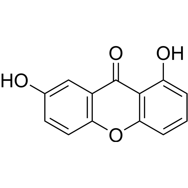
| SMILES | C1=CC(=C2C(=C1)OC3=C(C=C(C=C3)O)C2=O)O |
| InChi Key | KDXFPEKLLFWHMN-UHFFFAOYSA-N |
| InChi Code | InChI=1S/C13H8O4/c14-7-4-5-10-8(6-7)13(16)12-9(15)2-1-3-11(12)17-10/h1-6,14-15H |
| Chemical Name | 1,7-dihydroxyxanthen-9-one |
| Synonyms | Euxanthone; 529-61-3; 1,7-dihydroxy-9H-xanthen-9-one; 1,7-Dihydroxyxanthone; 1,7-dihydroxyxanthen-9-one; eyxanthone; purrenone; CHEBI:4946; |
| HS Tariff Code | 2934.99.9001 |
| Storage |
Powder-20°C 3 years 4°C 2 years In solvent -80°C 6 months -20°C 1 month |
| Shipping Condition | Room temperature (This product is stable at ambient temperature for a few days during ordinary shipping and time spent in Customs) |
Biological Activity
| Targets | Autophagy |
| ln Vitro | Euxanthone (10–20 μM; 24 h) strongly inhibits cell invasion and, in a dose-dependent manner, reduces OS cell migration. Adhesion to fibronectin is dramatically decreased by eauxanthone [1]. COX-2 expression is regulated by auxanthone (10–20 μM; 24 h) via the miR-21/PDCD4/c-jun signaling pathway. The antimetastatic action of euxanthone is mediated by its suppression of COX-2 [1]. |
| ln Vivo | Euxanthone (40–80 mg/kg) dramatically lowers the amount of metastatic nodules in lung tissue in models of lung metastasis [1]. In mice with bilateral common carotid artery blockage treated with Euxanthone (30–60 mg/kg; oral; once daily; for 7 days), normal Bnip3, Beclin1, Pink1, Parkin, p53, Bax, caspase-3, and LC3 II/I change. When mitochondrial stress is caused by mitochondrial fragmentation, euxanthone controls both mitophagy and apoptosis [2]. |
| Enzyme Assay | Mitophagy and apoptosis significantly contribute to the dynamics of mitochondria and their associated disorders. Euxanthone (EUX) is a xanthone derivative that exhibits several therapeutic effects at a preclinical level. However, its impact in a cerebral ischemia and reperfusion model has not been investigated. The investigation aimed to determine the protective effect of EUX in cerebral ischemia and cognitive impairment and explore its underlying mechanism. A bilateral common carotid artery occlusion (BCCAO) model was employed in the present work. Forty male ICR mice were divided into four groups - Sham, BCCAO, EUX30 (BCCAO + EUX 30 mg/kg) and EUX60 (BCCAO + EUX 60 mg/kg). The mice were then subjected to a Morris water maze study for investigation of learning and memorizing capabilities. The hippocampal specimens of mice were quantified for the presence of oxidative markers. Homogenized hippocampal fractions were determined for the levels of Beclin-1, LC3, p53, Bax, caspase-3, Bnip3, DRP1 and Nrf2. The present investigation revealed that BCCAO caused oxidative stress in mitochondria and led to mitochondrial breakdown. EUX administration markedly attenuated BCCAO triggered mitochondrial stress and related breakdown. EUX treatment normalized Bnip3, Beclin1, Pink1, Parkin, p53, Bax, caspase-3, and LC3 II/I. Altogether, EUX treatment modulated mitophagy and apoptosis induced by mitochondrial stress mediated by mitochondrial fragmentation, due to cerebral ischemia and reperfusion injury [2]. |
| Cell Assay |
Cell Migration Assay [1] Cell Types: Osteosarcoma (OS) cells Tested Concentrations: 10 μM, 20 μM Incubation Duration: 24 h Experimental Results: Inhibited cell migration at 24 hr. Western Blot Analysis[1] Cell Types: Osteosarcoma (OS) cells Tested Concentrations: 10 μM, 20 μM Incubation Duration: 24 h Experimental Results: Repressed both the mRNA and protein level of COX-2 in OS cells in a dose-dependent fashion. |
| Animal Protocol |
Animal/Disease Models: Forty male ICR mice (20 g) induced cerebral ischemia and reperfusion[2] Doses: 30 mg/kg, 60 mg/kg Route of Administration: po ;one time/day; for 7 days Experimental Results: Markedly attenuated BCCAO triggered mitochondrial stress and related breakdown. |
| References |
[1]. Euxanthone Impairs the Metastatic Potential of Osteosarcoma by Reducing COX-2 Expression. Anat Rec (Hoboken). 2019 Aug;302(8):1399-1408. [2]. Euxanthone improves cognitive impairment by attenuating mitochondrial fragmentation and suppressing oxidative stress. Cent Eur J Immunol. 2021;46(4):446-455. [3]. Euxanthone Attenuates Aβ1-42-Induced Oxidative Stress and Apoptosis by Triggering Autophagy. J Mol Neurosci. 2018 Dec;66(4):512-523. |
| Additional Infomation |
Euxanthone is a member of the class of xanthones that is 9H-xanthene substituted by hydroxy group at positions 1 and 7 and an oxo group at position 9. It has been isolated from Cratoxylum cochinchinense. It has a role as a plant metabolite and a metabolite. It is a member of xanthones and a member of phenols. Euxanthone has been reported in Hypericum ascyron, Hypericum erectum, and other organisms with data available. steosarcoma (OS) is one of the most common malignancies of bone. This study was aimed to explore the anti-metastatic effect of euxanthone on OS. Adhesion assay and Transwell assay were used to examine the effect of euxanthone on adhesion, migration and invasion of OS cells. COX-2-over-expressing plasmid was applied to transfect OS cells to assess whether COX-2 affects the anti-metastatic function of euxanthone. PDCD4 knockdown and miR-21 mimic were applied to assess whether euxanthone suppresses the transactivation of c-jun via modulating miR-21-PDCD4 signaling. The effect of euxanthone in vivo was also examined by lung metastasis assay. Euxanthone, a xanthone derivative extracted from Polygala caudata, has been found to exhibit anti-neoplastic activities. In present study, our results showed that euxanthone suppressed cell adhesion, migration, and invasion in OS cells. Our experimental data also showed that repression of COX-2 by euxanthone mediated its anti-metastatic activities. Moreover, our findings revealed that euxanthone modulated the COX-2 expression through the miR-21/PDCD4/c-jun signaling pathway. The anti-metastatic activities of euxanthone were also validated in a pulmonary metastasis model. Taken together, our results highlighted the potential of euxanthone to be used in the treatment of OS. [1] lzheimer's disease (AD) is the most common neurodegenerative disorder and is characterized by the deposition of β-Amyloid (Aβ) plaques which contribute to its pathology. The present study was aimed at exploring the protective effects of euxanthone against Aβ-induced neurotoxicity both in vivo and in vitro. We found that euxanthone significantly attenuated Aβ1-42-induced memory and spatial learning dysfunction and also significantly reversed Aβ1-42-induced neuronal apoptosis and autophagy in the hippocampal region. Euxanthone also protected the neuroblastic PC12 cells against Aβ1-42-induced oxidative stress and apoptosis by inducing autophagy. In conclusion, euxanthone exerts its neuroprotective effect against Aβ1-42 by inducing autophagy, indicating its potential therapeutic role in AD.[3] |
Solubility Data
| Solubility (In Vitro) | May dissolve in DMSO (in most cases), if not, try other solvents such as H2O, Ethanol, or DMF with a minute amount of products to avoid loss of samples |
| Solubility (In Vivo) |
Note: Listed below are some common formulations that may be used to formulate products with low water solubility (e.g. < 1 mg/mL), you may test these formulations using a minute amount of products to avoid loss of samples. Injection Formulations (e.g. IP/IV/IM/SC) Injection Formulation 1: DMSO : Tween 80: Saline = 10 : 5 : 85 (i.e. 100 μL DMSO stock solution → 50 μL Tween 80 → 850 μL Saline) *Preparation of saline: Dissolve 0.9 g of sodium chloride in 100 mL ddH ₂ O to obtain a clear solution. Injection Formulation 2: DMSO : PEG300 :Tween 80 : Saline = 10 : 40 : 5 : 45 (i.e. 100 μL DMSO → 400 μLPEG300 → 50 μL Tween 80 → 450 μL Saline) Injection Formulation 3: DMSO : Corn oil = 10 : 90 (i.e. 100 μL DMSO → 900 μL Corn oil) Example: Take the Injection Formulation 3 (DMSO : Corn oil = 10 : 90) as an example, if 1 mL of 2.5 mg/mL working solution is to be prepared, you can take 100 μL 25 mg/mL DMSO stock solution and add to 900 μL corn oil, mix well to obtain a clear or suspension solution (2.5 mg/mL, ready for use in animals). Injection Formulation 4: DMSO : 20% SBE-β-CD in saline = 10 : 90 [i.e. 100 μL DMSO → 900 μL (20% SBE-β-CD in saline)] *Preparation of 20% SBE-β-CD in Saline (4°C,1 week): Dissolve 2 g SBE-β-CD in 10 mL saline to obtain a clear solution. Injection Formulation 5: 2-Hydroxypropyl-β-cyclodextrin : Saline = 50 : 50 (i.e. 500 μL 2-Hydroxypropyl-β-cyclodextrin → 500 μL Saline) Injection Formulation 6: DMSO : PEG300 : castor oil : Saline = 5 : 10 : 20 : 65 (i.e. 50 μL DMSO → 100 μLPEG300 → 200 μL castor oil → 650 μL Saline) Injection Formulation 7: Ethanol : Cremophor : Saline = 10: 10 : 80 (i.e. 100 μL Ethanol → 100 μL Cremophor → 800 μL Saline) Injection Formulation 8: Dissolve in Cremophor/Ethanol (50 : 50), then diluted by Saline Injection Formulation 9: EtOH : Corn oil = 10 : 90 (i.e. 100 μL EtOH → 900 μL Corn oil) Injection Formulation 10: EtOH : PEG300:Tween 80 : Saline = 10 : 40 : 5 : 45 (i.e. 100 μL EtOH → 400 μLPEG300 → 50 μL Tween 80 → 450 μL Saline) Oral Formulations Oral Formulation 1: Suspend in 0.5% CMC Na (carboxymethylcellulose sodium) Oral Formulation 2: Suspend in 0.5% Carboxymethyl cellulose Example: Take the Oral Formulation 1 (Suspend in 0.5% CMC Na) as an example, if 100 mL of 2.5 mg/mL working solution is to be prepared, you can first prepare 0.5% CMC Na solution by measuring 0.5 g CMC Na and dissolve it in 100 mL ddH2O to obtain a clear solution; then add 250 mg of the product to 100 mL 0.5% CMC Na solution, to make the suspension solution (2.5 mg/mL, ready for use in animals). Oral Formulation 3: Dissolved in PEG400 Oral Formulation 4: Suspend in 0.2% Carboxymethyl cellulose Oral Formulation 5: Dissolve in 0.25% Tween 80 and 0.5% Carboxymethyl cellulose Oral Formulation 6: Mixing with food powders Note: Please be aware that the above formulations are for reference only. InvivoChem strongly recommends customers to read literature methods/protocols carefully before determining which formulation you should use for in vivo studies, as different compounds have different solubility properties and have to be formulated differently. (Please use freshly prepared in vivo formulations for optimal results.) |