Physicochemical Properties
| Molecular Formula | C19H24O6 |
| Molecular Weight | 348.3903 |
| Exact Mass | 348.157 |
| CAS # | 23062-24-0 |
| PubChem CID | 441793 |
| Appearance | White to off-white solid powder |
| Density | 1.3±0.1 g/cm3 |
| Boiling Point | 588.1±50.0 °C at 760 mmHg |
| Melting Point | 268-270 °C |
| Flash Point | 213.1±23.6 °C |
| Vapour Pressure | 0.0±3.7 mmHg at 25°C |
| Index of Refraction | 1.576 |
| LogP | -0.43 |
| Hydrogen Bond Donor Count | 2 |
| Hydrogen Bond Acceptor Count | 6 |
| Rotatable Bond Count | 0 |
| Heavy Atom Count | 25 |
| Complexity | 725 |
| Defined Atom Stereocenter Count | 9 |
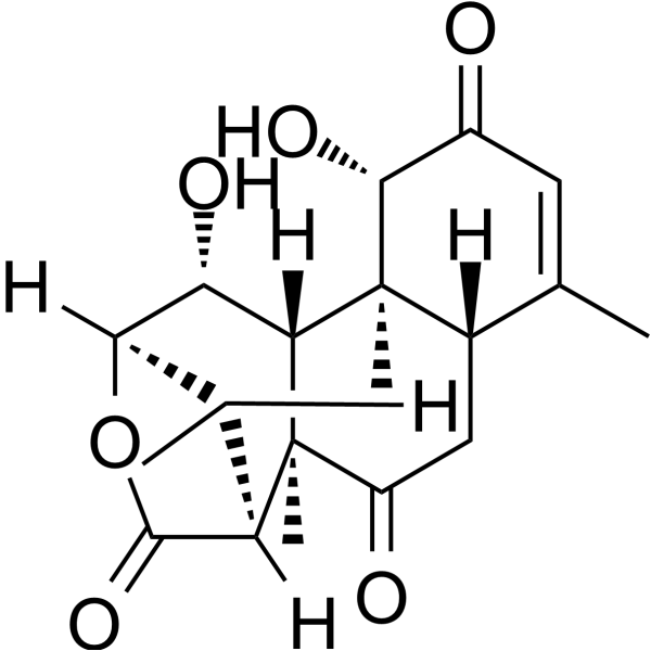
| SMILES | C[C@H]1[C@@H]2[C@@H]([C@@H]3[C@@]4([C@@H](CC(=O)[C@]3([C@H]1C(=O)O2)C)C(=CC(=O)[C@H]4O)C)C)O |
| InChi Key | OGHYZHNTIINXEO-MGBQOKOWSA-N |
| InChi Code | InChI=1S/C19H24O6/c1-7-5-10(20)16(23)18(3)9(7)6-11(21)19(4)12-8(2)14(25-17(12)24)13(22)15(18)19/h5,8-9,12-16,22-23H,6H2,1-4H3/t8-,9+,12-,13+,14-,15-,16-,18+,19+/m1/s1 |
| Chemical Name | (1S,2R,5S,9S,10S,11R,12R,13R,16R)-9,12-dihydroxy-2,6,10,16-tetramethyl-14-oxatetracyclo[11.2.1.02,11.05,10]hexadec-6-ene-3,8,15-trione |
| HS Tariff Code | 2934.99.9001 |
| Storage |
Powder-20°C 3 years 4°C 2 years In solvent -80°C 6 months -20°C 1 month Note: This product requires protection from light (avoid light exposure) during transportation and storage. |
| Shipping Condition | Room temperature (This product is stable at ambient temperature for a few days during ordinary shipping and time spent in Customs) |
Biological Activity
| Targets |
Eurycomalactone targets AKT/NF-κB signaling pathway (inhibits AKT phosphorylation and NF-κB activation) [2] Eurycomalactone targets DNA double-strand break (DSB) repair pathway (delays repair process,) [1] |
| ln Vitro |
A549 and COR-L23 cell viability is specifically inhibited by tongkat alilactone (24, 48, and 72 hours). The viability of A549 cells is inhibited by tongkat alilactone, with IC50 values of 20.17 and 3.77 for 24, 48, and 72 hours, respectively. and 1.90 μM. COR-L23 cell viability is inhibited by tongkat alilactone at IC50 values of 25.02, 2.74, and 1.80 μM for 24, 48, and 72 hours, respectively [1]. Non-small cell lung cancer (NSCLC) cells are stimulated to undergo apoptosis by tongkat alilactone (2.29–156.3-26.6 μM; 24 hours; A549 and Calu-1 cells) [2]. Tongkat alilactone (0-25.05 μM; 24 hours; A549 and COR-L23 cells) causes irradiated non-small cell lung cancer (NSCLC) cells to undergo apoptosis and triggers cell cycle arrest in the radiosensitive G2/M phase. When non-small cell lung cancer (NSCLC) cells are exposed to radiation, tongkat alilactone downregulates important G2/M regulatory proteins [1]. Tongkat alilactone (2.5–25 μM; 24 hours; A549 cells) prevents DNA double-strand breaks caused by radiation from healing [1]. In non-small cell lung cancer (NSCLC) cells, erycomalactone (2.29–156.3-25.6 μM; 24 hours; A549 and Calu-1 cells) suppresses AKT/NF-κB activation [2]. - Radiosensitizing activity: Eurycomalactone enhanced radiosensitivity of human NSCLC cells (A549, H1299) with IC50 values of 8.5 μM and 10.2 μM respectively. It induced G₂/M cell cycle arrest (G₂/M phase cells increased from 15% to 42% at 10 μM) and delayed DNA DSB repair, as evidenced by persistent γ-H2AX foci (3.2-fold more foci at 24 hours post-irradiation with 10 μM treatment) [1] - Antitumor activity against NSCLC: Eurycomalactone inhibited proliferation of A549, H1299, and PC-9 NSCLC cells with IC50 values of 7.8 μM, 9.5 μM, and 8.9 μM respectively. It suppressed AKT phosphorylation (inhibition rate 65% at 10 μM) and NF-κB activation, reducing Bcl-2 expression and increasing Bax levels to induce apoptosis (apoptotic rate 35% at 10 μM in A549 cells) [2] - Chemosensitizing activity: Eurycomalactone improved cisplatin sensitivity in A549 cells, reducing cisplatin IC50 from 8.2 μM to 3.1 μM when combined with 5 μM Eurycomalactone. The combination enhanced apoptosis by 2.8-fold compared to cisplatin alone [2] - Inhibition of endothelial adhesion molecules: Eurycomalactone suppressed TNF-α-induced expression of E-selectin, VCAM-1, and ICAM-1 in HUVECs at the post-transcriptional level. At 10 μM, it reduced E-selectin and VCAM-1 protein levels by 68% and 72% respectively, without affecting their mRNA levels [3] |
| ln Vivo |
- Radiosensitizing efficacy: In nude mice bearing A549 xenografts, intraperitoneal administration of Eurycomalactone (10 mg/kg) 1 hour before irradiation (8 Gy) significantly enhanced tumor growth inhibition. The combination group showed a tumor growth inhibition rate of 75%, compared to 42% in the irradiation alone group [1] - Antitumor and chemosensitizing efficacy: In A549 xenograft-bearing nude mice, intraperitoneal injection of Eurycomalactone (10 mg/kg, once daily) combined with cisplatin (5 mg/kg, once weekly) resulted in a tumor growth inhibition rate of 82%, which was significantly higher than cisplatin alone (53%) or drug alone (45%). No significant body weight loss was observed [2] |
| Enzyme Assay |
- AKT kinase activity assay: Recombinant AKT kinase was mixed with ATP (10 μM), substrate peptide, and Eurycomalactone (2.5-20 μM) in kinase buffer. The mixture was incubated at 37°C for 1 hour, and phosphorylated substrate was detected by HTRF assay. The inhibition of AKT phosphorylation was quantified by comparing with vehicle control [2] - NF-κB activity assay: NF-κB-luciferase reporter plasmid-transfected A549 cells were treated with Eurycomalactone (5-20 μM) for 24 hours, then stimulated with TNF-α (10 ng/mL) for 6 hours. Luciferase activity was measured using a luminometer to evaluate NF-κB activation inhibition [2] - DNA DSB repair assay: A549 cells were treated with Eurycomalactone (10 μM) for 24 hours, then irradiated with 4 Gy. At 0, 6, 12, and 24 hours post-irradiation, cells were fixed, stained with γ-H2AX antibody, and foci were counted under fluorescence microscopy to assess DNA repair efficiency [1] |
| Cell Assay |
Apoptosis analysis[2] Cell Types: A549 and Calu-1 Cell Tested Concentrations: 2.29, 10.14, 12.02, 20.81, 80.77 and 156.3 μM Incubation Duration: 24 hrs (hours) Experimental Results: Apoptosis rate increased in a dose-dependent manner. Cell cycle analysis[1] Cell Types: A549 and COR-L23 Cell Tested Concentrations: 1.57, 2.57, 20.17 and 25.05 μM Incubation Duration: 24 hrs (hours) Experimental Results: Induced cell cycle arrest in G2/M phase and increased sub-G1 phase in irradiated cells population. A549 and COR-L23 cells Western Blot analysis [2] Cell Types: A549 and Calu-1 Cell Tested Concentrations: 2.29, 10.14, 12.02, 20.81, 80.77 and 156.3 μM Incubation Duration: 24 hrs (hours) Experimental Results: Expression level of induced active caspase-3 and active PARP (cleaved form), while Bcl-xL and survival were diminished. Western Blot Analysis[1] Cell Types: A549 and COR-L23 Cell Tested Concentrations: 1.57, 2.57, 20.17 and 25.05 μM Incubation Duration: 24 hrs (hours) Experimental Results: The expression of two G2/M regulatory proteins was downregulated in a dose-dependent manner. Western Blot Analysis[2] Ce - Cell viability and radiosensitivity assay: NSCLC cells (A549, H1299) were seeded into 96-well plates, treated with Eurycomalactone (2.5-40 μM) for 24 hours, then irradiated with 0-8 Gy. Cell viability was measured by tetrazolium salt assay 72 hours later to calculate IC50 and radiosensitization ratio [1] - Cell cycle and apoptosis assay: A549 cells were treated with Eurycomalactone (5-15 μM) for 24 hours. For cell cycle analysis, cells were fixed, stained with PI, and analyzed by flow cytometry. For apoptosis, cells were stained with Annexin V-FITC/PI and detected by flow cytometry [1][2] - Western blot and PCR assay: NSCLC cells or HUVECs were treated with Eurycomalactone (5-10 μM) for 24-48 hours. Proteins (AKT, p-AKT, NF-κB, Bcl-2, Bax, E-selectin, VCAM-1, γ-H2AX) were detected by western blot. For HUVECs, E-selectin/VCAM-1 mRNA levels were measured by RT-PCR [1][2][3] - Clonogenic assay: A549 cells were treated with Eurycomalactone (2.5-10 μM) for 24 hours, irradiated with 4 Gy, then seeded into 6-well plates and incubated for 14 days. Colonies were stained and counted to calculate survival fraction [1] |
| Animal Protocol |
- NSCLC xenograft radiosensitization model: Nude mice (6-8 weeks old) were subcutaneously injected with A549 cells (5×10⁶ cells/mouse). When tumors reached ~100 mm³, mice were randomly divided into four groups: control, Eurycomalactone alone (10 mg/kg, ip, once), irradiation alone (8 Gy, local), and combination. Eurycomalactone was administered 1 hour before irradiation. Tumor volume was measured every 3 days for 21 days [1] - NSCLC xenograft chemosensitization model: Nude mice were subcutaneously injected with A549 cells (5×10⁶ cells/mouse). When tumors reached ~100 mm³, mice were divided into four groups: control, Eurycomalactone alone (10 mg/kg, ip, once daily), cisplatin alone (5 mg/kg, ip, once weekly), and combination. Treatment lasted for 21 days, with tumor volume and body weight monitored weekly [2] |
| Toxicity/Toxicokinetics |
- In vitro toxicity: Eurycomalactone showed low cytotoxicity to normal human lung fibroblasts (MRC-5) with IC50 > 40 μM, indicating a favorable therapeutic index [2] - In vivo toxicity: In xenograft-bearing mice treated with Eurycomalactone (10 mg/kg, 21 days), no significant body weight loss (≤ 6%) or histopathological abnormalities were observed in major organs (heart, liver, spleen, lungs, kidneys). Serum ALT, AST, creatinine, and urea nitrogen levels were within normal ranges [1][2] |
| References |
[1]. Enhancement of Radiosensitivity by Eurycomalactone in Human NSCLC Cells Through G₂/M Cell Cycle Arrest and Delayed DNA Double-Strand Break Repair. Oncol Res. 2020 Mar 27;28(2):161-175. [2]. Inactivation of AKT/NF κB signaling by eurycomalactone decreases human NSCLC cell viability and improves the chemosensitivity to cisplatin. Oncol Rep. 2020 Oct;44(4):1441-1454. [3]. Eurycomalactone Inhibits Expression of Endothelial Adhesion Molecules at a Post-Transcriptional Level. J Nat Prod. 2017 Dec 22;80(12):3186-3193. |
| Additional Infomation |
Eurycomalactone is a steroid lactone. Eurycomalactone has been reported in Eurycoma longifolia with data available. - Natural source: Eurycomalactone is a bioactive diterpenoid isolated from the roots of Eurycoma longifolia Jack (Simaroubaceae family) [1][2][3] - Chemical classification: It belongs to the diterpenoid class, characterized by a rearranged ent-kaurane skeleton [3] - Mechanism of action: Eurycomalactone exerts multi-functional activities via distinct pathways: inducing G₂/M arrest and delaying DNA DSB repair to enhance radiosensitivity [1]; inhibiting AKT/NF-κB signaling to reduce NSCLC cell viability and improve cisplatin sensitivity [2]; suppressing endothelial adhesion molecule expression at the post-transcriptional level by promoting their proteasomal degradation [3] |
Solubility Data
| Solubility (In Vitro) | DMSO : ~100 mg/mL (~287.03 mM) |
| Solubility (In Vivo) |
Note: Listed below are some common formulations that may be used to formulate products with low water solubility (e.g. < 1 mg/mL), you may test these formulations using a minute amount of products to avoid loss of samples. Injection Formulations (e.g. IP/IV/IM/SC) Injection Formulation 1: DMSO : Tween 80: Saline = 10 : 5 : 85 (i.e. 100 μL DMSO stock solution → 50 μL Tween 80 → 850 μL Saline) *Preparation of saline: Dissolve 0.9 g of sodium chloride in 100 mL ddH ₂ O to obtain a clear solution. Injection Formulation 2: DMSO : PEG300 :Tween 80 : Saline = 10 : 40 : 5 : 45 (i.e. 100 μL DMSO → 400 μLPEG300 → 50 μL Tween 80 → 450 μL Saline) Injection Formulation 3: DMSO : Corn oil = 10 : 90 (i.e. 100 μL DMSO → 900 μL Corn oil) Example: Take the Injection Formulation 3 (DMSO : Corn oil = 10 : 90) as an example, if 1 mL of 2.5 mg/mL working solution is to be prepared, you can take 100 μL 25 mg/mL DMSO stock solution and add to 900 μL corn oil, mix well to obtain a clear or suspension solution (2.5 mg/mL, ready for use in animals). Injection Formulation 4: DMSO : 20% SBE-β-CD in saline = 10 : 90 [i.e. 100 μL DMSO → 900 μL (20% SBE-β-CD in saline)] *Preparation of 20% SBE-β-CD in Saline (4°C,1 week): Dissolve 2 g SBE-β-CD in 10 mL saline to obtain a clear solution. Injection Formulation 5: 2-Hydroxypropyl-β-cyclodextrin : Saline = 50 : 50 (i.e. 500 μL 2-Hydroxypropyl-β-cyclodextrin → 500 μL Saline) Injection Formulation 6: DMSO : PEG300 : castor oil : Saline = 5 : 10 : 20 : 65 (i.e. 50 μL DMSO → 100 μLPEG300 → 200 μL castor oil → 650 μL Saline) Injection Formulation 7: Ethanol : Cremophor : Saline = 10: 10 : 80 (i.e. 100 μL Ethanol → 100 μL Cremophor → 800 μL Saline) Injection Formulation 8: Dissolve in Cremophor/Ethanol (50 : 50), then diluted by Saline Injection Formulation 9: EtOH : Corn oil = 10 : 90 (i.e. 100 μL EtOH → 900 μL Corn oil) Injection Formulation 10: EtOH : PEG300:Tween 80 : Saline = 10 : 40 : 5 : 45 (i.e. 100 μL EtOH → 400 μLPEG300 → 50 μL Tween 80 → 450 μL Saline) Oral Formulations Oral Formulation 1: Suspend in 0.5% CMC Na (carboxymethylcellulose sodium) Oral Formulation 2: Suspend in 0.5% Carboxymethyl cellulose Example: Take the Oral Formulation 1 (Suspend in 0.5% CMC Na) as an example, if 100 mL of 2.5 mg/mL working solution is to be prepared, you can first prepare 0.5% CMC Na solution by measuring 0.5 g CMC Na and dissolve it in 100 mL ddH2O to obtain a clear solution; then add 250 mg of the product to 100 mL 0.5% CMC Na solution, to make the suspension solution (2.5 mg/mL, ready for use in animals). Oral Formulation 3: Dissolved in PEG400 Oral Formulation 4: Suspend in 0.2% Carboxymethyl cellulose Oral Formulation 5: Dissolve in 0.25% Tween 80 and 0.5% Carboxymethyl cellulose Oral Formulation 6: Mixing with food powders Note: Please be aware that the above formulations are for reference only. InvivoChem strongly recommends customers to read literature methods/protocols carefully before determining which formulation you should use for in vivo studies, as different compounds have different solubility properties and have to be formulated differently. (Please use freshly prepared in vivo formulations for optimal results.) |
| Preparing Stock Solutions | 1 mg | 5 mg | 10 mg | |
| 1 mM | 2.8703 mL | 14.3517 mL | 28.7035 mL | |
| 5 mM | 0.5741 mL | 2.8703 mL | 5.7407 mL | |
| 10 mM | 0.2870 mL | 1.4352 mL | 2.8703 mL |