Epertinib hydrochloride, the salt form of Epertinib (S22611), is an orally bioavailable and reversible dual EGFR and HER2 inhibitor with antitumor activities.
Physicochemical Properties
| Molecular Formula | C30H28CL2FN5O3 |
| Molecular Weight | 596.479428291321 |
| Exact Mass | 595.155 |
| CAS # | 2071195-74-7 |
| Related CAS # | Epertinib;908305-13-5 |
| PubChem CID | 139290994 |
| Appearance | Light yellow to yellow solid powder |
| Hydrogen Bond Donor Count | 3 |
| Hydrogen Bond Acceptor Count | 9 |
| Rotatable Bond Count | 10 |
| Heavy Atom Count | 41 |
| Complexity | 899 |
| Defined Atom Stereocenter Count | 1 |
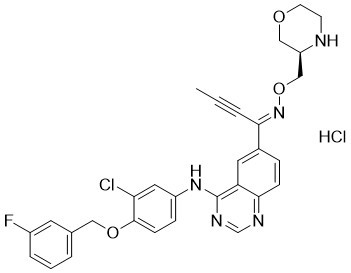
| SMILES | ClC1=C(C=CC(=C1)NC1=C2C(C=CC(=C2)/C(/C#CC)=N/OC[C@H]2COCCN2)=NC=N1)OCC1C=CC=C(C=1)F.Cl |
| InChi Key | NVWJPQQXBPWOOA-PLXRJBJVSA-N |
| InChi Code | InChI=1S/C30H27ClFN5O3.ClH/c1-2-4-27(37-40-18-24-17-38-12-11-33-24)21-7-9-28-25(14-21)30(35-19-34-28)36-23-8-10-29(26(31)15-23)39-16-20-5-3-6-22(32)13-20;/h3,5-10,13-15,19,24,33H,11-12,16-18H2,1H3,(H,34,35,36);1H/b37-27+;/t24-;/m1./s1 |
| Chemical Name | N-[3-chloro-4-[(3-fluorophenyl)methoxy]phenyl]-6-[(Z)-N-[[(3R)-morpholin-3-yl]methoxy]-C-prop-1-ynylcarbonimidoyl]quinazolin-4-amine;hydrochloride |
| HS Tariff Code | 2934.99.9001 |
| Storage |
Powder-20°C 3 years 4°C 2 years In solvent -80°C 6 months -20°C 1 month Note: Please store this product in a sealed and protected environment, avoid exposure to moisture. |
| Shipping Condition | Room temperature (This product is stable at ambient temperature for a few days during ordinary shipping and time spent in Customs) |
Biological Activity
| Targets |
- Epertinib hydrochloride (also known as S-222611) acts on epidermal growth factor receptor (EGFR), human epidermal growth factor receptor 2 (HER2), and human epidermal growth factor receptor 4 (HER4), functioning as a potent, reversible, and selective tyrosine kinase inhibitor [1] - Epertinib hydrochloride (designated as S-222611 in this study) selectively inhibits the kinase activity of EGFR and HER2, with half-maximal inhibitory concentrations (IC₅₀) below 10 nmol/L for both kinases [2] |
| ln Vitro |
In NCI-N87 cells, ipatinib hydrochloride suppresses EGFR and HER2 phosphorylation with IC50 values of 4.5 and 1.6 nM, respectively [2]. With an IC50 of 26.5 nM, ipatinib hydrochloride has inhibitory effect on MDA-MB-361 cells[1]. EGFR and/or HER2-expressing cancer cell lines can be specifically inhibited from proliferating when exposed to ipatinib hydrochloride (0–10 μM) for 72 hours [2]. - In cell lines derived from HER2-positive breast cancer (MDA-MB-361-luc-BR2/BR3) and T790M-EGFR-positive lung cancer (NCI-H1975-luc), immunohistochemical (IHC) staining was performed to detect HER2 expression, and no significant differences in HER2 staining intensity were observed across different tumor lesions [1] - Fluorescently labeled dextran (3 kDa, Texas-Red®) was used to assess blood-tumor barrier (BTB) permeability. In the NCI-H1975 lung cancer intraventricular injection mouse model (IVM), dextran fluorescence intensity was significantly higher in brain metastatic regions than in brain parenchyma; however, in MDA-MB-361 breast cancer IVMs, dextran fluorescence intensity in brain metastatic regions was comparable to that in brain parenchyma, indicating that the BTB remained largely intact in breast cancer brain metastases [1] - Epertinib hydrochloride (S-222611) inhibited the intracellular kinase activity of EGFR and HER2, as demonstrated by reduced phosphorylation levels of EGFR and HER2 in NCI-N87 gastric cancer cells following drug treatment. Western blot analysis showed that after oral administration of 50 mg/kg Epertinib hydrochloride, the relative phosphorylation levels of EGFR and HER2 in tumors were significantly lower than those in vehicle-treated groups, and this inhibitory effect was more potent than that of lapatinib [2] - Epertinib hydrochloride (S-222611) exhibited antiproliferative activity against a variety of cancer cell lines, including EGFR-expressing and HER2-expressing cells. For example, it inhibited the growth of NCI-N87 (gastric cancer), BT-474 (breast cancer), MDA-MB-361 (breast cancer), A431 (epidermoid carcinoma), and HT115 (colon cancer) cell lines. A short-time pulse treatment experiment showed that the fold increase in IC₅₀ of Epertinib hydrochloride after short-time exposure (compared to 72-h continuous exposure) was lower than that of lapatinib, indicating more sustained antiproliferative effects [2] |
| ln Vivo |
Ipetinib hydrochloride exhibits anti-tumor effect when taken orally once daily for 28 days at a dose of 0–100 mg/kg [1]. A brain tumor's growth can be considerably decreased by taking 50 mg/kg of imipetinib hydrochloride orally once a day for 30 days [1]. Oral administration of petinib hydrochloride (0-50 mg/kg) once day for 10–28 days substantially and dose-dependently suppresses the development of tumors [2]. - In the MDA-MB-361-luc-BR2/BR3 HER2-positive breast cancer IVM, oral administration of Epertinib hydrochloride at a dose of 50 mg/kg (as epertinib) resulted in a concentration of Epertinib hydrochloride in brain metastatic regions that was >10 times higher than that of lapatinib at 4 h and 8 h post-administration; additionally, the tumor-to-normal brain ratio of Epertinib hydrochloride was ~4 times higher than that of lapatinib. In the NCI-H1975-luc T790M-EGFR-positive lung cancer IVM, the concentration of Epertinib hydrochloride in brain metastases was comparable to that of lapatinib when administered at a dose of 100 mg/kg (as epertinib) [1] - In the in vivo antitumor activity assay using the MDA-MB-361-luc-BR3 breast cancer IVM, Epertinib hydrochloride was orally administered daily at a dose of 50 mg/kg for 30 days. Tumor growth was monitored by measuring photons emitted from luciferase-expressing tumor cells, and the results showed that Epertinib hydrochloride significantly inhibited tumor growth compared to the vehicle group (P < 0.05) [1] - In orthotopic implantation models of human breast cancer cells (MDA-MB-361 and MDA-MB-361-luc-BR2) into the mammary fat pad of mice, Epertinib hydrochloride was orally administered daily for 28 days, and it effectively inhibited tumor growth compared to the vehicle group [1] - In patient-oriented preclinical models, including intrafemoral and intracranial implantation models of luciferase-expressing BT474-luc breast cancer cells, Epertinib hydrochloride (S-222611) was orally administered daily for 21–28 days. In the intracranial model, 50 mg/kg Epertinib hydrochloride showed significantly stronger antitumor activity than 50 mg/kg lapatinib (P < 0.01), as monitored by bioluminescent imaging. In the survival study of mice with intracranially implanted BT474 cells, Epertinib hydrochloride treatment prolonged survival compared to vehicle and lapatinib groups [2] - In xenograft models of various cancers (NCI-N87 gastric cancer, BT-474 breast cancer, A431 epidermoid carcinoma, HT115 colon cancer), Epertinib hydrochloride (S-222611) was orally administered daily for 11–28 days, and it exhibited potent antitumor activity with lower effective doses (ED₅₀) than lapatinib [2] |
| Enzyme Assay |
- Kinase activity inhibition assays were conducted to evaluate the selectivity of Epertinib hydrochloride for EGFR, HER2, and HER4. The assay measured the ability of Epertinib hydrochloride to inhibit the kinase activity of these targets, confirming its potent and selective inhibitory effects [1] - In vitro kinase inhibition assays were performed to determine the IC₅₀ values of Epertinib hydrochloride (S-222611) against EGFR and HER2. The assay involved incubating the kinases with appropriate substrates in the presence of different concentrations of Epertinib hydrochloride, followed by detection of kinase activity to calculate the IC₅₀ values, which were found to be below 10 nmol/L for both targets [2] |
| Cell Assay |
Cell proliferation assay[2] Cell Types: NCI-N87 (gastric), BT-474 (breast), SK-BR-3 (breast), MDA-MB-453 (breast), MDA-MB-175VII (breast), HT115 (colon), Calu-3 (lung), fR2 (breast) and MRC-5 (lung) Tested Concentrations: 0-10 μM Incubation Duration: 72 hrs (hours) Experimental Results: Inhibition of growth of NCI-N87, BT-474, SK-BR -3. MDA-MB-453, MDA-MB-175VII, HT115, Calu-3, fR2 and MRC-5, IC50 values are 8.3 ± 2.6, 9.9 ± 0.8, 14.0 ± 3.6, 48.6 ± 3.1, 21.6±4.3, 53.3±8.6, 241.5±29.2, 5366.7±65.2 and 4964.6±340.3. - HER2 expression detection: Cancer cells (MDA-MB-361-luc-BR2/BR3, NCI-H1975-luc) were cultured, and immunohistochemical staining was performed on cell-derived tumor lesions. The staining intensity was analyzed to assess HER2 expression levels, with no significant differences observed between lesions [1] - BTB permeability assessment: Cancer cells were implanted to form brain metastases, and fluorescently labeled dextran (3 kDa) was administered. Fluorescence intensity in brain metastatic regions and brain parenchyma was detected and compared to evaluate BTB integrity [1] - Antiproliferative activity assay: Various cancer cell lines (NCI-N87, BT-474, MDA-MB-361, A431, HT115) were seeded in culture plates and treated with different concentrations of Epertinib hydrochloride (S-222611) or lapatinib. Cell growth was monitored for 72 h, and IC₅₀ values were calculated. A short-time pulse treatment was also conducted, where cells were exposed to the drug for a short period and then cultured without the drug, followed by growth assessment to compare with continuous 72-h treatment [2] - Kinase phosphorylation assay: NCI-N87 cells were implanted subcutaneously to form tumors. After oral administration of Epertinib hydrochloride (S-222611) or lapatinib, tumors were collected at 6 h and 24 h post-administration. Western blot analysis was performed to measure the relative phosphorylation levels of EGFR and HER2, comparing the inhibitory effects of the drugs [2] |
| Animal Protocol |
Animal/Disease Models: Nude mice (BALB/cAJcl-nu/nu, intracranial implantation of MDA-MB-361 or BR2 cells) [1] Doses: 12.5, 25, 50, 100 mg/kg Route of Administration: Orally, one time/day , lasted 28 days. Experimental Results: demonstrated antitumor activity in a mammary fat pad implantation model using both cell lines with comparable ED50 values (24.1 mg/kg for MDA-MB-361 and 26.5 mg/kg for BR2 (MDA- MB-361-luc-BR2) ), respectively). Animal/Disease Models: Nude mice (BALB/cAJcl-nu/nu, intracranial implantation of MDA-MB-361 or BR2 cells) [1] Doses: 50 mg/kg Route of Administration: Orally, one time/day for 30 days Experimental Results: The brain Dramatically diminished tumor volume, indicating that erpotinib can have potent anti-tumor activity in brain metastases even in the presence of an intact BTB (blood-tumor barrier). Animal/Disease Models: Nude mice (BALB/cAJcl-nu/nu, prepared by subcutaneously (sc) (sc) implanting human gastric cancer cell NCI-N87 cells on the back of nude mice) [2] Doses: 0, 6.25, 12.5, 25, 50 mg/kg Route of Administration: po (oral gavage), daily - Breast cancer brain metastasis IVM: HER2-positive breast cancer cells (MDA-MB-361-luc-BR2/BR3) were injected intraventricularly into mice to establish brain metastasis models. Epertinib hydrochloride was formulated as a solution and orally administered at a dose of 50 mg/kg (as epertinib) once. Mice were sacrificed at 4 h or 8 h post-administration, and brain sections were prepared for imaging mass spectrometry analysis of drug concentrations [1] - Lung cancer brain metastasis IVM: T790M-EGFR-positive lung cancer cells (NCI-H1975-luc) were injected intraventricularly into mice. Epertinib hydrochloride was orally administered at a dose of 100 mg/kg (as epertinib) once, and mice were sacrificed at 4 h post-administration for drug concentration analysis in brain metastases [1] - Orthotopic breast cancer model: MDA-MB-361 or MDA-MB-361-luc-BR2 human breast cancer cells were implanted orthotopically into the mammary fat pad of mice. Once tumors formed, Epertinib hydrochloride was orally administered daily at a specified dose for 28 days. Tumor volume was measured twice or thrice weekly to assess antitumor activity [1] - Intracranial breast cancer model: BT474-luc human breast cancer cells were implanted intracranially into mice. Epertinib hydrochloride (S-222611) was orally administered daily at doses of 50 mg/kg, 100 mg/kg, or 200 mg/kg for 28 days. Tumor growth was monitored by bioluminescent imaging (photons/sec) once or twice weekly. For the survival study, BT474 cells were implanted intracranially, and Epertinib hydrochloride was orally administered daily until death or the end of the study (106 days post-implantation) [2] - Intrafemoral breast cancer model: BT474-luc human breast cancer cells were implanted into the bone marrow of the left femur of mice. Epertinib hydrochloride (S-222611) was orally administered daily at doses of 50 mg/kg, 100 mg/kg, or 200 mg/kg for 21 days, with tumor growth monitored by bioluminescent imaging [2] - Subcutaneous xenograft models: Various human cancer cells (NCI-N87, BT-474, A431, HT115) were implanted subcutaneously into the back of nude mice or SCID mice. After randomization 3–12 days post-implantation, Epertinib hydrochloride (S-222611) was orally administered daily at multiple doses for 11–21 days. Tumor volume was measured twice or thrice weekly to calculate ED₅₀ values [2] |
| ADME/Pharmacokinetics |
- Distribution: In MDA-MB-361-luc-BR2/BR3 breast cancer IVM, Epertinib hydrochloride showed high distribution to brain metastatic regions, with concentrations >10 times higher than lapatinib at 4 h and 8 h post-oral administration (50 mg/kg). The tumor-to-normal brain ratio of Epertinib hydrochloride was ~4 times higher than that of lapatinib. In NCI-H1975-luc lung cancer IVM, the concentration of Epertinib hydrochloride in brain metastases was comparable to lapatinib after oral administration of 100 mg/kg [1] |
| Toxicity/Toxicokinetics |
- Compared to irreversible EGFR/HER2 inhibitors (neratinib, afatinib), Epertinib hydrochloride (S-222611) showed a safer toxicity profile. In the NCI-N87 xenograft model, mice treated with 25 mg/kg Epertinib hydrochloride showed no remarkable histopathological changes in the colon or eyeball. In contrast, neratinib (25 mg/kg) and afatinib (12.5 mg/kg) caused regeneration of colonic epithelium and atrophy of corneal epithelium [2] |
| References |
[1]. Distribution analysis of epertinib in brain metastasis of HER2-positive breast cancer by imaging mass spectrometry and prospect for antitumor activity. Sci Rep. 2018 Jan 10;8(1):343. [2]. Preclinical antitumor activity of S-222611, an oral reversible tyrosine kinase inhibitor of epidermal growth factor receptor and human epidermal growth factor receptor 2. Cancer Sci. 2014 Aug;105(8):1040-8. |
| Additional Infomation |
- Epertinib hydrochloride is a promising therapeutic agent for HER2-positive breast cancer with brain metastasis, especially due to its high distribution to brain metastatic regions even when the BTB is largely intact [1] - Epertinib hydrochloride (S-222611) is an oral reversible tyrosine kinase inhibitor, and its dual inhibition of EGFR and HER2 provides a therapeutic advantage in treating cancers expressing these receptors. It exhibits more potent antitumor activity than lapatinib in preclinical models and a better safety profile than irreversible inhibitors, making it a potential option for cancer therapy [2] |
Solubility Data
| Solubility (In Vitro) |
DMSO : ~125 mg/mL (~209.56 mM) H2O : ~33.33 mg/mL (~55.88 mM) |
| Solubility (In Vivo) |
Solubility in Formulation 1: ≥ 2.08 mg/mL (3.49 mM) (saturation unknown) in 10% DMSO + 40% PEG300 + 5% Tween80 + 45% Saline (add these co-solvents sequentially from left to right, and one by one), clear solution. For example, if 1 mL of working solution is to be prepared, you can add 100 μL of 20.8 mg/mL clear DMSO stock solution to 400 μL PEG300 and mix evenly; then add 50 μL Tween-80 to the above solution and mix evenly; then add 450 μL normal saline to adjust the volume to 1 mL. Preparation of saline: Dissolve 0.9 g of sodium chloride in 100 mL ddH₂ O to obtain a clear solution. Solubility in Formulation 2: ≥ 2.08 mg/mL (3.49 mM) (saturation unknown) in 10% DMSO + 90% (20% SBE-β-CD in Saline) (add these co-solvents sequentially from left to right, and one by one), clear solution. For example, if 1 mL of working solution is to be prepared, you can add 100 μL of 20.8 mg/mL clear DMSO stock solution to 900 μL of 20% SBE-β-CD physiological saline solution and mix evenly. Preparation of 20% SBE-β-CD in Saline (4°C,1 week): Dissolve 2 g SBE-β-CD in 10 mL saline to obtain a clear solution. (Please use freshly prepared in vivo formulations for optimal results.) |
| Preparing Stock Solutions | 1 mg | 5 mg | 10 mg | |
| 1 mM | 1.6765 mL | 8.3825 mL | 16.7650 mL | |
| 5 mM | 0.3353 mL | 1.6765 mL | 3.3530 mL | |
| 10 mM | 0.1677 mL | 0.8383 mL | 1.6765 mL |