Physicochemical Properties
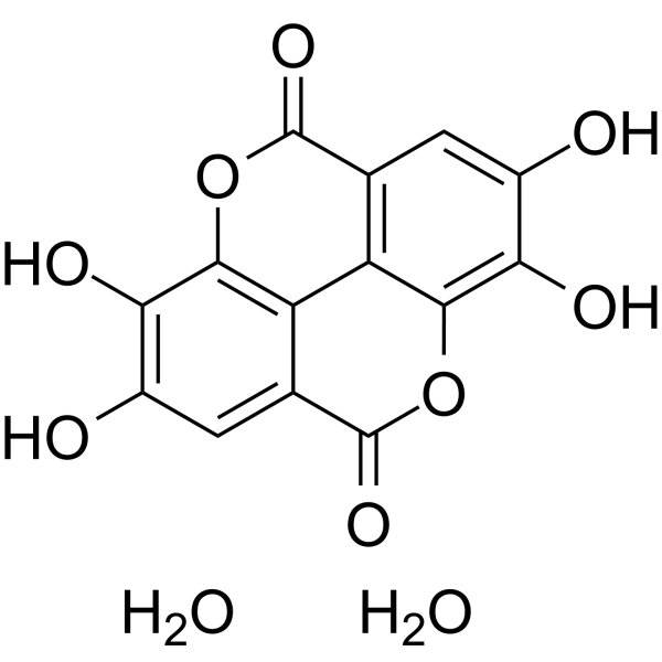
| Molecular Formula | C14H10O10 |
| Molecular Weight | 338.22 |
| Exact Mass | 338.027 |
| CAS # | 133039-73-3 |
| Related CAS # | Ellagic acid;476-66-4 |
| PubChem CID | 16760409 |
| Appearance | Typically exists as solid at room temperature |
| Melting Point | 450 °C |
| Hydrogen Bond Donor Count | 6 |
| Hydrogen Bond Acceptor Count | 10 |
| Rotatable Bond Count | 0 |
| Heavy Atom Count | 24 |
| Complexity | 475 |
| Defined Atom Stereocenter Count | 0 |
| SMILES | O1C(C2=C([H])C(=C(C3=C2C2=C1C(=C(C([H])=C2C(=O)O3)O[H])O[H])O[H])O[H])=O.O([H])[H].O([H])[H] |
| InChi Key | ZEPCRIPMALGRJR-UHFFFAOYSA-N |
| InChi Code | InChI=1S/C14H6O8.2H2O/c15-5-1-3-7-8-4(14(20)22-11(7)9(5)17)2-6(16)10(18)12(8)21-13(3)19;;/h1-2,15-18H;2*1H2 |
| Chemical Name | 6,7,13,14-tetrahydroxy-2,9-dioxatetracyclo[6.6.2.04,16.011,15]hexadeca-1(15),4,6,8(16),11,13-hexaene-3,10-dione;dihydrate |
| HS Tariff Code | 2934.99.9001 |
| Storage |
Powder-20°C 3 years 4°C 2 years In solvent -80°C 6 months -20°C 1 month |
| Shipping Condition | Room temperature (This product is stable at ambient temperature for a few days during ordinary shipping and time spent in Customs) |
Biological Activity
| Targets |
- Ellagic acid targets protein kinase CK2 (CK2); Ki value for CK2 inhibition was 0.8 μM (competitive inhibition against ATP, detected via radiometric kinase assay) [1] - Ellagic acid targets reactive oxygen species (ROS), tumor necrosis factor-α (TNF-α), interleukin-1β (IL-1β), and caspase-3 (neuroprotective-related targets); no IC50, Ki, or EC50 values were reported [2] - Ellagic acid targets apoptotic proteins (caspase-3, caspase-9, Bcl-2, Bax) and γ-radiation-induced DNA damage response proteins; no IC50, Ki, or EC50 values were reported [3] - Ellagic acid targets Src homology phosphotyrosyl phosphatase 2 (SHP2); IC50 for SHP2 phosphatase inhibition was 2.1 μM, and Ki value (competitive against substrate) was 1.7 μM (detected via fluorometric phosphatase assay) [4] |
| ln Vitro |
1. In recombinant human CK2 enzyme assays, Ellagic acid (concentrations: 0.1 μM, 0.5 μM, 1 μM, 5 μM) dose-dependently inhibited CK2 activity. At 1 μM, it inhibited CK2-mediated casein phosphorylation by 85% (radiometric assay, 32P-ATP as tracer). In HeLa cells (human cervical cancer cells) treated with Ellagic acid (5 μM, 10 μM) for 24 h, Western blot showed it reduced CK2 substrate phosphorylation (e.g., phospho-CDC37 at Ser13, reduced by 62% at 10 μM) without affecting total CK2 protein levels, confirming intracellular CK2 inhibition [1] 2. In human breast cancer cell lines (MCF-7, estrogen receptor-positive; MDA-MB-231, triple-negative), Ellagic acid (concentrations: 5 μM, 10 μM, 20 μM) enhanced apoptotic sensitivity to γ-radiation (2 Gy, 4 Gy). Pre-treatment with 20 μM Ellagic acid for 24 h followed by 4 Gy γ-radiation increased apoptotic rate from 18% (radiation alone) to 45% in MCF-7 cells (Annexin V-FITC/PI staining) and from 22% to 48% in MDA-MB-231 cells. It also upregulated cleaved caspase-3 (2.8-fold in MCF-7) and cleaved caspase-9 (2.3-fold in MCF-7), downregulated anti-apoptotic Bcl-2 (reduced by 65% in MCF-7), and increased pro-apoptotic Bax (1.9-fold in MCF-7) (Western blot). Clone formation assay showed 20 μM Ellagic acid + 4 Gy reduced colony survival rate by 72% vs radiation alone in MCF-7 [3] 3. In recombinant human SHP2 enzyme assays, Ellagic acid (concentrations: 0.5 μM, 1 μM, 2 μM, 5 μM) competitively inhibited SHP2 phosphatase activity (against fluorogenic substrate DiFMUP). At 2 μM, it inhibited SHP2 activity by 52% (fluorometric assay, excitation: 360 nm, emission: 460 nm). In A549 lung cancer cells and HCT116 colon cancer cells treated with Ellagic acid (10 μM, 20 μM, 30 μM) for 48 h, Western blot revealed reduced phosphorylation of SHP2 downstream targets (p-ERK1/2: reduced by 68% in A549 at 20 μM; p-AKT: reduced by 59% in HCT116 at 20 μM). MTT assay showed IC50 values for inhibiting A549 and HCT116 proliferation were 25 μM and 22 μM, respectively [4] |
| ln Vivo |
In male Wistar rats (180-220 g) with doxorubicin-induced neurotoxicity, Ellagic acid (doses: 50 mg/kg, 100 mg/kg, oral gavage) exerted prophylactic neuroprotective effects. Rats were divided into 4 groups: control (saline), doxorubicin (DOX) group (2.5 mg/kg, intraperitoneal injection (ip), once weekly for 4 weeks), DOX + Ellagic acid 50 mg/kg, DOX + Ellagic acid 100 mg/kg. Ellagic acid was administered once daily for 21 days, starting 3 days before the first DOX injection. - Neurobehavioral tests: Tail-flick latency (thermal pain threshold) was increased from 3.2 s (DOX group) to 5.8 s (100 mg/kg group) at day 21; gait score (0-4 scale) was reduced from 3.1 (DOX group) to 1.2 (100 mg/kg group). - Spinal cord biochemistry: 100 mg/kg group reduced malondialdehyde (MDA, oxidative stress marker) by 62% and increased glutathione peroxidase (GSH-Px) activity by 2.3-fold vs DOX group; ELISA showed reduced TNF-α (by 58%) and IL-1β (by 55%). - Histopathology: TUNEL assay showed 100 mg/kg group reduced spinal cord motor neuron apoptosis by 71% vs DOX group; immunohistochemistry showed increased Bcl-2 and decreased Bax expression [2] |
| Enzyme Assay |
1. CK2 kinase activity assay: Recombinant human CK2 holoenzyme (α2β2) was incubated with reaction buffer containing 100 μM ATP (including [γ-32P]ATP), 1 mg/mL casein (substrate), and serial concentrations of Ellagic acid (0.05 μM-10 μM) at 30°C for 30 min. The reaction was stopped by adding trichloroacetic acid (TCA) to precipitate proteins. Precipitates were spotted on filter paper, washed with TCA and ethanol, and radioactivity was measured via liquid scintillation counting. Non-specific activity was determined in the absence of CK2. Competitive inhibition against ATP was confirmed by measuring Ki at different ATP concentrations (10 μM, 50 μM, 100 μM), with Ki = 0.8 μM calculated via Lineweaver-Burk plot [1] 2. SHP2 phosphatase activity assay: Recombinant human SHP2 (catalytic domain) was incubated with reaction buffer containing 50 μM fluorogenic substrate DiFMUP (6,8-difluoro-4-methylumbelliferyl phosphate) and Ellagic acid (0.1 μM-10 μM) at 37°C for 60 min. Fluorescence intensity (excitation: 360 nm, emission: 460 nm) was measured to quantify hydrolyzed DiFMU (indicator of SHP2 activity). Competitive inhibition against substrate was confirmed by testing different DiFMUP concentrations (25 μM, 50 μM, 100 μM), with Ki = 1.7 μM calculated via Dixon plot [4] |
| Cell Assay |
1. HeLa cell CK2 inhibition assay: HeLa cells were maintained in DMEM supplemented with 10% fetal bovine serum (FBS) and 1% penicillin-streptomycin at 37°C in 5% CO2. Cells were seeded in 6-well plates (3×10⁵ cells/well) and treated with Ellagic acid (5 μM, 10 μM) for 24 h. Cells were lysed, total protein was extracted, and Western blot was performed using antibodies against phospho-CDC37 (Ser13, CK2 substrate), total CDC37, and total CK2α (loading control). Band intensity was quantified via ImageJ to calculate phospho-CDC37/total CDC37 ratio [1] 2. Breast cancer cell radiation sensitivity assay: MCF-7 and MDA-MB-231 cells were maintained in RPMI 1640 (MCF-7) or DMEM (MDA-MB-231) supplemented with 10% FBS at 37°C in 5% CO2. - Apoptosis detection: Cells were seeded in 6-well plates (2×10⁵ cells/well), pre-treated with Ellagic acid (5 μM, 10 μM, 20 μM) for 24 h, then exposed to γ-radiation (2 Gy, 4 Gy). After 48 h, cells were harvested, stained with Annexin V-FITC/PI, and analyzed via flow cytometry. - Clone formation assay: Cells were seeded in 6-well plates (1×10³ cells/well), pre-treated with Ellagic acid (20 μM) for 24 h, irradiated, and cultured for 14 days. Colonies (>50 cells) were stained with crystal violet and counted [3] 3. SHP2-expressing cancer cell assay: A549 and HCT116 cells were maintained in RPMI 1640 supplemented with 10% FBS at 37°C in 5% CO2. Cells were treated with Ellagic acid (10 μM, 20 μM, 30 μM) for 48 h. - Cell viability: MTT assay (5 mg/mL MTT, 4 h incubation, DMSO dissolution, absorbance at 570 nm) was used to calculate IC50 values. - Western blot: Cells were lysed, and proteins were probed with anti-p-ERK1/2, anti-total ERK1/2, anti-p-AKT, anti-total AKT, and anti-GAPDH (internal control) antibodies [4] |
| Animal Protocol |
Rat DOX-induced neurotoxicity model protocol: Male Wistar rats (180-220 g) were acclimated for 7 days before experiment. Rats were randomly divided into 4 groups (n=6/group): 1. Control group: Oral gavage of 0.5% carboxymethyl cellulose sodium (CMC-Na) once daily for 21 days; ip injection of saline once weekly for 4 weeks. 2. DOX group: Oral gavage of 0.5% CMC-Na (same as control); ip injection of DOX (2.5 mg/kg) once weekly for 4 weeks. 3. DOX + Ellagic acid 50 mg/kg group: Oral gavage of Ellagic acid (dissolved in 0.5% CMC-Na, 50 mg/kg) once daily for 21 days; ip injection of DOX (same as DOX group). 4. DOX + Ellagic acid 100 mg/kg group: Oral gavage of Ellagic acid (100 mg/kg, dissolved in 0.5% CMC-Na) once daily for 21 days; ip injection of DOX (same as DOX group). At day 7, 14, and 21, neurobehavioral tests (tail-flick, gait score) were performed. At day 21, rats were anesthetized with isoflurane, blood was collected for biochemical analysis (ALT, AST, BUN, Cr), and spinal cord (L4-L6 segments) was dissected for TUNEL assay, immunohistochemistry, and MDA/GSH-Px detection [2] |
| Toxicity/Toxicokinetics |
1. In HeLa cells, Ellagic acid (5 μM, 10 μM) for 24 h did not cause significant cytotoxicity (MTT assay: cell viability >90% vs control group) [1] 2. In normal human breast epithelial cells (MCF-10A), Ellagic acid (20 μM) for 48 h showed no obvious toxicity (cell viability >85% vs control group), while it inhibited breast cancer cell proliferation (as shown in In Vitro section) [3] 3. In A549 and HCT116 cancer cells, Ellagic acid exhibited selective toxicity: IC50 for normal human liver cells (LO2) was >50 μM, which was higher than IC50 for A549 (25 μM) and HCT116 (22 μM) [4] 4. In Wistar rats, Ellagic acid (50 mg/kg, 100 mg/kg, oral gavage for 21 days) showed no systemic toxicity: no weight loss, normal food/water intake; serum ALT, AST, BUN, and Cr levels were within normal ranges (no significant difference vs control group); HE staining of liver and kidney showed no pathological changes [2] |
| References |
[1]. Identification of ellagic acid as potent inhibitor of protein kinase CK2: a successful example of a virtual screening application. J Med Chem. 2006 Apr 20;49(8):2363-6. [2]. Prophylactic effects of ellagic acid and rosmarinic acid on doxorubicin-induced neurotoxicity in rats. J Biochem Mol Toxicol. 2017 Dec;31(12). [3]. Ellagic Acid Enhances Apoptotic Sensitivity of Breast Cancer Cells to γ-Radiation. Nutr Cancer. 2017 Aug-Sep;69(6):904-910. [4]. Discovery of ellagic acid as a competitive inhibitor of Src homology phosphotyrosyl phosphatase 2 (SHP2) for cancer treatment: In vitro and in silico study. Int J Biol Macromol. 2023 Nov 5;254(Pt 2):127845. |
| Additional Infomation |
1. Ellagic acid is a natural polyphenolic compound found in pomegranate, strawberries, raspberries, and walnuts. It was identified as a potent CK2 inhibitor via virtual screening of a natural product library, representing a successful example of in silico drug discovery for kinase targets [1] 2. Ellagic acid exerts neuroprotection against DOX-induced neurotoxicity through multiple mechanisms: scavenging ROS (reducing MDA, increasing GSH-Px), inhibiting pro-inflammatory cytokines (TNF-α, IL-1β), and suppressing neuronal apoptosis (regulating Bcl-2/Bax, reducing TUNEL-positive cells). It provides a safe adjunct to DOX chemotherapy to mitigate neurotoxic side effects [2] 3. Ellagic acid enhances cancer cell sensitivity to γ-radiation by promoting radiation-induced apoptosis, which is mediated by upregulating pro-apoptotic caspases and downregulating anti-apoptotic Bcl-2. Its low toxicity to normal cells supports its potential as a radio-sensitizer in breast cancer therapy [3] 4. Ellagic acid is the first reported natural competitive inhibitor of SHP2, a proto-oncogenic phosphatase frequently mutated in human cancers. Its ability to inhibit SHP2 activity and downstream ERK/AKT signaling pathways makes it a promising lead compound for cancer treatment [4] |
Solubility Data
| Solubility (In Vitro) | May dissolve in DMSO (in most cases), if not, try other solvents such as H2O, Ethanol, or DMF with a minute amount of products to avoid loss of samples |
| Solubility (In Vivo) |
Note: Listed below are some common formulations that may be used to formulate products with low water solubility (e.g. < 1 mg/mL), you may test these formulations using a minute amount of products to avoid loss of samples. Injection Formulations (e.g. IP/IV/IM/SC) Injection Formulation 1: DMSO : Tween 80: Saline = 10 : 5 : 85 (i.e. 100 μL DMSO stock solution → 50 μL Tween 80 → 850 μL Saline) *Preparation of saline: Dissolve 0.9 g of sodium chloride in 100 mL ddH ₂ O to obtain a clear solution. Injection Formulation 2: DMSO : PEG300 :Tween 80 : Saline = 10 : 40 : 5 : 45 (i.e. 100 μL DMSO → 400 μLPEG300 → 50 μL Tween 80 → 450 μL Saline) Injection Formulation 3: DMSO : Corn oil = 10 : 90 (i.e. 100 μL DMSO → 900 μL Corn oil) Example: Take the Injection Formulation 3 (DMSO : Corn oil = 10 : 90) as an example, if 1 mL of 2.5 mg/mL working solution is to be prepared, you can take 100 μL 25 mg/mL DMSO stock solution and add to 900 μL corn oil, mix well to obtain a clear or suspension solution (2.5 mg/mL, ready for use in animals). Injection Formulation 4: DMSO : 20% SBE-β-CD in saline = 10 : 90 [i.e. 100 μL DMSO → 900 μL (20% SBE-β-CD in saline)] *Preparation of 20% SBE-β-CD in Saline (4°C,1 week): Dissolve 2 g SBE-β-CD in 10 mL saline to obtain a clear solution. Injection Formulation 5: 2-Hydroxypropyl-β-cyclodextrin : Saline = 50 : 50 (i.e. 500 μL 2-Hydroxypropyl-β-cyclodextrin → 500 μL Saline) Injection Formulation 6: DMSO : PEG300 : castor oil : Saline = 5 : 10 : 20 : 65 (i.e. 50 μL DMSO → 100 μLPEG300 → 200 μL castor oil → 650 μL Saline) Injection Formulation 7: Ethanol : Cremophor : Saline = 10: 10 : 80 (i.e. 100 μL Ethanol → 100 μL Cremophor → 800 μL Saline) Injection Formulation 8: Dissolve in Cremophor/Ethanol (50 : 50), then diluted by Saline Injection Formulation 9: EtOH : Corn oil = 10 : 90 (i.e. 100 μL EtOH → 900 μL Corn oil) Injection Formulation 10: EtOH : PEG300:Tween 80 : Saline = 10 : 40 : 5 : 45 (i.e. 100 μL EtOH → 400 μLPEG300 → 50 μL Tween 80 → 450 μL Saline) Oral Formulations Oral Formulation 1: Suspend in 0.5% CMC Na (carboxymethylcellulose sodium) Oral Formulation 2: Suspend in 0.5% Carboxymethyl cellulose Example: Take the Oral Formulation 1 (Suspend in 0.5% CMC Na) as an example, if 100 mL of 2.5 mg/mL working solution is to be prepared, you can first prepare 0.5% CMC Na solution by measuring 0.5 g CMC Na and dissolve it in 100 mL ddH2O to obtain a clear solution; then add 250 mg of the product to 100 mL 0.5% CMC Na solution, to make the suspension solution (2.5 mg/mL, ready for use in animals). Oral Formulation 3: Dissolved in PEG400 Oral Formulation 4: Suspend in 0.2% Carboxymethyl cellulose Oral Formulation 5: Dissolve in 0.25% Tween 80 and 0.5% Carboxymethyl cellulose Oral Formulation 6: Mixing with food powders Note: Please be aware that the above formulations are for reference only. InvivoChem strongly recommends customers to read literature methods/protocols carefully before determining which formulation you should use for in vivo studies, as different compounds have different solubility properties and have to be formulated differently. (Please use freshly prepared in vivo formulations for optimal results.) |
| Preparing Stock Solutions | 1 mg | 5 mg | 10 mg | |
| 1 mM | 2.9567 mL | 14.7833 mL | 29.5666 mL | |
| 5 mM | 0.5913 mL | 2.9567 mL | 5.9133 mL | |
| 10 mM | 0.2957 mL | 1.4783 mL | 2.9567 mL |