Physicochemical Properties
| Molecular Formula | C20H30O2 |
| Molecular Weight | 302.451 |
| Exact Mass | 324.207 |
| CAS # | 73167-03-0 |
| Related CAS # | Eicosapentaenoic Acid;10417-94-4 |
| PubChem CID | 23679014 |
| Appearance | Typically exists as solid at room temperature |
| Density | 0.943 g/mL at 25ºC(lit.) |
| Boiling Point | 439.3ºC at 760 mmHg |
| Melting Point | -54--53ºC(lit.) |
| Flash Point | 336ºC |
| Index of Refraction | n20/D 1.4977(lit.) |
| LogP | 4.658 |
| Hydrogen Bond Donor Count | 0 |
| Hydrogen Bond Acceptor Count | 2 |
| Rotatable Bond Count | 13 |
| Heavy Atom Count | 23 |
| Complexity | 404 |
| Defined Atom Stereocenter Count | 0 |
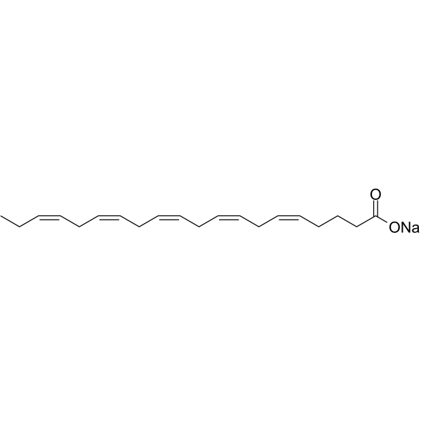
| SMILES | CC/C=C\C/C=C\C/C=C\C/C=C\C/C=C\CCCC(=O)[O-].[Na+] |
| InChi Key | RBZYGQJEMWGTOH-RSDXMDNYSA-M |
| InChi Code | InChI=1S/C20H30O2.Na/c1-2-3-4-5-6-7-8-9-10-11-12-13-14-15-16-17-18-19-20(21)22;/h3-4,6-7,9-10,12-13,15-16H,2,5,8,11,14,17-19H2,1H3,(H,21,22);/q;+1/p-1/b4-3-,7-6-,10-9-,13-12-,16-15-; |
| Chemical Name | sodium;(5Z,8Z,11Z,14Z,17Z)-icosa-5,8,11,14,17-pentaenoate |
| HS Tariff Code | 2934.99.9001 |
| Storage |
Powder-20°C 3 years 4°C 2 years In solvent -80°C 6 months -20°C 1 month |
| Shipping Condition | Room temperature (This product is stable at ambient temperature for a few days during ordinary shipping and time spent in Customs) |
Biological Activity
| Targets | Endogenous Metabolite |
| ln Vitro | In cells treated with sodium eicosapentaenoic acid (EPA; 100 µM; 24 h), the phosphorylated form of C/EBPβ was clearly visible, but it was hardly noticeable in normal and OA- or LA-treated U937 cells [1]. After 1 and 3 hours of conditioning, there was a considerable rise in H-Ras and N-Ras mRNA levels caused by sodium eicosapentaenoic acid (100 µM; 1, 3, 24 hours). Sodium eicosapentaenoate has no effect on the levels of K-Ras mRNA [1]. |
| ln Vivo |
Two hundred forty-one studies were identified, of which 28 met the above inclusion criteria and were therefore included in the subsequent meta-analysis. Using a random effects model, overall standardized mean depression scores were reduced in response to omega3 LC-PUFA supplementation as compared with placebo (standardized mean difference = -0.291, 95% CI = -0.463 to -0.120, z = -3.327, p = 0.001). However, significant heterogeneity and evidence of publication bias were present. Meta-regression studies showed a significant effect of higher levels of baseline depression and lower supplement DHAEPA ratio on therapeutic efficacy. Subgroup analyses showed significant effects for: (1) diagnostic category (bipolar disorder and major depression showing significant improvement with omega3 LC-PUFA supplementation versus mild-to-moderate depression, chronic fatigue and non-clinical populations not showing significant improvement); (2) therapeutic as opposed to preventive intervention; (3) adjunctive treatment as opposed to monotherapy; and (4) supplement type. Symptoms of depression were not significantly reduced in 3 studies using pure DHA (standardized mean difference 0.001, 95% CI -0.330 to 0.332, z = 0.004, p = 0.997) or in 4 studies using supplements containing greater than 50% DHA (standardized mean difference = 0.141, 95% CI = -0.195 to 0.477, z = 0.821, p = 0.417). In contrast, symptoms of depression were significantly reduced in 13 studies using supplements containing greater than 50% EPA (standardized mean difference = -0.446, 95% CI = -0.753 to -0.138, z = -2.843, p = 0.005) and in 8 studies using pure ethyl-EPA (standardized mean difference = -0.396, 95% CI = -0.650 to -0.141, z = -3.051, p = 0.002). However, further meta-regression studies showed significant inverse associations between efficacy and study methodological quality, study sample size, and duration, thus limiting the confidence of these findings. Conclusions: The current meta-analysis provides evidence that EPA may be more efficacious than DHA in treating depression. However, owing to the identified limitations of the included studies, larger, well-designed, randomized controlled trials of sufficient duration are needed to confirm these findings [4]. Myopia is a leading cause of visual impairment and blindness worldwide. However, a safe and accessible approach for myopia control and prevention is currently unavailable. Here, we investigated the therapeutic effect of dietary supplements of omega-3 polyunsaturated fatty acids (ω-3 PUFAs) on myopia progression in animal models and on decreases in choroidal blood perfusion (ChBP) caused by near work, a risk factor for myopia in young adults. We demonstrated that daily gavage of ω-3 PUFAs (300 mg docosahexaenoic acid [DHA] plus 60 mg eicosapentaenoic acid [EPA]) significantly attenuated the development of form deprivation myopia in guinea pigs and mice, as well as of lens-induced myopia in guinea pigs. Peribulbar injections of DHA also inhibited myopia progression in form-deprived guinea pigs. The suppression of myopia in guinea pigs was accompanied by inhibition of the "ChBP reduction-scleral hypoxia cascade." Additionally, treatment with DHA or EPA antagonized hypoxia-induced myofibroblast transdifferentiation in cultured human scleral fibroblasts. In human subjects, oral administration of ω-3 PUFAs partially alleviated the near-work-induced decreases in ChBP. Therefore, evidence from these animal and human studies suggests ω-3 PUFAs are potential and readily available candidates for myopia control [2]. |
| Cell Assay |
DNA isolation and quantitative DNA methylation analysis of C/EBPβ and H-Ras CpG islands [1] Genomic DNA from control U937 cells or U937 grown for 24 hours with 100 µM OA or 100 µM Eicosapentaenoic Acid/EPA was extracted using FlexiGene DNA Kit. EMBOSS and MethPrimer on-line software programs were used to identify potential CpG islands for C/EBPβ, N-Ras, and H-Ras genes. DNA methylation levels were quantified for human C/EBPβ (MePH25981-3A) and H-Ras (MePH14574-1A) using Methyl-Profiler qPCR Primer Assay. qRT-PCR program was performed as indicated in the manual instructions. Analysis of DNA methylation status of CpG islands was carried out using restriction enzyme digestion (DNA Methylation Enzyme kit MeA-03) followed by SYBR Green-based real time PCR detection as previously described. The relative amount of each DNA fraction (methylated and unmethylated) was calculated using ΔCt method. Bisulfite modification of genomic DNA and sequencing [1] Genomic DNA was obtained from U937 cells, control or grown for 24 h with 100 µM OA or 100 µM Eicosapentaenoic Acid/EPA, using FlexiGene DNA Kit. The bisulfite reaction to determine DNA methylation status was performed as previously described. The DNA fragments covering N-Ras CpG island (−29/+171) and H-Ras CpG island B (640/882) were amplified by PCR using the following primers. N-Ras: for, 5′-AAAGTTTTATTGATTTTTGAGATATTAGTA-3′; rev, 5′-TTTAAACAAATTTAAAACCACAACC-3′. H-Ras: for, 5′-AGTTTTTTGTGGTTGAAAGATGTT-3′; rev, 5′-ACACCCAAATTAAAAACTACTAAATC -3′. The PCR products were cloned into pCR2.1 TOPO and six clones randomly picked from each of two independent PCRs were sequenced using T7 primer at the Genechron-Ylichron Laboratory. Chromatin immunoprecipitation [1] ChIP assays were performed on U937 cells (about 106), control or grown with 100 µM OA or Eicosapentaenoic Acid/EPA for 24 hours, using the EZ-Chip kit. Cells were cross-linked and cell lysates sonicated until chromatin fragments became 200–1.000 bp in size. Mouse RNAPII 8WG16 monoclonal antibody MMS-126R or rabbit p53 antibody #928 were used for immunoprecipitation. Mouse or rabbit IgG were used as a negative control. After immunoprecipitation, recovered chromatin samples were subject to qRT-PCR with Brilliant SYBR Green qPCR Master Mix. In RNAPII assays the H-Ras sequences amplified were within i) exon 1 (1/+135), ii) intron 1 region C (+136/+639), iii) CpG island B (+640/+882), iv) intron 1 region D (+883/+1167), and v) exon 2 (+1168/+1331). The following primers were used. i) Exon 1: for, 5′-TGCCCTGCGCCCGCAACCCGAG-3′; rev, 5′-CGTTCACAGGCGCGACTGCC-3′. ii) Intron 1 region C: for, 5′-GTGAACGGTGAGTGCGGGCA-3′; rev, 5′-CGCGCCGCGCGTATTGCTGC-3′. iii) CpG island B: for, 5′-CCTGTTCTGGAGGACGGTAA-3′; rev, 5′-GTCGGCAGAAAGGCTAAAGG-3′. iv) Intron 1 region D: for, 5′-TCAGATGGCCCTGCCAGCAG-3′; rev, 5′-TCCTCCTACAGGGTCTCCTG-3′. v) Exon 2: for, 5′CAGGAGACCCTGTAGGAGGA-3′; rev, 5′-GGATCAGCTGGATGGTCAGC-3′. In p53 assays the sequence containing the p53 element of CpG island B was amplified using the following primers: for, 5′-CGCTCAGCAAATACTTGTCGG-3′; rev, 5′-TTACCGTCCTCCAGAACAGG-3′. Data were analyzed quantitatively according to the formula 2−Δ[C(IP)−C(input)]−2−Δ[C(control IgG)−C(input)]. |
| Animal Protocol | Six-week-old male C57BL/6J mice were used. After one week of acclimatization with free access to standard mouse chow (commercial diet, 17.14% of energy from fat, 5.05 g/100 g) and water, the mice were randomly divided into nine groups each containing six mice and fed ALA series diets (1, 2.5, 5, or 7.5 wt%), 5% ALA and EPA series diets (0.25, 0.5, 1 wt%), EPA diet (2 wt%), or the control diet (Ctl diet: depleted in ω-3 PUFA) for seven weeks. The diet ingredients were shown in ESI Tables S1 and S2.† All animals were maintained in barrier cages and fed with the appropriate special diet restricted to 10 g per mouse per day.[3] |
| References |
[1]. Eicosapentaenoic acid activates RAS/ERK/C/EBPβ pathway through H-Ras intron 1 CpG island demethylation in U937 leukemia cells. PLoS One. 2014 Jan 13;9(1):e85025. [2]. Dietary ω-3 polyunsaturated fatty acids are protective for myopia. Proc Natl Acad Sci U S A. 2021 Oct 26;118(43):e2104689118. [3]. Endogenous omega-3 long-chain fatty acid biosynthesis from alpha-linolenic acid is affected by substrate levels, gene expression, and product inhibition. RSC Adv., 2017, 7, 40946-40951. [4]. EPA but not DHA appears to be responsible for the efficacy of omega-3 long chain polyunsaturated fatty acid supplementation in depression: evidence from a meta-analysis of randomized controlled trials. J Am Coll Nutr, 2009. 28(5): p. 525-42. |
| Additional Infomation | Epigenetic alterations, including aberrant DNA methylation, contribute to tumor development and progression. Silencing of tumor suppressor genes may be ascribed to promoter DNA hypermethylation, a reversible phenomenon intensely investigated as potential therapeutic target. Previously, we demonstrated that eicosapentaenoic acid (EPA) exhibits a DNA demethylating action that promotes the re-expression of the tumor suppressor gene CCAAT/enhancer-binding protein δ (C/EBPδ). The C/EBPβ/C/EBPδ heterodimer formed appears essential for the monocyte differentiation commitment. The present study aims to evaluate the effect of EPA on RAS/extracellular signal regulated kinases (ERK1/2)/C/EBPβ pathway, known to be induced during the monocyte differentiation program. We found that EPA conditioning of U937 leukemia cells activated RAS/ERK/C/EBPβ pathway, increasing the C/EBPβ and ERK1/2 active phosphorylated forms. Transcriptional induction of the upstream activator H-Ras gene resulted in increased expression of H-Ras protein in the active pool of non raft membrane fraction. H-Ras gene analysis identified an hypermethylated CpG island in intron 1 that can affect the DNA-protein interaction modifying RNA polymerase II (RNAPII) activity. EPA treatment demethylated almost completely this CpG island, which was associated with an enrichment of active RNAPII. The increased binding of the H-Ras transcriptional regulator p53 to its consensus sequence within the intronic CpG island further confirmed the effect of EPA as demethylating agent. Our results provide the first evidence that an endogenous polyunsaturated fatty acid (PUFA) promotes a DNA demethylation process responsible for the activation of RAS/ERK/C/EBPβ pathway during the monocyte differentiation commitment. The new role of EPA as demethylating agent paves the way for studying PUFA action when aberrant DNA methylation is involved. [1] |
Solubility Data
| Solubility (In Vitro) | May dissolve in DMSO (in most cases), if not, try other solvents such as H2O, Ethanol, or DMF with a minute amount of products to avoid loss of samples |
| Solubility (In Vivo) |
Note: Listed below are some common formulations that may be used to formulate products with low water solubility (e.g. < 1 mg/mL), you may test these formulations using a minute amount of products to avoid loss of samples. Injection Formulations (e.g. IP/IV/IM/SC) Injection Formulation 1: DMSO : Tween 80: Saline = 10 : 5 : 85 (i.e. 100 μL DMSO stock solution → 50 μL Tween 80 → 850 μL Saline) *Preparation of saline: Dissolve 0.9 g of sodium chloride in 100 mL ddH ₂ O to obtain a clear solution. Injection Formulation 2: DMSO : PEG300 :Tween 80 : Saline = 10 : 40 : 5 : 45 (i.e. 100 μL DMSO → 400 μLPEG300 → 50 μL Tween 80 → 450 μL Saline) Injection Formulation 3: DMSO : Corn oil = 10 : 90 (i.e. 100 μL DMSO → 900 μL Corn oil) Example: Take the Injection Formulation 3 (DMSO : Corn oil = 10 : 90) as an example, if 1 mL of 2.5 mg/mL working solution is to be prepared, you can take 100 μL 25 mg/mL DMSO stock solution and add to 900 μL corn oil, mix well to obtain a clear or suspension solution (2.5 mg/mL, ready for use in animals). Injection Formulation 4: DMSO : 20% SBE-β-CD in saline = 10 : 90 [i.e. 100 μL DMSO → 900 μL (20% SBE-β-CD in saline)] *Preparation of 20% SBE-β-CD in Saline (4°C,1 week): Dissolve 2 g SBE-β-CD in 10 mL saline to obtain a clear solution. Injection Formulation 5: 2-Hydroxypropyl-β-cyclodextrin : Saline = 50 : 50 (i.e. 500 μL 2-Hydroxypropyl-β-cyclodextrin → 500 μL Saline) Injection Formulation 6: DMSO : PEG300 : castor oil : Saline = 5 : 10 : 20 : 65 (i.e. 50 μL DMSO → 100 μLPEG300 → 200 μL castor oil → 650 μL Saline) Injection Formulation 7: Ethanol : Cremophor : Saline = 10: 10 : 80 (i.e. 100 μL Ethanol → 100 μL Cremophor → 800 μL Saline) Injection Formulation 8: Dissolve in Cremophor/Ethanol (50 : 50), then diluted by Saline Injection Formulation 9: EtOH : Corn oil = 10 : 90 (i.e. 100 μL EtOH → 900 μL Corn oil) Injection Formulation 10: EtOH : PEG300:Tween 80 : Saline = 10 : 40 : 5 : 45 (i.e. 100 μL EtOH → 400 μLPEG300 → 50 μL Tween 80 → 450 μL Saline) Oral Formulations Oral Formulation 1: Suspend in 0.5% CMC Na (carboxymethylcellulose sodium) Oral Formulation 2: Suspend in 0.5% Carboxymethyl cellulose Example: Take the Oral Formulation 1 (Suspend in 0.5% CMC Na) as an example, if 100 mL of 2.5 mg/mL working solution is to be prepared, you can first prepare 0.5% CMC Na solution by measuring 0.5 g CMC Na and dissolve it in 100 mL ddH2O to obtain a clear solution; then add 250 mg of the product to 100 mL 0.5% CMC Na solution, to make the suspension solution (2.5 mg/mL, ready for use in animals). Oral Formulation 3: Dissolved in PEG400 Oral Formulation 4: Suspend in 0.2% Carboxymethyl cellulose Oral Formulation 5: Dissolve in 0.25% Tween 80 and 0.5% Carboxymethyl cellulose Oral Formulation 6: Mixing with food powders Note: Please be aware that the above formulations are for reference only. InvivoChem strongly recommends customers to read literature methods/protocols carefully before determining which formulation you should use for in vivo studies, as different compounds have different solubility properties and have to be formulated differently. (Please use freshly prepared in vivo formulations for optimal results.) |
| Preparing Stock Solutions | 1 mg | 5 mg | 10 mg | |
| 1 mM | 3.3063 mL | 16.5317 mL | 33.0633 mL | |
| 5 mM | 0.6613 mL | 3.3063 mL | 6.6127 mL | |
| 10 mM | 0.3306 mL | 1.6532 mL | 3.3063 mL |