EHT-1610 is a novel, potent and selective DYRK kinase inhibitor with IC50s of 0.22, 0.28 nM for DYRK1A and DYRK1B, respectively. DYRK1B, also called minibrain-related kinase (Mirk) is a protein that is highly expressed in quiescent cancer cells but expressed at very low levels in normal tissue, making it a potentially appealing cancer target.
Physicochemical Properties
| Molecular Formula | C18H14FN5O2S |
| Molecular Weight | 383.399465084076 |
| Exact Mass | 383.09 |
| Elemental Analysis | C, 56.39; H, 3.68; F, 4.96; N, 18.27; O, 8.35; S, 8.36 |
| CAS # | 1425945-60-3 |
| Related CAS # | 1425945-60-3 |
| PubChem CID | 71529602 |
| Appearance | Light yellow to yellow solid powder |
| LogP | 4.2 |
| Hydrogen Bond Donor Count | 2 |
| Hydrogen Bond Acceptor Count | 9 |
| Rotatable Bond Count | 5 |
| Heavy Atom Count | 27 |
| Complexity | 544 |
| Defined Atom Stereocenter Count | 0 |
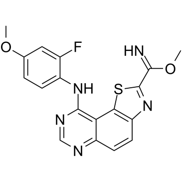
| SMILES | S1C(C(=N)OC)=NC2=CC=C3C(C(=NC=N3)NC3C=CC(=CC=3F)OC)=C12 |
| InChi Key | RYBNARZBIXTFJS-UHFFFAOYSA-N |
| InChi Code | InChI=1S/C18H14FN5O2S/c1-25-9-3-4-11(10(19)7-9)23-17-14-12(21-8-22-17)5-6-13-15(14)27-18(24-13)16(20)26-2/h3-8,20H,1-2H3,(H,21,22,23) |
| Chemical Name | methyl 9-(2-fluoro-4-methoxyanilino)-[1,3]thiazolo[5,4-f]quinazoline-2-carboximidate |
| Synonyms | EHT-1610; EHT1610; EHT 1610; EHT 5372; 1425945-60-3; methyl 9-((2-fluoro-4-methoxyphenyl)amino)thiazolo[5,4-f]quinazoline-2-carbimidate; Methyl 9-[(2-Fluoro-4-methoxyphenyl)amino]thiazolo[5,4-f]quinazoline-2-carbimidate; methyl 9-(2-fluoro-4-methoxyanilino)-[1,3]thiazolo[5,4-f]quinazoline-2-carboximidate; MFCD31922711; EHT-5372; EHT5372 |
| HS Tariff Code | 2934.99.03.00 |
| Storage |
Powder-20°C 3 years 4°C 2 years In solvent -80°C 6 months -20°C 1 month Note: (1). This product requires protection from light (avoid light exposure) during transportation and storage.(2). Please store this product in a sealed and protected environment (e.g. under nitrogen), avoid exposure to moisture.(3). This product is not stable in solution, please use freshly prepared working solution for optimal results. |
| Shipping Condition | Room temperature (This product is stable at ambient temperature for a few days during ordinary shipping and time spent in Customs) |
Biological Activity
| Targets |
DYRK1B; DYRK1A; EHT-1610 is a potent and selective inhibitor of dual-specificity tyrosine phosphorylation-regulated kinases (DYRKs), with IC₅₀ values of 0.36 nM for DYRK1A and 0.59 nM for DYRK1B [1][4]. It exhibits minimal activity against CLK kinases (CLK1 IC₅₀ > 10 μM) and GSK-3β (IC₅₀ = 221 nM), confirming high selectivity for DYRK family kinases [1][4].
EHT-1610 is a potent and selective inhibitor of dual-specificity tyrosine phosphorylation-regulated kinase 1A (DYRK1A), with an IC₅₀ of 0.36 nM against DYRK1A [3]. |
| ln Vitro |
Arabinoblast batches can be assembled by primary ALL cells induced by EHT 1610 [2]. In primary human pediatric cells and B and T cell lines, EHT 1610 dose-sensitively induces Frankfurt [2]. B cells are specifically killed when 1610 (72 hours) suppresses DYRK1A, which causes the loss of DYRK1A-mediated FOXO1 and STAT3 signaling [3]. EHT 1610 (2.5-10 μM; 4-5 hours) inhibits the phosphorylation of cyclin D3, STAT3, and FOXO1, consequently controlling DNA damage, mitochondrial ROS, and late cell cycle progression, in that order [3].
EHT-1610 (1 μM) significantly inhibited proliferation of primary B-cell acute lymphoblastic leukemia (B-ALL) cells and cell lines (e.g., SEM, RS4;11) by 60–80% after 72 hours (MTT assay). It induced apoptosis (Annexin V⁺ cells increased by 40–50%) and reduced phosphorylation of FOXO1 (Ser329) and STAT3 (Tyr705) by >70% (Western blot) [3]. |
| ln Vivo |
As EHT 1610 and AS1842856 are well tolerated and have on-target effects in vivo, we transplanted NOD.Cg-PrkdcscidIl2rgtm1Wjl/SzJ (NSG) mice with human B-ALL cells to determine whether they could be treated effectively. We transplanted MHH-CALL-4 cells into 1 cohort of NSG mice and treated them with 40 mg/kg/day EHT 1610, and then transplanted Nalm-6 cells into a separate cohort of NSG mice followed by treatment with 10 mg/kg/day AS1842856. Note that the 2 different lines were used on the basis of our determined sensitivities (Supplemental Table 1). In both cases, the mice were treated for 2 weeks following detection of 1% or more human CD45+CD19+ cells in the peripheral blood. Both cohorts displayed highly significant survival advantages (Figure 6, A and B). We also evaluated the degree of antileukemia activity in 2 PDX models of HSA21 aneuploidy (22). First, we transplanted NSG mice with luciferase-expressing leukemic blasts from a patient with DS-ALL (sample DS-ALL-03) to follow leukemia progression by noninvasive in vivo imaging. When the recipient mice reached 107 photons/second (p/s) in total flux, we treated the animals for 3 weeks with 40 mg/kg/day EHT 1610 or 30 mg/kg/day AS1842856. EHT 1610 treatment alone conferred a 20% decrease in leukemic burden compared with the control group versus a more than 100-fold decrease in the AS1842856 group as determined by bioluminescence (Figure 6C). These reductions in leukemic burden correlated with significant survival advantages compared with vehicle treatment, with AS1842856 providing a greater survival benefit than EHT 1610 (Figure 6D). Last, we also evaluated the efficacy of EHT 1610 and AS1842856 in a highly aggressive luciferase-expressing PDX model of HeH-ALL (sample HeH-ALL-09). After detection of 107 p/s in total flux, we treated transplanted mice for 3 weeks with 40 mg/kg/day EHT 1610 or 30 mg/kg/day AS1842856. EHT 1610 treatment in this aggressive model reduced leukemic burden by approximately 8% and conferred a modest survival advantage (Figure 6, E and F); by contrast, AS1842856 treatment reduced the leukemic burden by approximately 10-fold at endpoint analysis and provided an even stronger survival benefit. Thus, these data demonstrate that DYRK1A and FOXO1 are both efficacious targets in B-ALL and may be of particular therapeutic value in models of disease with chromosome 21 aneuploidy. [3] In a B-ALL xenograft mouse model (NOD/SCID mice injected with SEM cells), intraperitoneal administration of EHT-1610 (10 mg/kg daily for 21 days) reduced tumor volume by 65% vs. vehicle control (p<0.001) and prolonged survival (median survival: 45 vs. 32 days). Tumor tissues showed reduced p-FOXO1 and p-STAT3 levels (immunohistochemistry) [3]. |
| Enzyme Assay |
Kinase inhibition assay: Recombinant DYRK1A, DYRK1B, and other kinases were incubated with EHT-1610 (0.01–100 nM) in kinase buffer containing ATP and substrate. Reactions were stopped by adding EDTA, and phosphorylation levels were quantified using a time-resolved fluorescence resonance energy transfer (TR-FRET) assay. Dose-response curves were plotted to calculate IC₅₀ values [1]. Binding mode analysis: Co-crystallization of EHT-1610 with DYRK2 was performed. The compound was soaked into DYRK2 crystals, and structures were resolved by X-ray diffraction (1.8 Å resolution), revealing a noncanonical binding mode involving hydrogen bonding with Glu239 and hydrophobic interactions with Leu241 and Val307 [1]. Kinase inhibition assay: Recombinant human DYRK1A protein was incubated with EHT-1610 (0.01–100 nM) and ATP in kinase buffer. Reactions were terminated by adding EDTA, and phosphorylation levels of the substrate (e.g., FOXO1-derived peptide) were quantified using a time-resolved fluorescence resonance energy transfer (TR-FRET) system. Dose-response curves were plotted to calculate IC₅₀ [3]. |
| Cell Assay |
Western Blot analysis[3] Cell Types: MHH-CALL-4 cells Tested Concentrations: 0, 2.5, 5, 10 μM Incubation Duration: 4, 5 hrs (hours) Experimental Results: p-cyclin D3 (Thr283) and p-FOXO1 protein levels were dose-dependent diminished sexual behavior. Tau phosphorylation assay: HEK293 cells were treated with EHT-1610 (0.1–10 μM) for 24 hours. Lysates were analyzed by Western blot using anti-pS396-Tau and total Tau antibodies. pS396-Tau levels decreased dose-dependently (IC₅₀ = 1.7 μM), while cell viability remained >87% (MTT assay) [1][3]. Anti-leukemic activity: Primary B-cell acute lymphoblastic leukemia (B-ALL) cells were treated with EHT-1610 (0.1–1 μM) for 48 hours. Apoptosis was measured via Annexin V/PI staining, showing 40–60% cell death at 1 μM. STAT3 phosphorylation (Tyr705) was suppressed by >50% (Western blot) [3]. Anti-proliferation assay: Primary B-ALL cells or cell lines were treated with EHT-1610 (0.1–10 μM) for 72 hours. Cell viability was measured by MTT assay (absorbance at 570 nm). Apoptosis assay: Cells treated with EHT-1610 (1 μM) for 48 hours were stained with Annexin V-FITC and propidium iodide (PI), followed by flow cytometry analysis. Western blot: Cells lysed after EHT-1610 treatment (1 μM, 6 hours) were subjected to SDS-PAGE, transferred to PVDF membranes, and probed with antibodies against p-FOXO1 (Ser329), p-STAT3 (Tyr705), and total proteins [3]. |
| Animal Protocol |
Animal/Disease Models: Mouse B-ALL xenograft model (12-14 weeks old) [3] Doses: 20 mg/kg Route of Administration: intraperitoneal (ip) injection; twice a day for 5 days, 2 days off; 3-week Experimental Results: The leukemia burden is diminished by about 8% and has a certain survival advantage. Animal studies were performed to determine the genetic requirement of Dyrk1a in murine models of B-ALL, the on-target effects of EHT 1610 and AS1842856, and the efficacy of EHT 1610 and AS1842856 in treating mouse models of B-ALL. The sample size of the animal experiments, the specific transplantation protocols, and the dosing are described in the figure legends and in the appropriate Methods sections, respectively. For all in vivo experiments, mice were randomly assigned to transplant groups (genetic models) or treatment groups (inhibitor studies). All experiments were performed in an unblinded manner. [3] Xenograft model: NOD/SCID mice were subcutaneously injected with SEM B-ALL cells (5×10⁶ cells/mouse). When tumors reached 100 mm³, mice received daily intraperitoneal injections of EHT-1610 (10 mg/kg dissolved in 10% DMSO + 90% corn oil) or vehicle for 21 days. Tumor size was measured every 3 days, and survival was monitored [3]. |
| References |
[1]. An Unusual Binding Model of the Methyl 9-Anilinothiazolo[5,4-f] quinazoline-2-carbimidates (EHT 1610 and EHT 5372) Confers High Selectivity for Dual-Specificity Tyrosine Phosphorylation-Regulated Kinases. J Med Chem. 2016 Nov 23;59(22):10315-10321. [2]. The Chromosome 21 Kinase DYRK1A Controls Cell Cycle Exit and Survival During Lymphoid Development and Is a Novel Therapeutic Target In Acute Lymphoblastic Leukemia[C]// Ash Meeting & Exposition. 2013. l. https://ashpublications.org/blood/article/122/21/814/70866/The-Chromosome-21-Kinase-DYRK1A-Controls-Cell [3]. DYRK1A regulates B cell acute lymphoblastic leukemia through phosphorylation of FOXO1 and STAT3. J Clin Invest. 2021 Jan 4;131(1):e135937. [4]. Design and synthesis of thiazolo[5,4-f]quinazolines as DYRK1A inhibitors, part II. Molecules. 2014 Sep 26;19(10):15411-39. |
| Additional Infomation |
EHT-1610 belongs to the methyl 9-anilinothiazolo[5,4-f]quinazoline-2-carbimidate class. Its unique binding mode exploits a hydrophobic pocket in DYRKs, conferring >100-fold selectivity over CLK1/2 [1].
Mechanism: Inhibits DYRK1A-mediated phosphorylation of FOXO1 and STAT3, inducing cell cycle arrest and apoptosis in B-ALL [3].
DYRK1A is a serine/threonine kinase encoded on human chromosome 21 (HSA21) that has been implicated in several pathologies of Down syndrome (DS), including cognitive deficits and Alzheimer’s disease. Although children with DS are predisposed to developing leukemia, especially B cell acute lymphoblastic leukemia (B-ALL), the HSA21 genes that contribute to malignancies remain largely undefined. Here, we report that DYRK1A is overexpressed and required for B-ALL. Genetic and pharmacologic inhibition of DYRK1A decreased leukemic cell expansion and suppressed B-ALL development in vitro and in vivo. Furthermore, we found that FOXO1 and STAT3, transcription factors that are indispensable for B cell development, are critical substrates of DYRK1A. Loss of DYRK1A-mediated FOXO1 and STAT3 signaling disrupted DNA damage and ROS regulation, respectively, leading to preferential cell death in leukemic B cells. Thus, we reveal a DYRK1A/FOXO1/STAT3 axis that facilitates the development and maintenance of B-ALL. [3] |
Solubility Data
| Solubility (In Vitro) | DMSO: ~5 mg/mL (~13.0 mM) |
| Solubility (In Vivo) |
Note: Listed below are some common formulations that may be used to formulate products with low water solubility (e.g. < 1 mg/mL), you may test these formulations using a minute amount of products to avoid loss of samples. Injection Formulations (e.g. IP/IV/IM/SC) Injection Formulation 1: DMSO : Tween 80: Saline = 10 : 5 : 85 (i.e. 100 μL DMSO stock solution → 50 μL Tween 80 → 850 μL Saline) *Preparation of saline: Dissolve 0.9 g of sodium chloride in 100 mL ddH ₂ O to obtain a clear solution. Injection Formulation 2: DMSO : PEG300 :Tween 80 : Saline = 10 : 40 : 5 : 45 (i.e. 100 μL DMSO → 400 μLPEG300 → 50 μL Tween 80 → 450 μL Saline) Injection Formulation 3: DMSO : Corn oil = 10 : 90 (i.e. 100 μL DMSO → 900 μL Corn oil) Example: Take the Injection Formulation 3 (DMSO : Corn oil = 10 : 90) as an example, if 1 mL of 2.5 mg/mL working solution is to be prepared, you can take 100 μL 25 mg/mL DMSO stock solution and add to 900 μL corn oil, mix well to obtain a clear or suspension solution (2.5 mg/mL, ready for use in animals). Injection Formulation 4: DMSO : 20% SBE-β-CD in saline = 10 : 90 [i.e. 100 μL DMSO → 900 μL (20% SBE-β-CD in saline)] *Preparation of 20% SBE-β-CD in Saline (4°C,1 week): Dissolve 2 g SBE-β-CD in 10 mL saline to obtain a clear solution. Injection Formulation 5: 2-Hydroxypropyl-β-cyclodextrin : Saline = 50 : 50 (i.e. 500 μL 2-Hydroxypropyl-β-cyclodextrin → 500 μL Saline) Injection Formulation 6: DMSO : PEG300 : castor oil : Saline = 5 : 10 : 20 : 65 (i.e. 50 μL DMSO → 100 μLPEG300 → 200 μL castor oil → 650 μL Saline) Injection Formulation 7: Ethanol : Cremophor : Saline = 10: 10 : 80 (i.e. 100 μL Ethanol → 100 μL Cremophor → 800 μL Saline) Injection Formulation 8: Dissolve in Cremophor/Ethanol (50 : 50), then diluted by Saline Injection Formulation 9: EtOH : Corn oil = 10 : 90 (i.e. 100 μL EtOH → 900 μL Corn oil) Injection Formulation 10: EtOH : PEG300:Tween 80 : Saline = 10 : 40 : 5 : 45 (i.e. 100 μL EtOH → 400 μLPEG300 → 50 μL Tween 80 → 450 μL Saline) Oral Formulations Oral Formulation 1: Suspend in 0.5% CMC Na (carboxymethylcellulose sodium) Oral Formulation 2: Suspend in 0.5% Carboxymethyl cellulose Example: Take the Oral Formulation 1 (Suspend in 0.5% CMC Na) as an example, if 100 mL of 2.5 mg/mL working solution is to be prepared, you can first prepare 0.5% CMC Na solution by measuring 0.5 g CMC Na and dissolve it in 100 mL ddH2O to obtain a clear solution; then add 250 mg of the product to 100 mL 0.5% CMC Na solution, to make the suspension solution (2.5 mg/mL, ready for use in animals). Oral Formulation 3: Dissolved in PEG400 Oral Formulation 4: Suspend in 0.2% Carboxymethyl cellulose Oral Formulation 5: Dissolve in 0.25% Tween 80 and 0.5% Carboxymethyl cellulose Oral Formulation 6: Mixing with food powders Note: Please be aware that the above formulations are for reference only. InvivoChem strongly recommends customers to read literature methods/protocols carefully before determining which formulation you should use for in vivo studies, as different compounds have different solubility properties and have to be formulated differently. (Please use freshly prepared in vivo formulations for optimal results.) |
| Preparing Stock Solutions | 1 mg | 5 mg | 10 mg | |
| 1 mM | 2.6082 mL | 13.0412 mL | 26.0824 mL | |
| 5 mM | 0.5216 mL | 2.6082 mL | 5.2165 mL | |
| 10 mM | 0.2608 mL | 1.3041 mL | 2.6082 mL |