Dp44mT is a novel and potent iron chelator with DNA-damaging activity mediated by top2a inhibition and with selective antitumor activity. In the human breast cancer cell line MDA-MB-231, Dp44mT specifically inhibits topoisomerase IIα, with a GI50 value of ~100 nmol/L. In comparison to wild type cells, Nalm-6 leukemic top2α+/- cells expressed ~57% more top2α enzyme in control experiments. Comparatively to top2α+/+ cells, treated with Dp44mT at 100 nmol/L, top2α+/- cells displayed partial resistance to the drug's cytotoxic effects. The top2α+/+ cells displayed 31.7% sub-G1 containing cells following exposure to Dp44mT at 100 nmol/L, compared to the top2α+/- cells' 9.4%.
Physicochemical Properties
| Molecular Formula | C14H15N5S | |
| Molecular Weight | 285.37 | |
| Exact Mass | 285.104 | |
| Elemental Analysis | C, 58.93; H, 5.30; N, 24.54; S, 11.23 | |
| CAS # | 152095-12-0 | |
| Related CAS # |
|
|
| PubChem CID | 10334137 | |
| Appearance | Light yellow to yellow solid powder | |
| Density | 1.2±0.1 g/cm3 | |
| Boiling Point | 438.4±43.0 °C at 760 mmHg | |
| Flash Point | 218.9±28.2 °C | |
| Vapour Pressure | 0.0±1.1 mmHg at 25°C | |
| Index of Refraction | 1.635 | |
| LogP | 1.21 | |
| Hydrogen Bond Donor Count | 1 | |
| Hydrogen Bond Acceptor Count | 4 | |
| Rotatable Bond Count | 3 | |
| Heavy Atom Count | 20 | |
| Complexity | 336 | |
| Defined Atom Stereocenter Count | 0 | |
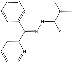
| SMILES | S=C(N/N=C(C1=NC=CC=C1)\C2=NC=CC=C2)N(C)C |
|
| InChi Key | XOBIGRNRXCAMJQ-UHFFFAOYSA-N | |
| InChi Code | InChI=1S/C24H20N4O4S/c1-30-24-26-23(16-7-12-20-21(13-16)32-15-31-20)28(27-24)18-10-8-17(9-11-18)25-22(29)14-33-19-5-3-2-4-6-19/h2-13H,14-15H2,1H3,(H,25,29) | |
| Chemical Name | 3-(dipyridin-2-ylmethylideneamino)-1,1-dimethylthiourea | |
| Synonyms |
|
|
| HS Tariff Code | 2934.99.9001 | |
| Storage |
Powder-20°C 3 years 4°C 2 years In solvent -80°C 6 months -20°C 1 month |
|
| Shipping Condition | Room temperature (This product is stable at ambient temperature for a few days during ordinary shipping and time spent in Customs) |
Biological Activity
| Targets | Iron chelator | ||
| ln Vitro | Dp44mT shows pronounced antiproliferative effects in SK-N-MC, SK-Mel-28, and MCF-7 cells with IC50 of 30 nM, 60 nM, and 60 nM, respectively, while no effects on normal MRC-5 fibroblasts. In SK-N-MC neuroepithelioma and M109 cells, Dp44mT reduces the amount of Fe that cells take up from Fe-Tf and triggers cell apoptosis. [1] When topoisomerase topo2 is specifically targeted by Dp44mT in MDA-MB-231 cells, DNA damage results. [2] By stealing control of lysosomal P-glycoprotein (Pgp), Dp44mT, a Pgp substrate, also defeats multidrug resistance. [3] | ||
| ln Vivo |
Dp44mT (0.4 mg/kg, i.v.) inhibits tumor growth in CD2F1 mice carrying M109 tumors in a dose-dependent manner. [1] Strikingly, Dp44mT significantly (P <.01) decreased tumor weight in mice to 47% of the weight in the control after only 5 days, whereas there was no marked change in animal weight or hematologic indices. Terminal deoxyribonucleotidyl transferase (TdT)-mediated dUTP nick end-labeling (TUNEL) staining demonstrated apoptosis in tumors taken from mice treated with Dp44mT.[1] |
||
| Enzyme Assay | For DNA topoisomerase II assays, the 5' ends of single stranded oligonucleotides or the 161-bp fragment from pBluescript SK(-) phagemid DNA are labeled with [32P]ATP and T4 polynucleotide kinase. To remove the unincorporated label, labeling mixtures are then centrifuged through Mini Quick Spin DNA columns (for pSK fragments) or Oligo columns (for oligonucleotides). The reaction mixture is heated to 95 °C and overnight cooled to room temperature in 10 mM Tris-HCl (pH 7.8), 100 mM NaCl, and 1 mM EDTA before being annealed to the complementary strand of the oligonucleotides. DNA substrates (10 pmol/reaction) are incubated with 500 ng of top2a or top2h in 10 L of reaction buffer at 25°C for the indicated times in the presence or absence of Dp44mT. SDS (final concentration of 0.5%) is added to stop reactions. Samples are separated on polyacrylamide denaturing gels at 16% (for pSK DNA) or 20% (for oligonucleotides) denaturing concentrations (7 M urea). A PhosphorImager[1] is used for imaging and quantitation. | ||
| Cell Assay |
Di-2-pyridylketone-4,4,-dimethyl-3-thiosemicarbazone (Dp44mT) is being developed as an iron chelator with selective anticancer activity. We investigated the mechanism whereby Dp44mT kills breast cancer cells, both as a single agent and in combination with doxorubicin. Dp44mT alone induced selective cell killing in the breast cancer cell line MDA-MB-231 when compared with healthy mammary epithelial cells (MCF-12A). It induces G(1) cell cycle arrest and reduces cancer cell clonogenic growth at nanomolar concentrations. Dp44mT, but not the iron chelator desferal, induces DNA double-strand breaks quantified as S139 phosphorylated histone foci (gamma-H2AX) and Comet tails induced in MDA-MB-231 cells. Doxorubicin-induced cytotoxicity and DNA damage were both enhanced significantly in the presence of low concentrations of Dp44mT. The chelator caused selective poisoning of DNA topoisomerase IIalpha (top2alpha) as measured by an in vitro DNA cleavage assay and cellular topoisomerase-DNA complex formation. Heterozygous Nalm-6 top2alpha knockout cells (top2alpha(+/-)) were partially resistant to Dp44mT-induced cytotoxicity compared with isogenic top2alpha(+/+) or top2beta(-/-) cells. Specificity for top2alpha was confirmed using top2alpha and top2beta small interfering RNA knockdown in HeLa cells. The results show that Dp44mT is cytotoxic to breast cancer cells, at least in part, due to selective inhibition of top2alpha. Thus, Dp44mT may serve as a mechanistically unique treatment for cancer due to its dual ability to chelate iron and inhibit top2alpha activity.[2] In this study, researchers investigated how the novel anti-tumor agent di-2-pyridylketone 4,4-dimethyl-3-thiosemicarbazone (Dp44mT) overcomes MDR. Four different cell types were utilized to evaluate the effect of Pgp-potentiated lysosomal targeting of drugs to overcome MDR. To assess the mechanism of how Dp44mT overcomes drug resistance, cellular studies utilized Pgp inhibitors, Pgp silencing, lysosomotropic agents, proliferation assays, immunoblotting, a Pgp-ATPase activity assay, radiolabeled drug uptake/efflux, a rhodamine 123 retention assay, lysosomal membrane permeability assessment, and DCF (2',7'-dichlorofluorescin) redox studies. Anti-tumor activity and selectivity of Dp44mT in Pgp-expressing, MDR cells versus drug-sensitive cells were studied using a BALB/c nu/nu xenograft mouse model. We demonstrate that Dp44mT is transported by the lysosomal Pgp drug pump, causing lysosomal targeting of Dp44mT and resulting in enhanced cytotoxicity in MDR cells. Lysosomal Pgp and pH were shown to be crucial for increasing Dp44mT-mediated lysosomal damage and subsequent cytotoxicity in drug-resistant cells, with Dp44mT being demonstrated to be a Pgp substrate. Indeed, Pgp-dependent lysosomal damage and cytotoxicity of Dp44mT were abrogated by Pgp inhibitors, Pgp silencing, or increasing lysosomal pH using lysosomotropic bases. In vivo, Dp44mT potently targeted chemotherapy-resistant human Pgp-expressing xenografted tumors relative to non-Pgp-expressing tumors in mice. This study highlights a novel Pgp hijacking strategy of the unique dipyridylthiosemicarbazone series of thiosemicarbazones that overcome MDR via utilization of lysosomal Pgp transport activity.[3] DFO, 311, 3-aminopyridine-2-carboxaldehyde thiosemicarbazone, doxorubicin, and the DpT series of chelators (0-25 μM) are incubated with and without the cells for 72 hours at 37°C. Using the MTT assay, the chelators' impact on cell proliferation is investigated. |
||
| Animal Protocol |
|
||
| References |
[1]. Blood . 2004 Sep 1;104(5):1450-8. [2]. Cancer Res . 2009 Feb 1;69(3):948-57. [3]. J Biol Chem . 2015 Apr 10;290(15):9588-603. |
||
| Additional Infomation |
Aroylhydrazone and thiosemicarbazone iron (Fe) chelators have potent antitumor activity. The aim of the current study was to examine the antitumor effects and mechanisms of action of a novel series of Fe chelators, the di-2-pyridyl thiosemicarbazones. Of 7 new chelators synthesized, 4 showed pronounced antiproliferative effects. The most active chelator was Dp44mT, which had marked and selective antitumor activity-for example, an IC(50) of 0.03 microM in neuroepithelioma cells compared with more than 25 microM in mortal fibroblasts. Indeed, this antiproliferative activity was the greatest yet observed for an Fe chelator. Efficacy was greater than it was for the cytotoxic ligand 311 and comparable to that of the antitumor agent doxorubicin. Strikingly, Dp44mT significantly (P <.01) decreased tumor weight in mice to 47% of the weight in the control after only 5 days, whereas there was no marked change in animal weight or hematologic indices. Terminal deoxyribonucleotidyl transferase (TdT)-mediated dUTP nick end-labeling (TUNEL) staining demonstrated apoptosis in tumors taken from mice treated with Dp44mT. This chelator caused a marked increase of caspase-3 activity in murine Madison-109 (M109) cells. Caspase activation was at least partially mediated by the release of mitochondrial holo-cytochrome c (h-cytc) after incubation with Dp44mT. In conclusion, Dp44mT is a novel, highly effective antitumor agent in vitro and in vivo that induces apoptosis.[1] Our investigation demonstrated that novel DpT analogs, in particular di-2-pyridylketone-4,4,-dimethyl-3-thiosemicarbazone (Dp44mT; Figure 1), showed selective antitumor activity. In addition, Dp44mT strikingly decreased tumor growth in mice while not markedly affecting animal weight or a range of hematologic indices. This chelator also induced tumor cell apoptosis that was at least partially mediated through the release of mitochondrial h-cytc into the cytosol and the activation of caspase-3, -8, and -9. The release of h-cytc could be mediated by the imbalance of Bcl-2 and Bax expression induced by incubation of cells with Dp44mT.[1] |
Solubility Data
| Solubility (In Vitro) |
|
|||
| Solubility (In Vivo) |
Solubility in Formulation 1: ≥ 2.5 mg/mL (8.76 mM) (saturation unknown) in 10% DMSO + 40% PEG300 + 5% Tween80 + 45% Saline (add these co-solvents sequentially from left to right, and one by one), clear solution. For example, if 1 mL of working solution is to be prepared, you can add 100 μL of 25.0 mg/mL clear DMSO stock solution to 400 μL PEG300 and mix evenly; then add 50 μL Tween-80 to the above solution and mix evenly; then add 450 μL normal saline to adjust the volume to 1 mL. Preparation of saline: Dissolve 0.9 g of sodium chloride in 100 mL ddH₂ O to obtain a clear solution. Solubility in Formulation 2: ≥ 2.5 mg/mL (8.76 mM) (saturation unknown) in 10% DMSO + 90% (20% SBE-β-CD in Saline) (add these co-solvents sequentially from left to right, and one by one), clear solution. For example, if 1 mL of working solution is to be prepared, you can add 100 μL of 25.0 mg/mL clear DMSO stock solution to 900 μL of 20% SBE-β-CD physiological saline solution and mix evenly. Preparation of 20% SBE-β-CD in Saline (4°C,1 week): Dissolve 2 g SBE-β-CD in 10 mL saline to obtain a clear solution. Solubility in Formulation 3: Propylene glycol : 1 mg/mL (Please use freshly prepared in vivo formulations for optimal results.) |
| Preparing Stock Solutions | 1 mg | 5 mg | 10 mg | |
| 1 mM | 3.5042 mL | 17.5211 mL | 35.0422 mL | |
| 5 mM | 0.7008 mL | 3.5042 mL | 7.0084 mL | |
| 10 mM | 0.3504 mL | 1.7521 mL | 3.5042 mL |