Physicochemical Properties
| Molecular Formula | C29H39NO5 |
| Molecular Weight | 481.62 |
| Exact Mass | 481.283 |
| CAS # | 39156-67-7 |
| PubChem CID | 6916220 |
| Appearance | White to off-white solid powder |
| Density | 1.194g/cm3 |
| Boiling Point | 727.474ºC at 760 mmHg |
| Melting Point | 203-205ºC |
| Flash Point | 393.762ºC |
| Index of Refraction | 1.584 |
| LogP | 4.044 |
| Hydrogen Bond Donor Count | 3 |
| Hydrogen Bond Acceptor Count | 5 |
| Rotatable Bond Count | 2 |
| Heavy Atom Count | 35 |
| Complexity | 818 |
| Defined Atom Stereocenter Count | 7 |
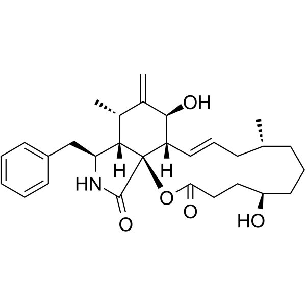
| SMILES | C[C@@H]1CCC[C@@H](O)CCC(O[C@]23[C@H]([C@H](O)C([C@H]([C@H]2[C@@H](NC3=O)CC4=CC=CC=C4)C)=C)C=CC1)=O |
| InChi Key | WIULKAASLBZREV-VXAQYHBESA-N |
| InChi Code | InChI=1S/C29H39NO5/c1-18-9-7-13-22(31)15-16-25(32)35-29-23(14-8-10-18)27(33)20(3)19(2)26(29)24(30-28(29)34)17-21-11-5-4-6-12-21/h4-6,8,11-12,14,18-19,22-24,26-27,31,33H,3,7,9-10,13,15-17H2,1-2H3,(H,30,34)/b14-8+/t18-,19+,22+,23-,24-,26?,27+,29+/m0/s1 |
| Chemical Name | (1S,6R,10S,12E,14S,15S,17S,19S)-19-benzyl-6,15-dihydroxy-10,17-dimethyl-16-methylidene-2-oxa-20-azatricyclo[12.7.0.01,18]henicos-12-ene-3,21-dione |
| Synonyms | dihydrocytochalasin B; 39156-67-7; Cytochalasin H2B; 21,22-Dihydrophomin; 21,22-Dihydrochalasin B; 21,22-Dihydrocytochalasan B; L9RT18CY85; MFCD12910542; |
| HS Tariff Code | 2934.99.9001 |
| Storage |
Powder-20°C 3 years 4°C 2 years In solvent -80°C 6 months -20°C 1 month |
| Shipping Condition | Room temperature (This product is stable at ambient temperature for a few days during ordinary shipping and time spent in Customs) |
Biological Activity
| Targets | Cytokinesis |
| ln Vitro | Dihydrocytochalasin B (H2CB) does not inhibit sugar uptake in BALB/c 3T3 cells. Excess H2CB does not affect inhibition of sugar uptake by cytochalasin B (CB), indicating that it does not compete with CB for binding to high-affinity sites. As in the case of CB, H2CB inhibits cytokinesis and changes the morphology of the cells. These results demonstrate that the effects of CB on sugar transport and on cell motility and morphology involve separate and independent sites. Comparison of the effects of H2CB, CB, and cytochalasin D (CD) indicates that treatment of cells with any one of the compounds results in the same series of morphological changes; the cells undergo zeiosis and elongation at 2-4 microM CB and become arborized and rounded up at 10-50 microM CB. H2CB is slightly less potent than CB, whereas CD is five to eight times more potent than CB in causing a given state of morphological change. These results indicate that the cytochalasin-induced changes in cell morphology are mediated by a specific site(s) which can distinguish the subtle differences in the structures of the three compounds. Competitive binding studies indicate that excess H2CB displaces essentially all of the high-affinity bound [3H]CB, but, at less than 5 x 10(-5) M H2CB is not so efficient as unlabeled CB in the displacement reaction. In contrast, excess CD displaces up to 40% of the bound [3H]CB. These results suggest that three different classes of high-affinity CB binding sites exist in 3T3 cells: sites related to sugar transport, sites related to cell motility and morphology, and sites with undetermined function[1]. |
| ln Vivo | In vivo calcium absorption was studied in normal and rachitic chicks. Cytochalasin B (CB) at a concentration of 25 microgram/ml added to the medium inside the duodenal lumen inhibited calcium absorption (20 min) from 82.5 +/- 1.9% of calcium absorbed in the controls to 59.2 +/- 3% in normal and from 70.0 +/- 2.3% to 47.0 +/- 2.1% in rachitic chicks. In vitro studies by everted ileal sacs of young rabbits also showed an inhibition of active transport of calcium due to CB. Whereas in the controls the ratio of 45Ca concentrations in serosal and mucosal media (60 min) was 7.2 +/- 0.32, the ratios were 5.24 +/- 0.52; 4.40 +/- 0.36; 3.40 +/- 0.42; 5.77 +/- 0.52; 1.38 +/- 0.08; and 1.06 +/- 0.02 in the presence of CB at concentrations of 5, 10 and 25 microgram/ml; colchicine 10(-4)M, Na citrate 0.02M, and heat-devitalized conditions, respectively. 45Ca concentration in the mucosal scrapings was also affected. It showed an increase from controls (15,101 +/- 404 cpm/mg) and correlated with CB concentration: 17,378 +/- 489, 19,015 +/- 1000, and 20,201 +/- 362 at 5, 10, and 25 microgram/ml, respectively. Dihydrocytochalasin B also inhibited active calcium transport and caused an increase in 45Ca concentration in the mucosal scrapings. Correlated electron microscopic studies showed certain changes in the brush border, especially in some actin microfilaments in the terminal web region. It seems that these morphological alterations may be related to transcytoplasmic movement of calcium[2]. |
| Cell Assay | Dihydrocytochalasin B (H2CB) disrupts the actin structure of Swiss/3T3 mouse fibroblasts and inhibits the ability of serum growth factors to stimulate DNA synthesis in quiescent cultures. Low doses of H2CB (2-10 X 10(-7) M) added to serum-arrested cells reversibly block initiation of DNA synthesis by serum; by epidermal growth factor and insulin; or by epidermal growth factor, fibroblast growth factor and insulin. H2CB is effective only when added to cells within 8-10 hr after stimulation. Low doses of H2CB cause cell rounding and a loss of actin microfilament bundles, but they do not interfere with glucose or thymidine transport. These results suggest that stimulation of 3T3 cells involves at least one obligatory actin-mediated step. Transformed cells appear to obviate this step, for H2CB does not inhibit the entry into S phase of SV40-transformed or Moloney murine sarcoma virus-transformed 3T3 cells synchronized by mitotic shake-off[3]. |
| References |
[1]. Dihydrocytochalasin B. Biological effects and binding to 3T3 cells. J Cell Biol. 1978 Feb;76(2):360-70. [2]. Effects of cytochalasin B and dihydrocytochalasin B on calcium transport by intestinal absorptive cells. Calcif Tissue Int. 1981;33(2):143-51. [3]. Dihydrocytochalasin B disorganizes actin cytoarchitecture and inhibits initiation of DNA synthesis in 3T3 cells. Cell. 1982 Aug;30(1):253-62. |
| Additional Infomation |
The differentiated phenotype of rabbit articular chondrocytes was modulated in primary culture by treatment with 1 microgram/ml retinoic acid (RA) and reexpressed in secondary culture by treatment with the microfilament-disruptive drug dihydrocytochalasin B (DHCB) in the absence of RA. Because the effective dose of DHCB (3 microM) did not elicit detectable cell rounding or retraction, the nature and extent of microfilament modification responsible for induction of reexpression was evaluated. The network of microfilament stress fibers detected with rhodamine-labeled phalloidin in primary control chondrocytes was altered by RA to a "cobblestone" pattern of circularly oriented fibers at the cell periphery. Subsequent treatment with DHCB resulted in rapid changes in this pattern before overt reexpression. Stress fibers decreased in number and were reoriented. Parallel arrays of long fibers that traversed the cell were evident, in addition to fiber fragments and focal condensations of staining. Immunofluorescent staining of intermediate filaments revealed a marked decrease in complexity and intensity during RA treatment but no change during reexpression. An extended microtubular architecture was present throughout the study. These results clearly identify microfilaments as the principal affected cytoskeletal element and demonstrate that their modification, rather than complete disruption, is sufficient for reexpression. The specificity of DHCB and the reorientation of these filaments before reexpression of the differentiated phenotype suggests a causative role in the mechanism of reexpression.J Cell Biol . 1988 Jan;106(1):171-9. Differentiation of preadipose 3T3-F442A cells into adipose cells is accelerated by the addition of dihydrocytochalasin B. The effect of the drug on 3T3-C2 cells is more marked: these cells are practically unable to differentiate in the absence of H2CB but a long-term exposure to the drug enables the cells to accumulate lipid droplets in medium supplemented with fetal calf serum and insulin. During their differentiation under these conditions the 3T3-C2 cells develop markers typical of adipose cells: glycerophosphate dehydrogenase, ATP-citrate lyase, fatty acid synthetase and glycerophosphate acyltransferase. Biol Cell . 1987;61(3):149-54. In vivo calcium absorption was studied in normal and rachitic chicks. Cytochalasin B (CB) at a concentration of 25 microgram/ml added to the medium inside the duodenal lumen inhibited calcium absorption (20 min) from 82.5 +/- 1.9% of calcium absorbed in the controls to 59.2 +/- 3% in normal and from 70.0 +/- 2.3% to 47.0 +/- 2.1% in rachitic chicks. In vitro studies by everted ileal sacs of young rabbits also showed an inhibition of active transport of calcium due to CB. Whereas in the controls the ratio of 45Ca concentrations in serosal and mucosal media (60 min) was 7.2 +/- 0.32, the ratios were 5.24 +/- 0.52; 4.40 +/- 0.36; 3.40 +/- 0.42; 5.77 +/- 0.52; 1.38 +/- 0.08; and 1.06 +/- 0.02 in the presence of CB at concentrations of 5, 10 and 25 microgram/ml; colchicine 10(-4)M, Na citrate 0.02M, and heat-devitalized conditions, respectively. 45Ca concentration in the mucosal scrapings was also affected. It showed an increase from controls (15,101 +/- 404 cpm/mg) and correlated with CB concentration: 17,378 +/- 489, 19,015 +/- 1000, and 20,201 +/- 362 at 5, 10, and 25 microgram/ml, respectively. Dihydrocytochalasin B also inhibited active calcium transport and caused an increase in 45Ca concentration in the mucosal scrapings. Correlated electron microscopic studies showed certain changes in the brush border, especially in some actin microfilaments in the terminal web region. It seems that these morphological alterations may be related to transcytoplasmic movement of calcium. Calcif Tissue Int . 1981;33(2):143-51. |
Solubility Data
| Solubility (In Vitro) | May dissolve in DMSO (in most cases), if not, try other solvents such as H2O, Ethanol, or DMF with a minute amount of products to avoid loss of samples |
| Solubility (In Vivo) |
Note: Listed below are some common formulations that may be used to formulate products with low water solubility (e.g. < 1 mg/mL), you may test these formulations using a minute amount of products to avoid loss of samples. Injection Formulations (e.g. IP/IV/IM/SC) Injection Formulation 1: DMSO : Tween 80: Saline = 10 : 5 : 85 (i.e. 100 μL DMSO stock solution → 50 μL Tween 80 → 850 μL Saline) *Preparation of saline: Dissolve 0.9 g of sodium chloride in 100 mL ddH ₂ O to obtain a clear solution. Injection Formulation 2: DMSO : PEG300 :Tween 80 : Saline = 10 : 40 : 5 : 45 (i.e. 100 μL DMSO → 400 μLPEG300 → 50 μL Tween 80 → 450 μL Saline) Injection Formulation 3: DMSO : Corn oil = 10 : 90 (i.e. 100 μL DMSO → 900 μL Corn oil) Example: Take the Injection Formulation 3 (DMSO : Corn oil = 10 : 90) as an example, if 1 mL of 2.5 mg/mL working solution is to be prepared, you can take 100 μL 25 mg/mL DMSO stock solution and add to 900 μL corn oil, mix well to obtain a clear or suspension solution (2.5 mg/mL, ready for use in animals). Injection Formulation 4: DMSO : 20% SBE-β-CD in saline = 10 : 90 [i.e. 100 μL DMSO → 900 μL (20% SBE-β-CD in saline)] *Preparation of 20% SBE-β-CD in Saline (4°C,1 week): Dissolve 2 g SBE-β-CD in 10 mL saline to obtain a clear solution. Injection Formulation 5: 2-Hydroxypropyl-β-cyclodextrin : Saline = 50 : 50 (i.e. 500 μL 2-Hydroxypropyl-β-cyclodextrin → 500 μL Saline) Injection Formulation 6: DMSO : PEG300 : castor oil : Saline = 5 : 10 : 20 : 65 (i.e. 50 μL DMSO → 100 μLPEG300 → 200 μL castor oil → 650 μL Saline) Injection Formulation 7: Ethanol : Cremophor : Saline = 10: 10 : 80 (i.e. 100 μL Ethanol → 100 μL Cremophor → 800 μL Saline) Injection Formulation 8: Dissolve in Cremophor/Ethanol (50 : 50), then diluted by Saline Injection Formulation 9: EtOH : Corn oil = 10 : 90 (i.e. 100 μL EtOH → 900 μL Corn oil) Injection Formulation 10: EtOH : PEG300:Tween 80 : Saline = 10 : 40 : 5 : 45 (i.e. 100 μL EtOH → 400 μLPEG300 → 50 μL Tween 80 → 450 μL Saline) Oral Formulations Oral Formulation 1: Suspend in 0.5% CMC Na (carboxymethylcellulose sodium) Oral Formulation 2: Suspend in 0.5% Carboxymethyl cellulose Example: Take the Oral Formulation 1 (Suspend in 0.5% CMC Na) as an example, if 100 mL of 2.5 mg/mL working solution is to be prepared, you can first prepare 0.5% CMC Na solution by measuring 0.5 g CMC Na and dissolve it in 100 mL ddH2O to obtain a clear solution; then add 250 mg of the product to 100 mL 0.5% CMC Na solution, to make the suspension solution (2.5 mg/mL, ready for use in animals). Oral Formulation 3: Dissolved in PEG400 Oral Formulation 4: Suspend in 0.2% Carboxymethyl cellulose Oral Formulation 5: Dissolve in 0.25% Tween 80 and 0.5% Carboxymethyl cellulose Oral Formulation 6: Mixing with food powders Note: Please be aware that the above formulations are for reference only. InvivoChem strongly recommends customers to read literature methods/protocols carefully before determining which formulation you should use for in vivo studies, as different compounds have different solubility properties and have to be formulated differently. (Please use freshly prepared in vivo formulations for optimal results.) |
| Preparing Stock Solutions | 1 mg | 5 mg | 10 mg | |
| 1 mM | 2.0763 mL | 10.3816 mL | 20.7633 mL | |
| 5 mM | 0.4153 mL | 2.0763 mL | 4.1527 mL | |
| 10 mM | 0.2076 mL | 1.0382 mL | 2.0763 mL |