Derazantinib Racemate is the racemic mixture of derazantinib which is formerly known as ARQ 087 and is a novel, orally bioavailable, ATP competitive, small molecule, multi-kinase inhibitor with potent in vitro and in vivo activity against FGFR (fibroblast growth factor receptor) addicted cell lines and tumors with IC50s of 4.5, 1.8, and 4.5 nM for FGFR1-3 respectively in biochemical assay, IC50 values of 1.8 nM for FGFR2, and 4.5 nM for FGFR1 and 3. The response to ARQ 087 treatment demonstrated that it inhibited the auto-phosphorylation of FGFR2 and other proteins downstream in the FGFR pathway (FRS2α, AKT, and ERK) in cells. Research on cell proliferation showed that ARQ 087 exhibited anti-proliferative activity in cell lines with FGFR dysregulation, encompassing mutations, fusions, and amplifications. Research on cell cycles in cell lines expressing high levels of FGFR2 protein revealed a positive correlation between the G1 cell cycle arrest induced by ARQ 087 and the subsequent induction of apoptosis. Furthermore, in FGFR2 modified SNU-16 and NCI-H716 xenograft tumor models with gene amplifications and fusions, ARQ 087 was successful in suppressing tumor growth in vivo. A subcohort of patients with intrahepatic cholangiocarcinoma who have been found to have FGFR2 gene fusions is part of the phase 1/2 clinical trial ongoing research on ARQ 087 (NCT01752920).
Physicochemical Properties
| Molecular Formula | C29H29FN4O |
| Molecular Weight | 468.5652 |
| Exact Mass | 468.232 |
| CAS # | 2309668-44-6 |
| Related CAS # | Derazantinib;1234356-69-4;Derazantinib dihydrochloride;1821329-75-2 |
| PubChem CID | 67541698 |
| Appearance | White to yellow solid |
| LogP | 5.3 |
| Hydrogen Bond Donor Count | 2 |
| Hydrogen Bond Acceptor Count | 6 |
| Rotatable Bond Count | 9 |
| Heavy Atom Count | 35 |
| Complexity | 638 |
| Defined Atom Stereocenter Count | 0 |
| InChi Key | KPJDVVCDVBFRMU-UHFFFAOYSA-N |
| InChi Code | InChI=1S/C29H29FN4O/c1-35-16-15-31-14-13-20-7-6-8-22(17-20)33-29-32-19-21-18-26(24-10-4-5-12-27(24)30)23-9-2-3-11-25(23)28(21)34-29/h2-12,17,19,26,31H,13-16,18H2,1H3,(H,32,33,34) |
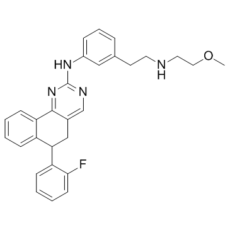
| Chemical Name | 6-(2-fluorophenyl)-N-[3-[2-(2-methoxyethylamino)ethyl]phenyl]-5,6-dihydrobenzo[h]quinazolin-2-amine |
| Synonyms | AR-Q087; ARQ 087; ARQ087; AR-Q087 racemate; Derazantinib Racemate |
| HS Tariff Code | 2934.99.9001 |
| Storage |
Powder-20°C 3 years 4°C 2 years In solvent -80°C 6 months -20°C 1 month |
| Shipping Condition | Room temperature (This product is stable at ambient temperature for a few days during ordinary shipping and time spent in Customs) |
Biological Activity
| Targets |
Fibroblast Growth Factor Receptor 1 (FGFR1) (IC₅₀=1.8 nM) [2] Fibroblast Growth Factor Receptor 2 (FGFR2) (IC₅₀=0.9 nM) [2] Fibroblast Growth Factor Receptor 3 (FGFR3) (IC₅₀=1.5 nM) [2] Fibroblast Growth Factor Receptor 4 (FGFR4) (IC₅₀=36 nM) [2] Vascular Endothelial Growth Factor Receptor 2 (VEGFR2) (IC₅₀=24 nM) [2] Platelet-Derived Growth Factor Receptor β (PDGFRβ) (IC₅₀=4.2 nM) [2] |
| ln Vitro |
Derazantinib, formerly known as ARQ 087, is a novel, orally bioavailable, ATP competitive, small molecule, multi-kinase inhibitor with strong in vivo and in vitro activity against tumors and cell lines addicted to the fibroblast growth factor receptor (FGFR). In a biochemical assay, the inhibitor's IC50 values were 1.8 nM for FGFR2, 4.5 nM for FGFR1, and 4.5 nM for FGFR3. The results of treating cells with ARQ 087 showed that it inhibited the auto-phosphorylation of FGFR2 and other proteins (FRS2α, AKT, and ERK) downstream in the FGFR pathway. ARQ 087 has been shown in cell lines driven by FGFR dysregulation, including amplifications, fusions, and mutations, to exhibit anti-proliferative activity. A positive correlation was observed between the induction of apoptosis and the G1 cell cycle arrest induced by ARQ 087 in cell lines that had high levels of FGFR2 protein, according to cell cycle studies. Furthermore, FGFR2 modified SNU-16 and NCI-H716 xenograft tumor models with gene amplifications and fusions showed that ARQ 087 was efficacious in suppressing tumor growth in vivo. A subcohort of patients with intrahepatic cholangiocarcinoma who have been found to have FGFR2 gene fusions is part of the phase 1/2 clinical trial ongoing research on ARQ 087 (NCT01752920). Derazantinib racemate inhibits RANKL-induced osteoclast differentiation in mouse bone marrow-derived macrophages (BMMs) in a dose-dependent manner; at 100 nM, it reduces the number of tartrate-resistant acid phosphatase (TRAP)-positive multinucleated osteoclasts by ~80% and downregulates the expression of osteoclast-specific genes including TRAP, cathepsin K (CTSK), matrix metalloproteinase 9 (MMP9), and nuclear factor of activated T-cells cytoplasmic 1 (NFATc1) [1] - It suppresses the phosphorylation of extracellular signal-regulated kinase (ERK) 1/2, Akt, and p38 mitogen-activated protein kinase (MAPK) in RANKL-stimulated BMMs, indicating inhibition of FGFR-mediated downstream signaling pathways [1] - In mouse calvarial osteoblasts, Derazantinib racemate promotes cell proliferation at concentrations of 10–100 nM and upregulates the expression of osteoblast-specific genes such as alkaline phosphatase (ALP), osteocalcin (OCN), and runt-related transcription factor 2 (Runx2) [1] - It exhibits potent antiproliferative activity against FGFR-dependent cancer cell lines: SNU-16 (FGFR2 amplification, GI₅₀=0.12 μM), NCI-H716 (FGFR2 fusion, GI₅₀=0.15 μM), and KG-1 (FGFR1 fusion, GI₅₀=0.18 μM) [2] - In FGFR-overexpressing COS-7 cells, it inhibits FGFR autophosphorylation and downstream FRS2α phosphorylation in a dose-dependent manner, with EC₅₀ values of 0.21 μM (FGFR1), 0.15 μM (FGFR2), and 0.32 μM (FGFR3) [2] |
| ln Vivo |
Derazantinib is efficient in preventing the growth of tumors in xenograft tumor models with gene fusions and amplifications that have FGFR2 altered, SNU-16, and NCI-H716. With Derazantinib-injected wings, the majority of embryos (81.3%) display aberrant external phenotypes, which may be caused by inhibition of limb bud mesenchyme proliferation. The ulna and radius are smaller or absent altogether, and the wings are shorter and thinner, with a skeletal phenotype characteristic of FGFR inhibition. In ovariectomized (OVX) mice (a model of postmenopausal osteoporosis), oral administration of Derazantinib racemate at 30 mg/kg and 60 mg/kg once daily for 8 weeks significantly increases lumbar spine bone mineral density (BMD) by 12.3% and 18.7%, respectively, compared to the OVX control group; it also improves trabecular bone volume fraction (BV/TV) and trabecular thickness (Tb.Th) while reducing trabecular separation (Tb.Sp) [1] - The drug reduces serum levels of bone resorption markers (C-terminal telopeptide of type I collagen, CTX-I) by 29.5% (30 mg/kg) and 41.2% (60 mg/kg) and increases serum bone formation markers (osteocalcin, OCN) by 15.3% (30 mg/kg) and 23.6% (60 mg/kg) in OVX mice [1] - In SNU-16 xenograft mice models, oral administration of Derazantinib racemate at 50 mg/kg and 75 mg/kg once daily for 14 days achieves tumor growth inhibition (TGI) of 72% and 85%, respectively, without significant weight loss [2] - It inhibits phosphorylation of FGFR2 and ERK1/2 in xenograft tumors, confirming suppression of FGFR signaling in vivo [2] |
| Enzyme Assay |
Derazantinib is titrated in DMSO using a three-fold dilution scheme, and the final DMSO concentration is achieved by diluting the mixture ten times more in deionized water. Each reaction plate well receives a volume (2.5 μL) of these dilutions or vehicle. Assay buffer containing FGFR1 or FGFR2 is added to each well in a volume of 17.5 μL, resulting in a final concentration of 0.50 or 0.25 nM, respectively. Following a 30-minute pre-incubation phase, ATP and substrate are added to assay buffer (5 μL) to achieve final concentrations of 80 nM biotinylated-PYK2 and 0-1,000 μM ATP in a final reaction volume of 25 μL. After incubating the plates for 60 minutes at room temperature, 10 μL of a stop/detection mixture made in assay buffer with EDTA is added to the plates to stop them in the dark. FGFR kinase activity assay: Recombinant FGFR1-4 kinases are diluted in assay buffer (50 mM Tris-HCl pH 7.5, 10 mM MgCl₂, 1 mM EGTA, 0.01% BSA, 1 mM DTT) and mixed with serial 3-fold dilutions of Derazantinib racemate (0.001–10 μM). The reaction is initiated by adding ATP (final concentration 10 μM) and biotinylated peptide substrate, followed by incubation at 37°C for 60 minutes. The reaction is stopped with EDTA-containing buffer, and streptavidin-conjugated beads and anti-phosphotyrosine antibody are added to detect phosphorylated substrate. Fluorescence intensity is measured, and IC₅₀ values are calculated using nonlinear regression analysis [2] - VEGFR2/PDGFRβ kinase activity assay: The assay is performed following the same protocol as FGFR kinase activity assay, using recombinant VEGFR2 or PDGFRβ kinase and corresponding specific substrates. Serial dilutions of Derazantinib racemate are tested, and IC₅₀ values are determined based on fluorescence signal inhibition [2] |
| Cell Assay |
Cells are seeded at 3000-5000 cells per well with 130 μL media in 96-well tissue culture treated plates. The cells are incubated overnight and subsequently treated with 3-fold serial dilutions of Derazantinib starting at 100 μM. The cells are returned to a 37°C humidified incubator for 72 hours. Cell proliferation is measured using MTS assay. Osteoclast differentiation assay: Mouse bone marrow-derived macrophages (BMMs) are isolated from femurs and tibias, seeded in 96-well plates (5×10³ cells/well), and cultured with M-CSF (30 ng/mL) for 3 days. Derazantinib racemate (0.1–100 nM) and RANKL (50 ng/mL) are added, and cells are cultured for another 4 days. Cells are fixed, stained with TRAP reagent, and TRAP-positive multinucleated cells (≥3 nuclei) are counted under a microscope [1] - Osteoblast proliferation and differentiation assay: Mouse calvarial osteoblasts are isolated, seeded in 96-well plates (3×10³ cells/well), and treated with Derazantinib racemate (1–1000 nM) for 72 hours. Cell proliferation is detected by MTS assay. For differentiation analysis, osteoblasts are cultured with the drug for 14 days, stained with ALP reagent, and ALP activity is measured by absorbance at 405 nm [1] - Cancer cell antiproliferation assay: FGFR-dependent cancer cells (SNU-16, NCI-H716, KG-1) are seeded in 96-well plates (5×10³ cells/well) and incubated overnight. Serial 3-fold dilutions of Derazantinib racemate (0.01–10 μM) are added, and cells are cultured for 72 hours. Cell viability is detected by MTT assay, and GI₅₀ values are calculated [2] - Western blot assay for signaling pathways: BMMs or cancer cells are treated with Derazantinib racemate for 2 hours, then stimulated with RANKL (for BMMs) or FGF2 (for cancer cells) for 15 minutes. Cells are lysed, and proteins are separated by SDS-PAGE, transferred to PVDF membranes, and probed with antibodies against phospho-ERK1/2, phospho-Akt, phospho-p38, phospho-FGFR, and total FGFR. β-actin is used as a loading control [1][2] - Quantitative real-time PCR (qPCR) assay: Total RNA is extracted from treated BMMs or osteoblasts using TRIzol reagent, reverse-transcribed into cDNA, and qPCR is performed with specific primers for TRAP, CTSK, MMP9, NFATc1 (for osteoclasts) and ALP, OCN, Runx2 (for osteoblasts). GAPDH is used as an internal reference, and relative gene expression is calculated by the 2⁻ΔΔCt method [1] |
| Animal Protocol |
25, 50,75, 100, 150 mg/kg Mouse with BaF3/FGFR2, SNU-16, and NCI-H716, xenograft models Ovariectomized (OVX) mouse model for osteoporosis: 8-week-old female C57BL/6 mice are subjected to bilateral ovariectomy or sham operation. After 1 week of recovery, mice are randomly divided into sham group, OVX control group, and Derazantinib racemate treatment groups (30 mg/kg and 60 mg/kg). The drug is formulated in 0.5% carboxymethylcellulose sodium (CMC-Na) with 0.1% Tween 80, administered orally once daily for 8 weeks. Body weight is measured weekly. At the end of treatment, mice are euthanized, serum is collected for bone marker detection, and lumbar vertebrae are isolated for BMD measurement by micro-CT and histological analysis [1] - Xenograft tumor model: 6-week-old female BALB/c nude mice are subcutaneously implanted with SNU-16 cells (5×10⁶ cells/mouse) in the right flank. When tumors reach 100–150 mm³, mice are randomized into vehicle control group and Derazantinib racemate treatment groups (50 mg/kg and 75 mg/kg). The drug is formulated in DMSO:cremophor EL:saline (10:10:80), administered orally once daily for 14 days. Tumor size is measured every 2 days with calipers, and tumor volume is calculated as length×width²×0.5. Body weight is monitored weekly. At the end of treatment, tumors are excised, weighed, and stored for Western blot analysis [2] |
| Toxicity/Toxicokinetics |
In OVX mice treated with Derazantinib racemate at 30 mg/kg or 60 mg/kg for 8 weeks, no significant changes in body weight, liver function (ALT, AST), or kidney function (BUN, creatinine) are observed compared to the control group [1] - In xenograft tumor mice, oral administration of Derazantinib racemate at doses up to 75 mg/kg for 14 days does not cause obvious weight loss (>10%) or other signs of toxicity (e.g., lethargy, diarrhea) [2] - Plasma protein binding rate of Derazantinib racemate is 92–95% in mouse plasma, as determined by equilibrium dialysis [2] |
| References |
[1]. Bone . 2017 Dec:105:57-66. [2]. PLoS One . 2016 Sep 14;11(9):e0162594. |
| Additional Infomation |
Derazantinib racemate is a racemic mixture of the ATP-competitive FGFR inhibitor Derazantinib, targeting FGFR1-3 with high potency and moderate activity against FGFR4, VEGFR2, and PDGFRβ [2] - It exerts dual effects on bone metabolism: inhibiting osteoclast differentiation and bone resorption, while promoting osteoblast proliferation and bone formation, making it a potential therapeutic agent for postmenopausal osteoporosis [1] - Its antiproliferative activity in FGFR-dependent cancer cells is attributed to the inhibition of FGFR-mediated ERK/Akt signaling pathways, leading to cell cycle arrest and reduced cell viability [2] - The drug shows good in vivo tolerability at effective doses, with no significant systemic toxicity observed in mouse models, supporting its potential for clinical development [1][2] |
Solubility Data
| Solubility (In Vitro) |
|
|||
| Solubility (In Vivo) |
Note: Listed below are some common formulations that may be used to formulate products with low water solubility (e.g. < 1 mg/mL), you may test these formulations using a minute amount of products to avoid loss of samples. Injection Formulations (e.g. IP/IV/IM/SC) Injection Formulation 1: DMSO : Tween 80: Saline = 10 : 5 : 85 (i.e. 100 μL DMSO stock solution → 50 μL Tween 80 → 850 μL Saline) *Preparation of saline: Dissolve 0.9 g of sodium chloride in 100 mL ddH ₂ O to obtain a clear solution. Injection Formulation 2: DMSO : PEG300 :Tween 80 : Saline = 10 : 40 : 5 : 45 (i.e. 100 μL DMSO → 400 μLPEG300 → 50 μL Tween 80 → 450 μL Saline) Injection Formulation 3: DMSO : Corn oil = 10 : 90 (i.e. 100 μL DMSO → 900 μL Corn oil) Example: Take the Injection Formulation 3 (DMSO : Corn oil = 10 : 90) as an example, if 1 mL of 2.5 mg/mL working solution is to be prepared, you can take 100 μL 25 mg/mL DMSO stock solution and add to 900 μL corn oil, mix well to obtain a clear or suspension solution (2.5 mg/mL, ready for use in animals). Injection Formulation 4: DMSO : 20% SBE-β-CD in saline = 10 : 90 [i.e. 100 μL DMSO → 900 μL (20% SBE-β-CD in saline)] *Preparation of 20% SBE-β-CD in Saline (4°C,1 week): Dissolve 2 g SBE-β-CD in 10 mL saline to obtain a clear solution. Injection Formulation 5: 2-Hydroxypropyl-β-cyclodextrin : Saline = 50 : 50 (i.e. 500 μL 2-Hydroxypropyl-β-cyclodextrin → 500 μL Saline) Injection Formulation 6: DMSO : PEG300 : castor oil : Saline = 5 : 10 : 20 : 65 (i.e. 50 μL DMSO → 100 μLPEG300 → 200 μL castor oil → 650 μL Saline) Injection Formulation 7: Ethanol : Cremophor : Saline = 10: 10 : 80 (i.e. 100 μL Ethanol → 100 μL Cremophor → 800 μL Saline) Injection Formulation 8: Dissolve in Cremophor/Ethanol (50 : 50), then diluted by Saline Injection Formulation 9: EtOH : Corn oil = 10 : 90 (i.e. 100 μL EtOH → 900 μL Corn oil) Injection Formulation 10: EtOH : PEG300:Tween 80 : Saline = 10 : 40 : 5 : 45 (i.e. 100 μL EtOH → 400 μLPEG300 → 50 μL Tween 80 → 450 μL Saline) Oral Formulations Oral Formulation 1: Suspend in 0.5% CMC Na (carboxymethylcellulose sodium) Oral Formulation 2: Suspend in 0.5% Carboxymethyl cellulose Example: Take the Oral Formulation 1 (Suspend in 0.5% CMC Na) as an example, if 100 mL of 2.5 mg/mL working solution is to be prepared, you can first prepare 0.5% CMC Na solution by measuring 0.5 g CMC Na and dissolve it in 100 mL ddH2O to obtain a clear solution; then add 250 mg of the product to 100 mL 0.5% CMC Na solution, to make the suspension solution (2.5 mg/mL, ready for use in animals). Oral Formulation 3: Dissolved in PEG400 Oral Formulation 4: Suspend in 0.2% Carboxymethyl cellulose Oral Formulation 5: Dissolve in 0.25% Tween 80 and 0.5% Carboxymethyl cellulose Oral Formulation 6: Mixing with food powders Note: Please be aware that the above formulations are for reference only. InvivoChem strongly recommends customers to read literature methods/protocols carefully before determining which formulation you should use for in vivo studies, as different compounds have different solubility properties and have to be formulated differently. (Please use freshly prepared in vivo formulations for optimal results.) |
| Preparing Stock Solutions | 1 mg | 5 mg | 10 mg | |
| 1 mM | 2.1342 mL | 10.6708 mL | 21.3415 mL | |
| 5 mM | 0.4268 mL | 2.1342 mL | 4.2683 mL | |
| 10 mM | 0.2134 mL | 1.0671 mL | 2.1342 mL |