Physicochemical Properties
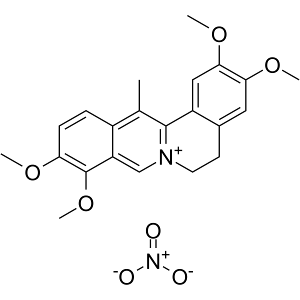
| Molecular Formula | C22H24NO4+.NO3- |
| Molecular Weight | 428.43516 |
| Exact Mass | 428.158 |
| CAS # | 13005-09-9 |
| Related CAS # | Dehydrocorydaline;30045-16-0;Dehydrocorydaline chloride;10605-03-5 |
| PubChem CID | 92043243 |
| Appearance | Yellow to orange solid powder |
| LogP | 3.977 |
| Hydrogen Bond Donor Count | 0 |
| Hydrogen Bond Acceptor Count | 7 |
| Rotatable Bond Count | 4 |
| Heavy Atom Count | 31 |
| Complexity | 522 |
| Defined Atom Stereocenter Count | 0 |
| InChi Key | CZZFYGAXGATDNG-UHFFFAOYSA-N |
| InChi Code | InChI=1S/C22H24NO4.NO3/c1-13-15-6-7-18(24-2)22(27-5)17(15)12-23-9-8-14-10-19(25-3)20(26-4)11-16(14)21(13)23;2-1(3)4/h6-7,10-12H,8-9H2,1-5H3;/q+1;-1 |
| Chemical Name | 2,3,9,10-tetramethoxy-13-methyl-5,6-dihydroisoquinolino[2,1-b]isoquinolin-7-ium;nitrate |
| HS Tariff Code | 2934.99.9001 |
| Storage |
Powder-20°C 3 years 4°C 2 years In solvent -80°C 6 months -20°C 1 month Note: Please store this product in a sealed and protected environment (e.g. under nitrogen), avoid exposure to moisture and light. |
| Shipping Condition | Room temperature (This product is stable at ambient temperature for a few days during ordinary shipping and time spent in Customs) |
Biological Activity
| Targets |
Dehydrocorydaline is an effective acetylcholinesterase (AChE) inhibitor with an IC50 of 0.62 µM. [1] p38 mitogen-activated protein kinases (MAPK) (activation)[2] |
| ln Vitro |
Dehydrocorydaline significantly inhibited the proliferation of human breast cancer MCF-7 cells in a dose-dependent manner, as determined by MTT assay. After 24-hour treatment, approximately 40% decrease in cell viability was observed at a concentration of 200 µM. [1] The cytotoxic effect induced by Dehydrocorydaline (200 µM) could be reversed by co-incubation with the caspase-8 inhibitor Z-IETD-FMK (10 µM). [1] Dehydrocorydaline induced apoptosis in MCF-7 cells, as evidenced by increased DNA fragmentation in a time-dependent manner (observed at 24, 48, and 72 hours), particularly prominent after 72 hours of treatment with 200 µM. [1] Hoechst 33342 staining revealed enhanced nuclear fluorescence and apoptotic morphological changes (condensed chromatin, shrunken nuclei) in MCF-7 cells treated with 200 µM Dehydrocorydaline in a time-dependent manner (24, 48, 72 h). [1] JC-1 staining assay showed that treatment with Dehydrocorydaline (0-200 µM for 1 hour) did not significantly alter the mitochondrial membrane potential (ΔΨm) in MCF-7 cells. [1] Western blot analysis demonstrated that Dehydrocorydaline treatment (0-200 µM for 24 hours) dose-dependently increased the protein expression of the pro-apoptotic factor Bax and decreased the protein expression of the anti-apoptotic factor Bcl-2. [1] Western blot analysis also showed that Dehydrocorydaline treatment induced the activation (cleavage) of caspase-7 and caspase-8, and the cleavage of PARP, but did not affect the cleavage of caspase-9. [1] The ratio of cleaved caspase-7 to total caspase-7 was dramatically increased by Dehydrocorydaline treatment. [1] Treatment of C2C12 myoblasts with dehydrocorydaline (125-500 nM) increased the expression levels of muscle-specific proteins, including MyoD, myogenin, and myosin heavy chain (MHC), in a dose-dependent manner.[2] Dehydrocorydaline treatment enhanced multinucleated myotube formation in C2C12 cells, with a significant increase in myotubes containing ≥10 nuclei compared to control.[2] Dehydrocorydaline (500 nM) elevated the levels of phosphorylated p38 MAPK (p-p38) in differentiating C2C12 myoblasts, indicating activation of the p38 MAPK pathway.[2] Dehydrocorydaline treatment enhanced the interaction (heterodimerization) between MyoD and the E protein E2A in C2C12 myoblasts, 10T1/2 fibroblasts, and 293T cells, as demonstrated by co-immunoprecipitation assays.[2] Dehydrocorydaline (500 nM) did not significantly affect the proliferation of C2C12 myoblasts, as assessed by BrdU incorporation assay.[2] In C2C12 cells depleted of the promyogenic receptor Cdo (via shRNA), dehydrocorydaline treatment restored the defective myoblast differentiation, as evidenced by rescued expression of MHC, MyoD, myogenin, p-p38, and improved myotube formation.[2] |
| Cell Assay |
MTT Assay: MCF-7 cells were seeded in 96-well plates at a density of 1 x 10⁴ cells per well. Cells were treated with various concentrations of Dehydrocorydaline (ranging from 0 to 200 µM) for 24 hours. After treatment, cell viability was determined using the MTT reagent. To investigate the role of caspase-8, cells were co-treated with Dehydrocorydaline (200 µM) and the caspase-8 inhibitor Z-IETD-FMK (10 µM). [1] JC-1 Staining for Mitochondrial Membrane Potential (ΔΨm): MCF-7 cells were treated with different concentrations of Dehydrocorydaline (0, 50, 100, 200 µM) for 1 hour. After treatment, cells were stained with the fluorescent probe JC-1 at a final concentration of 2.5 µg/mL for 10 minutes. The fluorescence (red/green) was observed and photographed using a fluorescence microscope to assess ΔΨm. [1] Hoechst 33342 Staining for Nuclear Morphology: MCF-7 cells were treated with 200 µM Dehydrocorydaline for 24, 48, or 72 hours. After treatment, the culture medium was removed, and cells were stained with Hoechst 33342 dye at a final concentration of 1 µg/mL for 20 minutes. Fluorescent images of stained nuclei were captured using a fluorescence microscope to observe chromatin condensation and apoptotic bodies. [1] DNA Fragmentation (DNA Ladder) Assay: MCF-7 cells (1 x 10⁶ cells) were harvested after treatment with various concentrations of Dehydrocorydaline (0-200 µM). Cell pellets were lysed in a buffer containing Tris-HCl, EDTA, Triton X-100, and proteinase K at 37°C for 2 hours. DNA was extracted using a salt and isopropanol precipitation method, incubated overnight at -20°C, centrifuged, washed with ethanol, and dissolved in buffer. The DNA was then treated with RNase, separated by 1% agarose gel electrophoresis, and stained with ethidium bromide to visualize the characteristic DNA ladder pattern indicative of apoptosis. [1] Western Blot Analysis: Dehydrocorydaline-treated cells were collected and lysed in RIPA buffer containing protease and phosphatase inhibitors. Lysates were centrifuged, and protein concentration was determined. Equal amounts of protein (40 µg) were separated by SDS-PAGE and transferred to a PVDF membrane. The membrane was incubated overnight at 4°C with primary antibodies against Bax, Bcl-2, total caspase-7, cleaved caspase-7, cleaved caspase-8, cleaved caspase-9, PARP, and β-actin (as a loading control). After incubation with appropriate secondary antibodies, protein bands were visualized using an enhanced chemiluminescence detection kit. Band intensities were quantified using densitometry software. [1] Myogenic Differentiation and Protein Expression: C2C12 myoblast cells were cultured in growth medium and then induced to differentiate by switching to differentiation medium containing 2% horse serum. Cells were treated with varying concentrations (125, 250, 500 nM) of dehydrocorydaline or vehicle (DMSO) during differentiation for 2 or 3 days. Cell lysates were harvested and analyzed by western blotting using antibodies against MHC, MyoD, myogenin, Cdo, phosphorylated p38 (p-p38), total p38, and pan-cadherin as a loading control.[2] Immunocytochemistry for Myotube Formation: C2C12 cells were treated with dehydrocorydaline (125, 250, 500 nM) or DMSO and induced to differentiate for 3 days. Cells were fixed, permeabilized, and immunostained with anti-MHC antibody, followed by an Alexa Fluor 594-conjugated secondary antibody. Nuclei were counterstained with DAPI. Myotube formation was quantified by counting the number of nuclei per MHC-positive cell, categorized as mononucleated, 2-5 nuclei, 6-9 nuclei, or ≥10 nuclei.[2] Immunocytochemistry for p-p38: C2C12 cells were treated with 500 nM dehydrocorydaline or DMSO and induced to differentiate for 1 day. Cells were fixed, permeabilized, blocked, and incubated with anti-p-p38 antibody, followed by an Alexa Fluor 594-conjugated secondary antibody. Nuclei were stained with DAPI. Fluorescent signals were captured by confocal microscopy and quantified.[2] Co-immunoprecipitation for MyoD-E2A Interaction: 293T or 10T1/2 cells were transfected with a MyoD expression vector. After 24 hours, cells were treated with 500 nM dehydrocorydaline or DMSO for an additional 24 hours. Whole cell extracts were prepared and subjected to immunoprecipitation using an anti-E2A antibody bound to protein G agarose beads. The immunoprecipitates were then analyzed by western blotting using anti-MyoD and anti-E2A antibodies.[2] BrdU Incorporation Assay for Proliferation: C2C12 cells were cultured in growth medium and treated with 500 nM dehydrocorydaline or DMSO for 24 hours. BrdU was added to the medium for 30 minutes. Cells were then fixed and immunostained with anti-BrdU antibody, followed by a FITC-conjugated secondary antibody. Nuclei were stained with DAPI. The percentage of BrdU-positive cells was quantified.[2] Rescue Experiment in Cdo-depleted Cells: C2C12 cells were transfected with control (pSuper) or Cdo-specific shRNA vectors to generate stable cell lines with reduced Cdo expression. These cells were then treated with 500 nM dehydrocorydaline or DMSO and induced to differentiate for 2-3 days. Differentiation was assessed by western blotting for muscle markers and p-p38, and by immunocytochemistry for MHC-positive myotube formation.[2] |
| References |
[1]. Dehydrocorydaline inhibits breast cancer cells proliferation by inducing apoptosis in MCF-7 cells. Am J Chin Med. 2012;40(1):177-85. [2]. Dehydrocorydaline promotes myogenic differentiation via p38 MAPK activation. Mol Med Rep. 2016 Oct;14(4):3029-36. [3]. Screening of a library of traditional Chinese medicines to identify anti-malarial compounds and extracts. Malar J. 2018 Jun 25;17(1):244. doi: 10.1186/s12936-018-2392-4. |
| Additional Infomation |
Dehydrocorydaline is an isoquinoline alkaloid isolated from the traditional Chinese herb Corydalis yanhusuo W.T. Wang. [1] It has been reported to possess various bioactivities, including adrenergic neuron blocking action, anti-inflammatory effects, aldose reductase inhibitory activity, anthelmintic potential, and AChE inhibitory activity. [1] This study is the first report on the anti-proliferative effect of Dehydrocorydaline on tumor cells (MCF-7 breast cancer cells). [1] The proposed mechanism for its anti-proliferative effect involves the induction of apoptosis through the extrinsic pathway, characterized by regulation of Bax/Bcl-2 ratio, activation of caspase-7 and caspase-8, cleavage of PARP, and without affecting mitochondrial membrane potential or caspase-9. [1] Dehydrocorydaline is an isoquinoline alkaloid isolated from the medicinal plant Corydalis tuber (Corydalis turtschaninovii).[2] It was identified through a screening of natural compounds from Corydalis tuber for activators of MyoD-responsive reporters and inducers of MHC expression in myoblasts.[2] The proposed mechanism of action is that dehydrocorydaline activates the p38 MAPK pathway. Activated p38 MAPK phosphorylates E proteins, enhancing their heterodimerization with the myogenic transcription factor MyoD. This active MyoD/E-protein complex then promotes the expression of muscle-specific genes, leading to enhanced myoblast differentiation and myotube formation.[2] The study suggests that dehydrocorydaline may have potential therapeutic application in improving muscle regeneration for conditions involving muscle atrophy.[2] |
Solubility Data
| Solubility (In Vitro) | DMSO : ~50 mg/mL (~116.70 mM) |
| Solubility (In Vivo) |
Solubility in Formulation 1: ≥ 2.5 mg/mL (5.84 mM) (saturation unknown) in 10% DMSO + 40% PEG300 + 5% Tween80 + 45% Saline (add these co-solvents sequentially from left to right, and one by one), clear solution. For example, if 1 mL of working solution is to be prepared, you can add 100 μL of 25.0 mg/mL clear DMSO stock solution to 400 μL PEG300 and mix evenly; then add 50 μL Tween-80 to the above solution and mix evenly; then add 450 μL normal saline to adjust the volume to 1 mL. Preparation of saline: Dissolve 0.9 g of sodium chloride in 100 mL ddH₂ O to obtain a clear solution. Solubility in Formulation 2: ≥ 2.5 mg/mL (5.84 mM) (saturation unknown) in 10% DMSO + 90% (20% SBE-β-CD in Saline) (add these co-solvents sequentially from left to right, and one by one), clear solution. For example, if 1 mL of working solution is to be prepared, you can add 100 μL of 25.0 mg/mL clear DMSO stock solution to 900 μL of 20% SBE-β-CD physiological saline solution and mix evenly. Preparation of 20% SBE-β-CD in Saline (4°C,1 week): Dissolve 2 g SBE-β-CD in 10 mL saline to obtain a clear solution. (Please use freshly prepared in vivo formulations for optimal results.) |
| Preparing Stock Solutions | 1 mg | 5 mg | 10 mg | |
| 1 mM | 2.3340 mL | 11.6702 mL | 23.3405 mL | |
| 5 mM | 0.4668 mL | 2.3340 mL | 4.6681 mL | |
| 10 mM | 0.2334 mL | 1.1670 mL | 2.3340 mL |