Physicochemical Properties
| Molecular Formula | C14H14O4 |
| Molecular Weight | 246.2586 |
| Exact Mass | 246.089 |
| CAS # | 23458-02-8 |
| PubChem CID | 442127 |
| Appearance | White to off-white solid powder |
| Density | 1.293g/cm3 |
| Boiling Point | 433.6ºC at 760mmHg |
| Flash Point | 167.9ºC |
| Vapour Pressure | 2.74E-08mmHg at 25°C |
| Index of Refraction | 1.592 |
| LogP | 1.867 |
| Hydrogen Bond Donor Count | 1 |
| Hydrogen Bond Acceptor Count | 4 |
| Rotatable Bond Count | 0 |
| Heavy Atom Count | 18 |
| Complexity | 387 |
| Defined Atom Stereocenter Count | 1 |
| SMILES | CC1([C@H](CC2=C(O1)C=C3C(=C2)C=CC(=O)O3)O)C |
| InChi Key | BGXFQDFSVDZUIW-LBPRGKRZSA-N |
| InChi Code | InChI=1S/C14H14O4/c1-14(2)12(15)6-9-5-8-3-4-13(16)17-10(8)7-11(9)18-14/h3-5,7,12,15H,6H2,1-2H3/t12-/m0/s1 |
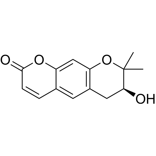
| Chemical Name | (3S)-3-hydroxy-2,2-dimethyl-3,4-dihydropyrano[3,2-g]chromen-8-one |
| HS Tariff Code | 2934.99.9001 |
| Storage |
Powder-20°C 3 years 4°C 2 years In solvent -80°C 6 months -20°C 1 month Note: This product requires protection from light (avoid light exposure) during transportation and storage. |
| Shipping Condition | Room temperature (This product is stable at ambient temperature for a few days during ordinary shipping and time spent in Customs) |
Biological Activity
| Targets |
Decursinol modulates μ-opioid receptor [1] Decursinol interacts with 5-hydroxytryptamine (5-HT) receptor [1] |
| ln Vitro |
1. Inhibition of colon cancer cell adhesion, migration, and invasion: - Decursinol (10, 20, 40 μM) inhibits adhesion of murine colon carcinoma cells (Colon26-M3.1) to extracellular matrix (ECM) components (fibronectin, laminin) in a concentration-dependent manner, with 40 μM showing 62% and 58% inhibition of adhesion to fibronectin and laminin, respectively [2] - It suppresses cell migration in wound-healing assay: 40 μM Decursinol reduces migration rate by 75% compared to control after 24 hours [2] - Transwell invasion assay shows 40 μM Decursinol inhibits invasive capacity of Colon26-M3.1 cells by 80% [2] 2. Inhibition of matrix metalloproteinase (MMP) activity: - Decursinol (10-40 μM) inhibits MMP-2 and MMP-9 activities in Colon26-M3.1 cells, with 40 μM reducing MMP-2 activity by 65% and MMP-9 activity by 70% (gelatin zymography) [2] |
| ln Vivo |
1. Antinociceptive activity in mice: - Decursinol (25, 50, 100 mg/kg, p.o.) exhibits dose-dependent antinociceptive effects in hot-plate test: 100 mg/kg group shows a significant increase in latency time from 10.2 s (control) to 28.5 s at 1 hour post-administration [1] - In acetic acid-induced writhing test, 100 mg/kg Decursinol reduces writhing number by 72% compared to control [1] - The antinociceptive effect is partially reversed by naloxone (μ-opioid receptor antagonist) and methysergide (5-HT receptor antagonist), confirming involvement of μ-opioid and 5-HT receptors [1] 2. Inhibition of lung metastasis of murine colon carcinoma: - Decursinol (25, 50 mg/kg, p.o., once daily for 21 days) inhibits lung metastasis of Colon26-M3.1 cells in BALB/c mice: 50 mg/kg group reduces the number of lung metastatic nodules from 42.6 (control) to 12.3 [2] - It decreases lung weight (a marker of metastatic burden) by 58% at 50 mg/kg, with no significant effect on primary tumor growth [2] |
| Enzyme Assay |
1. μ-opioid receptor binding assay: - Membrane fractions containing μ-opioid receptors are prepared from mouse brain tissues [1] - Decursinol is serially diluted (1 μM to 100 μM) and mixed with the membrane fraction and [3H]-dihydromorphine (specific μ-opioid receptor ligand) [1] - The mixture is incubated at 25°C for 60 minutes, then filtered through glass fiber filters to separate bound and free ligand [1] - Radioactivity of the bound ligand is measured using a liquid scintillation counter, and displacement rate is calculated to assess receptor interaction [1] 2. Matrix metalloproteinase (MMP) activity assay: - Conditioned medium from Colon26-M3.1 cells is collected after treatment with Decursinol (10-40 μM) for 24 hours [2] - MMP activity is analyzed by gelatin zymography: samples are loaded onto SDS-PAGE gels containing gelatin (substrate) and electrophoresed [2] - Gels are renatured, incubated at 37°C for 24 hours, stained with Coomassie brilliant blue, and destained to visualize clear bands corresponding to MMP activity [2] - Band intensity is quantified using image analysis software to calculate inhibition rate [2] |
| Cell Assay |
1. Cell adhesion assay: - 96-well plates are coated with fibronectin or laminin and incubated overnight at 4°C [2] - Colon26-M3.1 cells are treated with Decursinol (10, 20, 40 μM) for 1 hour, then seeded into coated wells at 5×10^4 cells per well [2] - Plates are incubated at 37°C with 5% CO2 for 1 hour, unbound cells are washed away, and adherent cells are fixed and stained with crystal violet [2] - Absorbance at 570 nm is measured to quantify adherent cells, and inhibition rate is calculated relative to control [2] 2. Wound-healing migration assay: - Colon26-M3.1 cells are seeded in 6-well plates and cultured until confluent [2] - A scratch is created in the cell monolayer using a pipette tip, and cells are treated with Decursinol (10, 20, 40 μM) [2] - Images are captured at 0 and 24 hours, and the wound closure rate is calculated by measuring the scratch width using image analysis software [2] 3. Transwell invasion assay: - Transwell inserts are coated with Matrigel and incubated at 37°C for 30 minutes to form a gel layer [2] - Colon26-M3.1 cells (1×10^5 cells) treated with Decursinol (10, 20, 40 μM) are added to the upper chamber, and medium containing chemoattractant is added to the lower chamber [2] - Plates are incubated at 37°C with 5% CO2 for 24 hours, non-invasive cells on the upper surface are removed, and invasive cells on the lower surface are fixed, stained, and counted [2] |
| Animal Protocol |
1. Antinociceptive activity mouse models: - ICR mice (6-8 weeks old) are randomly divided into control and Decursinol treatment groups (n=8 per group) [1] - Decursinol is dissolved in 0.5% carboxymethyl cellulose (CMC) and administered orally at doses of 25, 50, or 100 mg/kg [1] - Hot-plate test: Latency time to paw withdrawal is measured at 30, 60, 90, and 120 minutes post-administration [1] - Acetic acid-induced writhing test: 30 minutes after Decursinol administration, acetic acid is injected intraperitoneally, and the number of writhings is counted for 15 minutes [1] - For receptor antagonist studies: Naloxone (μ-opioid antagonist) or methysergide (5-HT antagonist) is injected intraperitoneally 15 minutes before Decursinol administration [1] 2. Murine colon carcinoma lung metastasis model: - BALB/c mice (6-8 weeks old) are intravenously injected with 1×10^6 Colon26-M3.1 cells to induce lung metastasis [2] - One day after cell injection, mice are randomized into control and Decursinol groups (n=10 per group) [2] - Decursinol is dissolved in 0.5% CMC and administered orally at 25 or 50 mg/kg once daily for 21 days [2] - Mice are euthanized on day 22, lungs are excised, and the number of metastatic nodules on the lung surface is counted under a stereomicroscope [2] - Lung weight is measured, and lung tissues are fixed for histopathological analysis [2] |
| Toxicity/Toxicokinetics |
- Acute toxicity: No mortality or obvious adverse effects (e.g., changes in behavior, food intake) observed in mice after oral administration of Decursinol at doses up to 200 mg/kg [1][2] - Subacute toxicity: Mice treated with 50 mg/kg/day Decursinol (p.o. for 21 days) show no significant changes in body weight, hematological parameters, or liver/kidney function markers [2] |
| References |
[1]. Antinociceptive mechanisms of orally administered decursinol in the mouse. Life Sci. 2003 Jun 13;73(4):471-85. [2]. Decursin and decursinol from Angelica gigas inhibit the lung metastasis of murine colon carcinoma. Phytother Res. 2011 Jul;25(7):959-64. |
| Additional Infomation |
Decursinol is an organic heterotricyclic compound that is 7,8-dihydro-2H,6H-pyrano[3,2-g]chromen-2-one substituted by a beta-hydroxy group at position 7 and two methyl groups at position 8. It is isolated from the roots of Angelica gigas and has been found to possess significant inhibitory activity against acetylcholinesterase enzyme (EC 3.1.1.7). It has a role as an EC 3.1.1.7 (acetylcholinesterase) inhibitor, an antineoplastic agent, an analgesic and a metabolite. It is an organic heterotricyclic compound, a delta-lactone, a secondary alcohol and a cyclic ether. Decursinol has been reported in Angelica gigas, Phlojodicarpus villosus, and other organisms with data available. - Decursinol is a natural coumarin derivative isolated from the roots of Angelica gigas Nakai (Korean angelica) [1][2] - Its antinociceptive mechanism involves dual modulation of μ-opioid and 5-HT receptors, contributing to synergistic pain relief [1] - The anti-metastatic effect is associated with inhibition of cancer cell adhesion, migration, invasion, and downregulation of MMP-2/MMP-9 activity, which are key processes in tumor metastasis [2] - It exhibits good oral bioavailability and low toxicity in animal models, supporting potential applications in pain management and metastatic cancer therapy [1][2] |
Solubility Data
| Solubility (In Vitro) | DMSO : ~100 mg/mL (~406.07 mM) |
| Solubility (In Vivo) |
Solubility in Formulation 1: ≥ 2.08 mg/mL (8.45 mM) (saturation unknown) in 10% DMSO + 40% PEG300 + 5% Tween80 + 45% Saline (add these co-solvents sequentially from left to right, and one by one), clear solution. For example, if 1 mL of working solution is to be prepared, you can add 100 μL of 20.8 mg/mL clear DMSO stock solution to 400 μL PEG300 and mix evenly; then add 50 μL Tween-80 to the above solution and mix evenly; then add 450 μL normal saline to adjust the volume to 1 mL. Preparation of saline: Dissolve 0.9 g of sodium chloride in 100 mL ddH₂ O to obtain a clear solution. Solubility in Formulation 2: ≥ 2.08 mg/mL (8.45 mM) (saturation unknown) in 10% DMSO + 90% (20% SBE-β-CD in Saline) (add these co-solvents sequentially from left to right, and one by one), clear solution. For example, if 1 mL of working solution is to be prepared, you can add 100 μL of 20.8 mg/mL clear DMSO stock solution to 900 μL of 20% SBE-β-CD physiological saline solution and mix evenly. Preparation of 20% SBE-β-CD in Saline (4°C,1 week): Dissolve 2 g SBE-β-CD in 10 mL saline to obtain a clear solution. Solubility in Formulation 3: ≥ 2.08 mg/mL (8.45 mM) (saturation unknown) in 10% DMSO + 90% Corn Oil (add these co-solvents sequentially from left to right, and one by one), clear solution. For example, if 1 mL of working solution is to be prepared, you can add 100 μL of 20.8 mg/mL clear DMSO stock solution to 900 μL of corn oil and mix evenly. (Please use freshly prepared in vivo formulations for optimal results.) |
| Preparing Stock Solutions | 1 mg | 5 mg | 10 mg | |
| 1 mM | 4.0607 mL | 20.3037 mL | 40.6075 mL | |
| 5 mM | 0.8121 mL | 4.0607 mL | 8.1215 mL | |
| 10 mM | 0.4061 mL | 2.0304 mL | 4.0607 mL |