DY131 (also known as GSK-9089) is a novel selective agonist at estrogen-related receptors ERRβ and ERRγ with minimal activity at ERRα, ERα and ERβ at concentrations up to 30 μM. DY131 stimulated the growth of ERα-negative endometrial cancer cells while inhibiting the growth of ERα-positive cancer cells. The structurally related receptors ERRα and the estrogen receptors alpha and beta (ERalpha/beta) were not affected by DY131. DY131 may be a cutting-edge treatment for prostate cancer since it targets ERRgamma.
Physicochemical Properties
| Molecular Formula | C18H21N3O2 | |
| Molecular Weight | 311.38 | |
| Exact Mass | 311.163 | |
| Elemental Analysis | C, 69.43; H, 6.80; N, 13.49; O, 10.28 | |
| CAS # | 95167-41-2 | |
| Related CAS # |
|
|
| PubChem CID | 5497124 | |
| Appearance | Light yellow to green yellow solid powder | |
| Density | 1.1±0.1 g/cm3 | |
| Index of Refraction | 1.574 | |
| LogP | 3.17 | |
| Hydrogen Bond Donor Count | 2 | |
| Hydrogen Bond Acceptor Count | 4 | |
| Rotatable Bond Count | 6 | |
| Heavy Atom Count | 23 | |
| Complexity | 380 | |
| Defined Atom Stereocenter Count | 0 | |
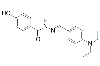
| SMILES | O=C(C1C=CC(O)=CC=1)NN=CC1C=CC(N(CC)CC)=CC=1 |
|
| InChi Key | WLKOCYWYAWBGKY-CPNJWEJPSA-N | |
| InChi Code | InChI=1S/C18H21N3O2/c1-3-21(4-2)16-9-5-14(6-10-16)13-19-20-18(23)15-7-11-17(22)12-8-15/h5-13,22H,3-4H2,1-2H3,(H,20,23)/b19-13+ | |
| Chemical Name | N-[(E)-[4-(diethylamino)phenyl]methylideneamino]-4-hydroxybenzamide | |
| Synonyms |
|
|
| HS Tariff Code | 2934.99.9001 | |
| Storage |
Powder-20°C 3 years 4°C 2 years In solvent -80°C 6 months -20°C 1 month |
|
| Shipping Condition | Room temperature (This product is stable at ambient temperature for a few days during ordinary shipping and time spent in Customs) |
Biological Activity
| Targets |
ERRγ; ERRβ
DY131 is a selective agonist ligand for estrogen-related receptors ERRβ/γ; it has no effect on ERRα, estrogen receptors α and β (ERα/β) [1] DY131 exerts antimitotic effects in breast cancer cells via ERRβ2 splice variant [2] |
||
| ln Vitro |
DY131 is a ligand that is unique to ERRβ/γ that exhibits preferential selectivity for ERRγ at lower concentrations. The related receptors ERα, ERβ, and ERRα are unaffected by it [1]. In a variety of breast cancer cell lines, DY131 suppresses growth, leading to a bimodal cell cycle arrest and the p38 stress kinase pathway being involved in cell death. DY131 postpones the transition from prophase to anaphase, while ERRβ2 facilitates the block in G2/M. Mitotic spindle defects are caused by DY131 treatment, which causes multi- and monopolar spindles to appear. ERRβ2, a cytosolic protein that also localizes to centrosomes, is also involved[2]. 1. DY131 effectively and selectively activates ERRβ/γ, with no agonistic or antagonistic effects on structurally related ERRα, ERα, or ERβ [1] 2. DY131 inhibits growth in a diverse panel of breast cancer cell lines (including MDA-MB-231, MCF7, HCC1806) in a concentration-dependent manner, while its effect on non-transformed breast epithelial cell lines was not specified; crystal violet staining confirmed growth inhibition over time (n=6, two-way ANOVA with Bonferroni post-tests, significant vs. DMSO control at Day 10/11) [2] 3. DY131 induces apoptotic cell death in breast cancer cells: flow cytometry showed increased subG1 DNA content (fragmented DNA) after 24 h exposure (n=3–5, one-way ANOVA with Tukey's post-test); Annexin V/PI staining confirmed dose-dependent increase in apoptotic cells after 12–24 h treatment (n=3–5, one-way ANOVA with Tukey's post-test); Western blot detected PARP cleavage and γH2AX upregulation (doxorubicin as positive control) [2] 4. DY131 induces bimodal cell cycle arrest (G1 and G2/M phases) in breast cancer cells: flow cytometry showed significant changes in G1, S, and G2/M phase distribution after 24 h exposure (n=3–5, one-way ANOVA with Tukey's post-test); Western blot confirmed upregulation of p21 and phosphorylation of Histone H3 (Serine 10) [2] 5. DY131-induced cell death is dependent on p38 MAPK activation: Western blot detected increased phosphorylated p38 (p-p38) in treated cells (densitometry: n=3, one-way ANOVA with Tukey's post-test); pre-treatment with p38 inhibitor SB203580 abrogated DY131-induced subG1 DNA content (n=3, two-way ANOVA with Bonferroni post-test) but did not affect G2/M arrest [2] 6. ERRβ2 facilitates DY131-induced G2/M arrest: transient transfection of MCF7 cells with ERRβ2 cDNA enhanced DY131-mediated Histone H3 phosphorylation (Serine 10); DY131 delays progression from prophase to anaphase in MCF7 cells stably expressing GFP-H2B (n=4–11 cells, one-way ANOVA with Tukey's post-test) [2] 7. DY131 causes mitotic spindle defects in breast cancer cells (HCC1806, MDA-MB-231): immunostaining for γ-tubulin/β-tubulin/DAPI showed increased monopolar and multipolar spindles after 24 h treatment with 5 μM DY131 (n=3, chi squared test) [2] 8. DY131 does not induce conventional DNA damage response or bind DNA directly: Western blot showed no activation of ATM signaling pathway; surface plasmon resonance (BIAcore) confirmed no binding of DY131 to dsDNA or ssDNA (mitoxantrone as positive control, experiment performed twice) [2] 9. DY131 has no transcription factor activity on ERRβ2 in breast cancer cells: luciferase reporter assay (ERRE-luciferase) showed no significant change in luciferase activity in MDA-MB-231/MCF7 cells treated with DY131 (n=3, two-way ANOVA with Bonferroni post-test) [2] 10. Clonogenic survival assay: MCF7 and MDA-MB-231 cells exposed to DY131 for 24 h showed reduced colony formation after 13 d culture[2] |
||
| ln Vivo |
|
||
| Enzyme Assay |
1. Receptor activation assay (ERRβ/γ, ERRα, ERα/β): Cells were transfected with receptor cDNA and relevant reporter constructs, then treated with DY131 (concentration not specified) or DMSO control for 18–20 h; luciferase activity was measured to assess receptor activation, with GSK4716 as a reference compound (n=3, two-way ANOVA with Bonferroni post-test) [1,2] 2. Surface plasmon resonance (BIAcore) assay: DY131, GSK4716, or mitoxantrone (positive control) were injected over dsDNA/ssDNA immobilized on sensor chips; peak Relative Unit (RU) values were recorded after 60 s injection to evaluate direct DNA binding (experiment performed twice, no binding of DY131 detected) [2] |
||
| Cell Assay |
In four wells of a 12-well plastic tissue culture dish, 150 (MDA-MB-231) or 200 (MCF7) cells were seeded per well on day 0. The next day, 18–24 hours were spent adding DY131 at the indicated concentrations. Day 2 involved removing the media that contained drugs, cleaning the wells with 1X PBS, and adding new media to them that contained no drugs. After a further 13 days of drug-free culture and two media changes, the cells were stained with crystal violet solution. 1. Cell growth assay (crystal violet staining): Breast cancer (MDA-MB-231, MCF7, HCC1806) and non-transformed breast epithelial cell lines were cultured with varying concentrations of DY131 or DMSO control for up to 10/11 days; cells were fixed and stained with crystal violet, and staining intensity was quantified to assess cell growth (n=6, two-way ANOVA with Bonferroni post-tests) [2] 2. Apoptosis assay (flow cytometry): Breast cancer cells were treated with DY131 for 24 h, fixed, and stained with propidium iodide (PI) to detect subG1 DNA content (fragmented DNA); for live-cell apoptosis, cells were stained with Annexin V/PI and analyzed by flow cytometry (n=3–5, one-way ANOVA with Tukey's post-test) [2] 3. Western blot assay: Cells treated with DY131 (24 h) were lysed, and protein extracts were analyzed for PARP, γH2AX, total H2AX, p21, phosphorylated/total p38, phosphorylated/total Histone H3 (Ser10), ERRβ2, ERRβsf, and ERRγ; densitometry was performed for p-p38/total p38 ratio (normalized to β-actin, n=3, one-way ANOVA with Tukey's post-test) [2] 4. Cell cycle assay (flow cytometry): DY131-treated breast cancer cells (24 h) were fixed, stained with PI, and analyzed for cell cycle phase distribution (G1, S, G2/M); data were normalized to DMSO control (n=3–5, one-way ANOVA with Tukey's post-test) [2] 5. Clonogenic survival assay: MCF7 and MDA-MB-231 cells were seeded at low density, exposed to DY131 for 24 h, and cultured for 13 days; colonies were counted to evaluate clonogenic survival [2] 6. Live-cell confocal microscopy: MCF7 cells stably expressing GFP-H2B were arrested in G2 with nocodazole, then released into DY131 or DMSO control; time elapsed from chromatin condensation (prophase) to anaphase was quantified (n=4–11 cells, one-way ANOVA with Tukey's post-test) [2] 7. Immunofluorescence staining: HCC1806/MDA-MB-231 cells treated with 5 μM DY131 for 24 h were fixed and stained for γ-tubulin (centrosomal marker), β-tubulin (mitotic spindle), and DAPI (DNA); spindle morphology (monopolar/bipolar/multipolar) was scored (n=3, chi squared test) [2] 8. Subcellular fractionation (REAP assay): HCC1806 cells were fractionated into total cell lysate (TCL), nuclear (nuc), and cytoplasmic (cyto) fractions; Western blot was performed to detect subcellular localization of ERRβ2, ERRβsf, vinculin (cytoplasmic marker), and Histone H3 (nuclear marker) [2] 9. Luciferase reporter assay: MDA-MB-231/MCF7 cells were transiently co-transfected with promoter-reporter luciferase constructs (ERRE-luciferase) and receptor cDNA (ERRβ2, ERRβsf, AIB1); cells were treated with DY131, GSK4716, or DMSO for 18–20 h, and luciferase activity was measured (n=3, two-way ANOVA with Bonferroni post-test) [2] |
||
| Animal Protocol |
|
||
| References |
[1]. Bioorg Med Chem Lett. 2005 Mar 1;15(5):1311-3. [2]. Oncotarget . 2016 Jul 26;7(30):47201-47220. |
||
| Additional Infomation |
DY131 is a member of benzoic acids. 1. DY131 (N'-{(1E)-[4-(diethylamino)phenyl]methylene}-4-hydroxybenzohydrazide) is a hydrazone compound synthesized with a 4-hydroxy group on one phenyl ring and a 4-diethylamino moiety on the other; it is a novel and selective pharmacologic tool for studying ERRβ/γ biological activities [1] 2. DY131 exerts antimitotic activity in breast cancer (including triple-negative breast cancer, TNBC) by targeting ERRβ2 splice variant: ERRβ2 localizes to centrosomes, and DY131 causes mitotic spindle defects and delays chromosome segregation (prophase to anaphase) [2] 3. DY131-induced cell death in breast cancer cells is mediated by the p38 stress kinase pathway, while cell cycle arrest (G2/M) is independent of p38 MAPK [2] 4. ERRβ2 has no transcription factor activity in breast cancer cells, and DY131 does not act via transcriptional regulation of ERRβ2 [2] 5. DY131 does not induce DNA damage or bind DNA directly, distinguishing its mechanism from conventional DNA-targeting chemotherapeutics (e.g., doxorubicin, mitoxantrone) [2] |
Solubility Data
| Solubility (In Vitro) |
|
|||
| Solubility (In Vivo) |
Solubility in Formulation 1: ≥ 2.5 mg/mL (8.03 mM) (saturation unknown) in 10% DMSO + 40% PEG300 + 5% Tween80 + 45% Saline (add these co-solvents sequentially from left to right, and one by one), clear solution. For example, if 1 mL of working solution is to be prepared, you can add 100 μL of 25.0 mg/mL clear DMSO stock solution to 400 μL PEG300 and mix evenly; then add 50 μL Tween-80 to the above solution and mix evenly; then add 450 μL normal saline to adjust the volume to 1 mL. Preparation of saline: Dissolve 0.9 g of sodium chloride in 100 mL ddH₂ O to obtain a clear solution. Solubility in Formulation 2: ≥ 2.5 mg/mL (8.03 mM) (saturation unknown) in 10% DMSO + 90% (20% SBE-β-CD in Saline) (add these co-solvents sequentially from left to right, and one by one), clear solution. For example, if 1 mL of working solution is to be prepared, you can add 100 μL of 25.0 mg/mL clear DMSO stock solution to 900 μL of 20% SBE-β-CD physiological saline solution and mix evenly. Preparation of 20% SBE-β-CD in Saline (4°C,1 week): Dissolve 2 g SBE-β-CD in 10 mL saline to obtain a clear solution. Solubility in Formulation 3: ≥ 2.5 mg/mL (8.03 mM) (saturation unknown) in 10% DMSO + 90% Corn Oil (add these co-solvents sequentially from left to right, and one by one), suspension solution. For example, if 1 mL of working solution is to be prepared, you can add 100 μL of 25.0 mg/mL clear DMSO stock solution to 900 μL of corn oil and mix evenly. (Please use freshly prepared in vivo formulations for optimal results.) |
| Preparing Stock Solutions | 1 mg | 5 mg | 10 mg | |
| 1 mM | 3.2115 mL | 16.0576 mL | 32.1151 mL | |
| 5 mM | 0.6423 mL | 3.2115 mL | 6.4230 mL | |
| 10 mM | 0.3212 mL | 1.6058 mL | 3.2115 mL |