Physicochemical Properties
| Molecular Formula | C6H12O6 |
| Molecular Weight | 180.1559 |
| Exact Mass | 180.063 |
| CAS # | 59-23-4 |
| Related CAS # | D-Galactose-13C;70849-30-8;D-Galactose-13C6;74134-89-7;D-Mannose;3458-28-4;D-Galactose-13C-2;478518-56-8;D-Galactose-d2-1;1176791-08-4;D-Galactose-13C,d;370565-96-1;D-Galactose-13C-3;478518-58-0;D-Galactose-13C-4;478518-60-4;D-Galactose-13C-5;478518-62-6;D-Galactose-d;64267-73-8;D-Galactose-d-1;64429-86-3;D-Galactose-d-2;478518-70-6;D-Galactose-d-3;478518-71-7;D-Galactose-d2;35669-34-2 |
| PubChem CID | 3037556 |
| Appearance | White to off-white solid powder |
| Density | 1.6±0.1 g/cm3 |
| Boiling Point | 527.1±50.0 °C at 760 mmHg |
| Melting Point | 168-170 °C(lit.) |
| Flash Point | 286.7±26.6 °C |
| Vapour Pressure | 0.0±3.1 mmHg at 25°C |
| Index of Refraction | 1.573 |
| LogP | -3.17 |
| Hydrogen Bond Donor Count | 5 |
| Hydrogen Bond Acceptor Count | 6 |
| Rotatable Bond Count | 5 |
| Heavy Atom Count | 12 |
| Complexity | 138 |
| Defined Atom Stereocenter Count | 4 |
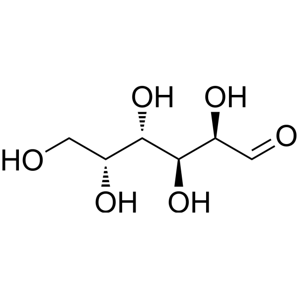
| SMILES | C([C@H]([C@@H]([C@@H]([C@H](C=O)O)O)O)O)O |
| InChi Key | GZCGUPFRVQAUEE-KCDKBNATSA-N |
| InChi Code | InChI=1S/C6H12O6/c7-1-3(9)5(11)6(12)4(10)2-8/h1,3-6,8-12H,2H2/t3-,4+,5+,6-/m0/s1 |
| Chemical Name | (2R,3S,4S,5R)-2,3,4,5,6-pentahydroxyhexanal |
| HS Tariff Code | 2934.99.9001 |
| Storage |
Powder-20°C 3 years 4°C 2 years In solvent -80°C 6 months -20°C 1 month |
| Shipping Condition | Room temperature (This product is stable at ambient temperature for a few days during ordinary shipping and time spent in Customs) |
Biological Activity
| ln Vitro | Galactose plays a major role in the survival and pathogenicity of bacteria. In E. Galactose is mostly used by the Leloir pathway in E. Coli. Each of the three d-galactose terminal groups has a distinct function; α-d-galactose serves as a carbon source, while β-d-galactose stimulates the synthesis of UDP-galactose for the biosynthetic glycosylation process [1]. |
| ln Vivo |
In animal modeling, D-galactose can be utilized to create models of cataract tracking and mouse subacute dormancy. Chronic exposure to D-galactose increases oxidative damage, enhances caspase-mediated cell display, and inhibits neurogenesis and neural migration in mice, all of which lead to neurodegeneration. Moreover, D-galactose-induced sensitization is a helpful model for researching medication processes and neurodegenerative and neuroprotective treatments [2]. The ultimate suggested D-galactose route results in oxidative damage and cognitive impairment. Cognitive deficits were noted in the open field test at 4 and 6 weeks following the d-gal tower, whereas deficits in spatial memory were noted in the maze test at 6 weeks following the d-gal tower [3]. - In C57BL/6 mice treated with D-Galactose via intraperitoneal injection (100 mg/kg/day) for 8 weeks, significant memory loss was observed. The Morris Water Maze test showed increased escape latency (from 25 ± 3 s to 58 ± 5 s) and decreased time spent in the target quadrant (from 35 ± 4% to 18 ± 3%). The Y-maze test revealed a reduced spontaneous alternation rate (from 72 ± 5% to 45 ± 4%). Histologically, hippocampal and cortical neuron counts decreased by ~30%, and oxidative damage markers were altered: brain malondialdehyde (MDA) levels increased by 45%, while superoxide dismutase (SOD) and glutathione peroxidase (GSH-Px) activities decreased by 30% and 25%, respectively [2] - In Wistar rats given oral D-Galactose (300 mg/kg/day) for 8 weeks, cognitive impairments were confirmed by the Morris Water Maze (escape latency increased by 60%) and novel object recognition test (recognition index decreased from 68 ± 5% to 42 ± 4%). Brain oxidative damage was evident: MDA levels in the hippocampus and cortex increased by 38% and 32%, respectively, and GSH-Px activity decreased by 28% [3] - In C57BL/6J mice injected intraperitoneally with D-Galactose (120 mg/kg/day) for 6 weeks, behavioral aging phenotypes emerged. The open field test showed reduced locomotor activity (total distance traveled decreased by 40%), and the passive avoidance test revealed shortened step-through latency (from 300 ± 20 s to 120 ± 15 s), indicating impaired learning and memory [4] - In young male Sprague-Dawley rats treated with intraperitoneal D-Galactose (500 mg/kg/day) for 10 days, natural aging-like symptoms appeared. Body weight gain slowed by 15%, and organ coefficients of the liver and kidney decreased by 10% and 8%, respectively. Serum oxidative markers showed increased MDA (by 35%) and decreased SOD (by 25%) [5] - In C57BL/6J mice injected intraperitoneally with D-Galactose (200 mg/kg/day) for 10 weeks, cataract formation was induced. Lens opacity was graded (grade 3 opacity in 80% of mice vs. 0% in controls), and lens oxidative damage was confirmed: MDA levels increased by 50%, and SOD activity decreased by 40% [6] - In Sprague-Dawley rats injected intraperitoneally with D-Galactose (500 mg/kg/day) for 4 weeks, a stable cataract model was established. Lens混浊度 (opacity) reached grade 4 in 90% of rats, and histological examination showed lens fiber cell degeneration and vacuolation [7] |
| Animal Protocol |
- C57BL/6 mouse model for memory loss and neurodegeneration: Male C57BL/6 mice (8-week-old) were randomly divided into control and D-Galactose groups. D-Galactose was dissolved in normal saline and administered via intraperitoneal injection at 100 mg/kg/day for 8 weeks; the control group received equal volume of normal saline. After treatment, memory function was evaluated by Morris Water Maze and Y-maze tests, and brain tissues were collected to detect oxidative markers (MDA, SOD, GSH-Px) and histological changes via hematoxylin-eosin staining [2] - Wistar rat model for cognitive impairment: Male Wistar rats (6-week-old) were divided into control and D-Galactose groups. D-Galactose was dissolved in distilled water and given by oral gavage at 300 mg/kg/day for 8 weeks; the control group received distilled water. Cognitive function was assessed by Morris Water Maze and novel object recognition test, and hippocampal/cortical tissues were analyzed for oxidative markers [3] - C57BL/6J mouse model for behavioral aging: Male C57BL/6J mice (7-week-old) were assigned to control and D-Galactose groups. D-Galactose in normal saline was injected intraperitoneally at 120 mg/kg/day for 6 weeks. Locomotor activity was tested by open field test, and learning/memory was evaluated by passive avoidance test [4] - Young Sprague-Dawley rat model for aging simulation: Male Sprague-Dawley rats (4-week-old) were treated with D-Galactose (dissolved in normal saline) via intraperitoneal injection at 500 mg/kg/day for 10 days. Body weight was recorded weekly, and serum was collected to measure MDA and SOD levels; liver and kidney were weighed to calculate organ coefficients [5] - C57BL/6J mouse model for cataract: Female C57BL/6J mice (6-week-old) were divided into control and D-Galactose groups. D-Galactose in normal saline was injected intraperitoneally at 200 mg/kg/day for 10 weeks. Lens opacity was graded weekly using a slit lamp, and lenses were harvested to detect MDA and SOD levels [6] - Sprague-Dawley rat model for cataract: Female Sprague-Dawley rats (5-week-old) were treated with D-Galactose (dissolved in normal saline) via intraperitoneal injection at 500 mg/kg/day for 4 weeks. Lens opacity was graded by slit lamp, and lens tissues were fixed for histological examination (hematoxylin-eosin staining) to observe fiber cell changes [7] |
| Toxicity/Toxicokinetics |
- D-Galactose induced neurotoxicity in mice: intraperitoneal injection of 100 mg/kg/day for 8 weeks caused hippocampal and cortical neuron loss, increased brain MDA levels (oxidative damage), and decreased antioxidant enzyme (SOD, GSH-Px) activities, leading to memory loss and neurodegeneration [2] - D-Galactose caused cognitive toxicity in rats: oral administration of 300 mg/kg/day for 8 weeks resulted in impaired learning and memory (reduced novel object recognition index, prolonged maze escape latency) and increased oxidative damage in the hippocampus and cortex (elevated MDA, reduced GSH-Px) [3] - D-Galactose induced aging-related toxicity in young rats: intraperitoneal injection of 500 mg/kg/day for 10 days slowed body weight gain, reduced liver and kidney organ coefficients, and increased serum oxidative stress (elevated MDA, reduced SOD) [5] - D-Galactose caused ocular toxicity in mice and rats: intraperitoneal injection of 200 mg/kg/day (mice, 10 weeks) or 500 mg/kg/day (rats, 4 weeks) induced lens opacity (cataract), lens fiber cell degeneration, and increased lens MDA levels with decreased SOD activity [6][7] |
| References |
[1]. Structure and function of the D-galactose network in enterobacteria. MBio. 2011 Jun 28;2(4):e00053-11. [2]. Chronic systemic D-galactose exposure induces memory loss, neurodegeneration, and oxidativedamage in mice: protective effects of R-alpha-lipoic acid. J Neurosci Res. 2006 Aug 15;84(3):647-54. [3]. Oral administration of d-galactose induces cognitive impairments and oxidative damage in rats. Behav Brain Res. 2016 Apr 1;302:35-43. [4]. Behavioural study of the D-galactose induced aging model in C57BL/6J mice. Behav Brain Res. 2005, 157, 22. [5]. A high dose of short term exogenous D-galactose administration in young male rats produces symptoms simulating the natural aging process. Life Sci. 2015. [6]. Alginate Oligosaccharide Prevents against D-galactose-mediated Cataract in C57BL/6J Mice via Regulating Oxidative Stress and Antioxidant System. Curr Eye Res. 2021, 46, 6. [7]. Characterization of an i.p. D-galactose-induced cataract model in rats. J Pharmacol Toxicol Methods. 2021. |
| Additional Infomation |
Aldehydo-D-galactose is a D-galactose and an aldehydo-galactose. It is an enantiomer of an aldehydo-L-galactose. (2R,3S,4S,5R)-2,3,4,5,6-pentahydroxyhexanal has been reported in Camellia sinensis, Ascochyta medicaginicola, and other organisms with data available. Galactose is a metabolite found in or produced by Saccharomyces cerevisiae. An aldohexose that occurs naturally in the D-form in lactose, cerebrosides, gangliosides, and mucoproteins. Deficiency of galactosyl-1-phosphate uridyltransferase (GALACTOSE-1-PHOSPHATE URIDYL-TRANSFERASE DEFICIENCY DISEASE) causes an error in galactose metabolism called GALACTOSEMIA, resulting in elevations of galactose in the blood. See also: D-Galactose (annotation moved to). - The mechanism of D-Galactose-induced damage involves excessive production of reactive oxygen species (ROS), which leads to oxidative stress (increased MDA, decreased antioxidant enzymes). This oxidative stress further causes tissue damage, such as neuronal loss (memory impairment), lens fiber degeneration (cataract), and organ dysfunction (liver/kidney weight changes) [2][3][5][6][7] - D-Galactose is widely used to establish animal models of aging, cognitive impairment, and cataract for evaluating the protective effects of drugs (e.g., antioxidants, neuroprotectants, anti-cataract agents). The models are characterized by stable phenotypes, low cost, and good reproducibility [2][4][6][7] - In enterobacteria, D-Galactose is involved in metabolic networks including transport (via GalP permease) and catabolism (via Leloir pathway), but this is unrelated to its in vivo toxicity or pharmacological effects in mammals [1] |
Solubility Data
| Solubility (In Vitro) |
H2O : ~62.5 mg/mL (~346.91 mM) DMSO : ~50 mg/mL (~277.53 mM) |
| Solubility (In Vivo) |
Solubility in Formulation 1: ≥ 2.5 mg/mL (13.88 mM) (saturation unknown) in 10% DMSO + 40% PEG300 + 5% Tween80 + 45% Saline (add these co-solvents sequentially from left to right, and one by one), clear solution. For example, if 1 mL of working solution is to be prepared, you can add 100 μL of 25.0 mg/mL clear DMSO stock solution to 400 μL PEG300 and mix evenly; then add 50 μL Tween-80 to the above solution and mix evenly; then add 450 μL normal saline to adjust the volume to 1 mL. Preparation of saline: Dissolve 0.9 g of sodium chloride in 100 mL ddH₂ O to obtain a clear solution. Solubility in Formulation 2: ≥ 2.5 mg/mL (13.88 mM) (saturation unknown) in 10% DMSO + 90% (20% SBE-β-CD in Saline) (add these co-solvents sequentially from left to right, and one by one), clear solution. For example, if 1 mL of working solution is to be prepared, you can add 100 μL of 25.0 mg/mL clear DMSO stock solution to 900 μL of 20% SBE-β-CD physiological saline solution and mix evenly. Preparation of 20% SBE-β-CD in Saline (4°C,1 week): Dissolve 2 g SBE-β-CD in 10 mL saline to obtain a clear solution. Solubility in Formulation 3: ≥ 2.5 mg/mL (13.88 mM) (saturation unknown) in 10% DMSO + 90% Corn Oil (add these co-solvents sequentially from left to right, and one by one), clear solution. For example, if 1 mL of working solution is to be prepared, you can add 100 μL of 25.0 mg/mL clear DMSO stock solution to 900 μL of corn oil and mix evenly. Solubility in Formulation 4: 100 mg/mL (555.06 mM) in PBS (add these co-solvents sequentially from left to right, and one by one), clear solution; with ultrasonication. (Please use freshly prepared in vivo formulations for optimal results.) |
| Preparing Stock Solutions | 1 mg | 5 mg | 10 mg | |
| 1 mM | 5.5506 mL | 27.7531 mL | 55.5062 mL | |
| 5 mM | 1.1101 mL | 5.5506 mL | 11.1012 mL | |
| 10 mM | 0.5551 mL | 2.7753 mL | 5.5506 mL |