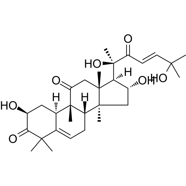
Physicochemical Properties
| Molecular Formula | C30H44O7 |
| Molecular Weight | 516.6662 |
| Exact Mass | 516.308 |
| CAS # | 3877-86-9 |
| PubChem CID | 5281318 |
| Appearance | White to off-white solid powder |
| Density | 1.2±0.1 g/cm3 |
| Boiling Point | 684.0±55.0 °C at 760 mmHg |
| Melting Point | 151-152ºC |
| Flash Point | 381.4±28.0 °C |
| Vapour Pressure | 0.0±4.8 mmHg at 25°C |
| Index of Refraction | 1.582 |
| LogP | 1.48 |
| Hydrogen Bond Donor Count | 4 |
| Hydrogen Bond Acceptor Count | 7 |
| Rotatable Bond Count | 4 |
| Heavy Atom Count | 37 |
| Complexity | 1100 |
| Defined Atom Stereocenter Count | 9 |
| SMILES | C[C@@]12C[C@H]([C@@H]([C@]1(CC(=O)[C@@]3([C@H]2CC=C4[C@H]3C[C@@H](C(=O)C4(C)C)O)C)C)[C@](C)(C(=O)/C=C/C(C)(C)O)O)O |
| InChi Key | SRPHMISUTWFFKJ-QJNWWGCFSA-N |
| InChi Code | InChI=1S/C30H44O7/c1-25(2,36)12-11-21(33)30(8,37)23-19(32)14-27(5)20-10-9-16-17(13-18(31)24(35)26(16,3)4)29(20,7)22(34)15-28(23,27)6/h9,11-12,17-20,23,31-32,36-37H,10,13-15H2,1-8H3/b12-11+/t17-,18+,19-,20+,23+,27+,28-,29+,30+/m1/s1 |
| Chemical Name | (2S,8S,9R,10R,13R,14S,16R,17R)-17-[(E,2R)-2,6-dihydroxy-6-methyl-3-oxohept-4-en-2-yl]-2,16-dihydroxy-4,4,9,13,14-pentamethyl-2,7,8,10,12,15,16,17-octahydro-1H-cyclopenta[a]phenanthrene-3,11-dione |
| HS Tariff Code | 2934.99.9001 |
| Storage |
Powder-20°C 3 years 4°C 2 years In solvent -80°C 6 months -20°C 1 month Note: This product requires protection from light (avoid light exposure) during transportation and storage. |
| Shipping Condition | Room temperature (This product is stable at ambient temperature for a few days during ordinary shipping and time spent in Customs) |
Biological Activity
| Targets |
The action target of Cucurbitacin D is heat shock protein 90 (HSP90), specifically inhibiting the ATPase activity of HSP90. The IC50 value for recombinant human HSP90α ATPase activity is 0.12 ± 0.02 μM [1] |
| ln Vitro |
1. Inhibition of HSP90 ATPase activity: Cucurbitacin D was tested for its effect on the ATPase activity of recombinant human HSP90α using the ADP-Glo ATPase assay. The compound exhibited concentration-dependent inhibition, with an IC50 of 0.12 ± 0.02 μM. At a concentration of 1 μM, it inhibited HSP90 ATPase activity by over 90%, which was comparable to the positive control geldanamycin (IC50: 0.05 μM) [1] 2. Disruption of HSP90 chaperone function: Human lung adenocarcinoma A549 cells and breast cancer MCF-7 cells were treated with Cucurbitacin D (0.01 μM, 0.05 μM, 0.1 μM, 0.5 μM) for 24 hours. Western blot analysis showed that Cucurbitacin D dose-dependently reduced the protein levels of HSP90 client proteins, including Akt, ErbB2, and Raf-1 (e.g., 0.1 μM reduced Akt protein by ~65% in A549 cells). Additionally, native PAGE analysis revealed that Cucurbitacin D inhibited the formation of HSP90 oligomers (a key form for chaperone activity) in A549 cells, with 0.5 μM completely blocking oligomer formation [1] 3. Antiproliferative activity against cancer cells: Cucurbitacin D was evaluated for antiproliferative activity in multiple human cancer cell lines (A549, MCF-7, HepG2, HCT116) using the MTT assay. The compound inhibited cell proliferation in a concentration-dependent manner, with IC50 values of 0.08 ± 0.01 μM (A549), 0.15 ± 0.03 μM (MCF-7), 0.11 ± 0.02 μM (HepG2), and 0.09 ± 0.01 μM (HCT116) after 72 hours of treatment [1] |
| Enzyme Assay |
HSP90 ATPase activity assay: The reaction system (25 μL total volume) contained 50 mM Tris-HCl buffer (pH 7.5), 5 mM MgCl2, 2 mM DTT, 0.2 μg recombinant human HSP90α, 100 μM ATP, and different concentrations of Cucurbitacin D (0.01 μM to 10 μM). The mixture was incubated at 37°C for 1 hour to allow ATP hydrolysis. Then, 25 μL of ADP-Glo reagent was added to terminate the reaction and deplete remaining ATP, followed by incubation at room temperature for 40 minutes. Next, 50 μL of kinase detection reagent was added to convert ADP to ATP and generate luminescence, which was measured using a microplate reader. The luminescence intensity was inversely proportional to the amount of ADP produced (i.e., HSP90 ATPase activity). The inhibition rate was calculated by comparing with the vehicle control, and the IC50 value was determined by nonlinear regression analysis of the concentration-inhibition curve [1] |
| Cell Assay |
1. Cancer cell proliferation assay (MTT assay): Human cancer cells (A549, MCF-7, HepG2, HCT116) were seeded in 96-well plates at a density of 3×10^3 cells per well and cultured in RPMI 1640 medium containing 10% fetal bovine serum overnight. The medium was replaced with fresh medium containing different concentrations of Cucurbitacin D (0.001 μM to 1 μM), and the cells were incubated for 72 hours at 37°C in a 5% CO2 incubator. After incubation, 20 μL of MTT solution (5 mg/mL) was added to each well, and the plates were incubated for another 4 hours. The supernatant was removed, and 150 μL of DMSO was added to dissolve the formazan crystals. The absorbance was measured at 570 nm using a microplate reader, and the IC50 value was calculated based on the cell viability curve [1] 2. Western blot analysis of HSP90 client proteins: A549 or MCF-7 cells were seeded in 6-well plates at a density of 5×10^5 cells per well and cultured overnight. The cells were treated with Cucurbitacin D (0.01 μM, 0.05 μM, 0.1 μM, 0.5 μM) for 24 hours, then washed with cold PBS and lysed with RIPA buffer containing protease inhibitors. The protein concentration was determined using a BCA kit. Equal amounts of protein (30 μg) were separated by SDS-PAGE, transferred to PVDF membranes, and incubated with primary antibodies against Akt, ErbB2, Raf-1, and β-actin (loading control) overnight at 4°C. After incubation with secondary antibodies, the bands were visualized using an enhanced chemiluminescence (ECL) system, and the band intensity was quantified using image analysis software [1] 3. Native PAGE analysis of HSP90 oligomers: A549 cells were treated with Cucurbitacin D (0.1 μM, 0.5 μM) for 24 hours, lysed with native lysis buffer (without SDS or reducing agents), and centrifuged to collect the supernatant. Equal amounts of protein were loaded onto a 4-12% native PAGE gel and electrophoresed at 4°C. The gel was transferred to a PVDF membrane and probed with an anti-HSP90 antibody to detect HSP90 monomers and oligomers. The results showed that Cucurbitacin D reduced the intensity of HSP90 oligomer bands in a concentration-dependent manner [1] |
| References |
[1]. Cucurbitacin D Is a Disruptor of the HSP90 Chaperone Machinery. J Nat Prod. 2015 Apr 24;78(4):873-9. |
| Additional Infomation |
Cucurbitacin D is a cucurbitacin in which a lanostane skeleton is multi-substituted with hydroxy, methyl and oxo substituents, with unsaturation at positions 5 and 23. It is a cucurbitacin, a secondary alpha-hydroxy ketone and a tertiary alpha-hydroxy ketone. It derives from a hydride of a lanostane. Cucurbitacin D has been reported in Trichosanthes tricuspidata, Elaeocarpus chinensis, and other organisms with data available. 1. Cucurbitacin D is a triterpenoid compound naturally occurring in plants of the Cucurbitaceae family (e.g., Cucurbita pepo , Momordica charantia ), which have been traditionally used in folk medicine for treating inflammation and tumors [1] 2. Mechanism of action: Cucurbitacin D disrupts the HSP90 chaperone machinery by two key ways: first, it directly inhibits the ATPase activity of HSP90 (a core step for chaperone function), and second, it prevents the formation of HSP90 oligomers (which are required for efficient client protein folding). This dual effect leads to the misfolding and degradation of HSP90 client proteins (most of which are oncogenic proteins, e.g., Akt, ErbB2), thereby inhibiting cancer cell proliferation [1] 3. Research significance: As HSP90 is a well-validated anticancer target, Cucurbitacin D shows potential as a lead compound for developing novel HSP90 inhibitors. Its distinct mechanism (inhibiting both ATPase activity and oligomerization) differentiates it from existing HSP90 inhibitors (e.g., geldanamycin, which only targets ATPase activity), providing a new direction for anticancer drug development [1] |
Solubility Data
| Solubility (In Vitro) | DMSO : ~110 mg/mL (~212.90 mM) |
| Solubility (In Vivo) |
Solubility in Formulation 1: 2.75 mg/mL (5.32 mM) in 10% DMSO + 90% (20% SBE-β-CD in Saline) (add these co-solvents sequentially from left to right, and one by one), suspension solution; with sonication. For example, if 1 mL of working solution is to be prepared, you can add 100 μL of 27.5 mg/mL clear DMSO stock solution to 900 μL of 20% SBE-β-CD physiological saline solution and mix evenly. Preparation of 20% SBE-β-CD in Saline (4°C,1 week): Dissolve 2 g SBE-β-CD in 10 mL saline to obtain a clear solution. Solubility in Formulation 2: ≥ 2.75 mg/mL (5.32 mM) (saturation unknown) in 10% DMSO + 90% Corn Oil (add these co-solvents sequentially from left to right, and one by one), clear solution. For example, if 1 mL of working solution is to be prepared, you can add 100 μL of 27.5 mg/mL clear DMSO stock solution to 900 μL of corn oil and mix evenly. (Please use freshly prepared in vivo formulations for optimal results.) |
| Preparing Stock Solutions | 1 mg | 5 mg | 10 mg | |
| 1 mM | 1.9355 mL | 9.6774 mL | 19.3547 mL | |
| 5 mM | 0.3871 mL | 1.9355 mL | 3.8709 mL | |
| 10 mM | 0.1935 mL | 0.9677 mL | 1.9355 mL |