Physicochemical Properties
| Molecular Formula | C9H10O4 |
| Molecular Weight | 182.1733 |
| Exact Mass | 182.057 |
| CAS # | 605-94-7 |
| PubChem CID | 69068 |
| Appearance | Brown to red solid powder |
| Density | 1.2±0.1 g/cm3 |
| Boiling Point | 331.4±42.0 °C at 760 mmHg |
| Melting Point | 58-60 °C(lit.) |
| Flash Point | 148.6±27.9 °C |
| Vapour Pressure | 0.0±0.7 mmHg at 25°C |
| Index of Refraction | 1.498 |
| LogP | 0.12 |
| Hydrogen Bond Donor Count | 0 |
| Hydrogen Bond Acceptor Count | 4 |
| Rotatable Bond Count | 2 |
| Heavy Atom Count | 13 |
| Complexity | 323 |
| Defined Atom Stereocenter Count | 0 |
| HS Tariff Code | 2934.99.9001 |
| Storage |
Powder-20°C 3 years 4°C 2 years In solvent -80°C 6 months -20°C 1 month |
| Shipping Condition | Room temperature (This product is stable at ambient temperature for a few days during ordinary shipping and time spent in Customs) |
Biological Activity
| ln Vitro | Coenzyme Q0 (0–40 μM; 24 h) prevents the growth and viability of human ovaries [1]. Coenzyme Q0 (CoQ0) (0-30 μM; 24 h; SKOV-3 cells) inhibits cell division by lowering cell cycle regulatory proteins and inducing G2/M cell cycle induction [1]. Coenzyme Q0 (CoQ0) (0-30 μM; 0-30 minutes; SKOV-3 cells) raises intracellular ROS levels to encourage the death of SKOV-3 cells [1]; via increasing the accumulation of LC3-II, GFP-LC3 pigment, AVO formation, and Beclin-1/Bcl-2 repair, Coenzyme Q0 (CoQ0) (0-30 μM; 24 hours; SKOV-3 cells) induces self-repair. Coenzyme Q0 (CoQ0) (0-30 μM; 24 hours; SKOV-3 cells) inhibits HER-2/AKT/mTOR signaling to improve cytochrome and autophagy mechanisms. Coenzyme Q0 (CoQ0) signals through mitochondria (caspase-3, PARP, and Bax/Bcl-2 corrector) and ER intermediates (caspase-12 and Hsp70) [1]. Q0 (CoQ0) (0-10 μM; 0.5-18 hours; RAW264.7 cells) improves Nrf2 stability and controls NFκB/AP-1 activation [2]. 5 μM Coenzyme Q0 (CoQ0); 0–12 hours; |
| ln Vivo | In SKOV-3 xenografted nude mice, intraperitoneal administration of Coenzyme Q0 (CoQ0) at 1.5 and 2.5 mg/kg every four days for 52 days inhibits the growth of tumors [1]. Through Nrf2 activation and NFκB inhibition, Coenzyme Q0 (CoQ0) (5 mg/kg; lateral wall; duration: 4 hours) has anti-inflammatory activity in the liver and the liver of mice treated with lipopolysaccharide (LPS) [2]. |
| Cell Assay |
Cell Viability Assay[1] Cell Types: SKOV-3, A2780 and A2870/CP70 cells Tested Concentrations: 0, 10, 20, 30 EA.hy 926 cells) in EA Anti-angiogenic activity in .hy 926 cells [3]. and 40 µM Incubation Duration: 24 hrs (hours) Experimental Results: The viability of SKOV-3, A2780 and A2870/CP70 cells was diminished, with IC50 values of 26.6 µM, 27.3 µM and 28.4 µM respectively. Cell cycle analysis[1] Cell Types: SKOV-3, A2780 and A2870/CP70 Cell Tested Concentrations: 0, 10, 20 and 30 µM Incubation Duration: 24 hrs (hours) Experimental Results: Cell cycle arrest in G2/M phase and diminished cyclin expression in in SKOV-3 cells. Apoptosis analysis[1] Cell Types: SKOV-3, A2780 and A2870/CP70 Cell Tested Concentrations: 0, 5, 15 and 30 µM Incubation Duration: 24 hrs (hours) Experimental Results: Promoted conversion of LC3–1 to LC3-II and increased LC3 -II accumulation. The Bax/Bcl-2 ratio increased in a dose-dependent manner. Apoptosis analysis[1] Cell Types: SKOV-3 Cell Tested Concentrations: 0, 10, 20 and 30 µM Incubation Duration: 24 hrs (hours) Experimental Results: Had the percentage of early apoptotic cells are 25.1%, 34% and 36% for 10, 20 and 30 µM, respectively. Western Blot Analysis[1] Cell Types: SKOV-3 cells Tested Concentrations: 0, 5, 15 and 30 µM Incubation Duration: 24 hrs (hours) Experimental Results: Activated of caspase-3 and cleavaged of PARP. Increased the expressions of caspase-12, HSP-70 and Bax in a dose-dependent manner, diminished the expressions of Bcl-2. Western Blot Analysis[1] Cell Types: SKOV-3 cells Tested Concentrations: 30 µM Incubation Duration: 24 hrs (hours) Experimental Results: diminished the phosphorylated HER-2 (Y1221) levels, p-AKT (Ser473) and p-mTOR (S2448) levels. Western Blot Analysis[2] Cell Types: RAW264.7 cells Tested Concentrations: 0, 2.5, 5 and 10 µM Incubation Duration: 0.5-18 hrs (hours) Experimental Results: Inhibited iNOS/COX-2 protein expressions with reductions of NO, PGE2, TNF-α and IL-1β secretions. Western Blot Analysis[3] Cell Types: EA.hy 926 cells Tested Concentrations: 5 µM Incubation Duration: 0, 1, 3, 6 and 12 hrs (hours) Experimental Results: Increased expressions of heme oxygenase-1 (HO-1) and γ-glutamylcysteine synthetase (γ-GCLC), inhibits protein expressions of matrix metalloproteinase-9 (MMP-9), reduces TNF-α-induced nuclear translocation and transcriptional activation of nuclear factor-κB (NF-κB). |
| Animal Protocol |
Animal/Disease Models: SKOV-3 xenograft nude mice [1] Doses: 1.5 and 2.5 mg/kg Route of Administration: intraperitoneal (ip) injection; once every four days for 52 days Experimental Results: 1.5 and 2.5 mg/kg inhibited tumor growth. Animal/Disease Models: LPS-treated female FVB mice [2] Doses: 5 mg/kg Route of Administration: po (po (oral gavage)) 4 hrs (hrs (hours)) Experimental Results: Down-regulated inflammatory genes in the liver and spleen tissues of LPS-injected mice. |
| References |
[1]. Coenzyme Q0 regulates NFκB/AP-1 activation and enhances Nrf2 stabilization in attenuation of LPS-induced inflammation and redox imbalance: Evidence from in vitro and in vivo studies. Biochim Biophys Acta. 2016 Feb;1859(2):246-61. [2]. Coenzyme Q0 regulates NFκB/AP-1 activation and enhances Nrf2 stabilization in attenuation of LPS-induced inflammation and redox imbalance: Evidence from in vitro and in vivo studies. Biochim Biophys Acta. 2016 Feb;1859(2):246-61. [3]. Anti-angiogenic properties of coenzyme Q0 through downregulation of MMP-9/NF-κB and upregulation of HO-1 signaling in TNF-α-activated human endothelial cells. Biochem Pharmacol. 2015 Nov 1;98(1):144-56. |
| Additional Infomation |
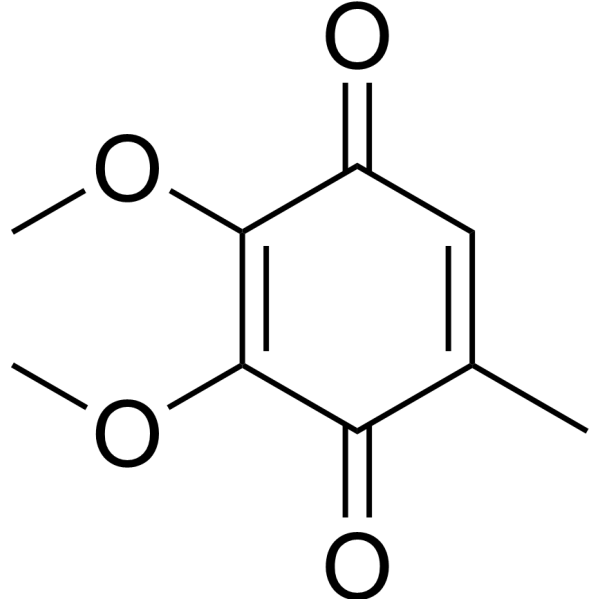
Ubiquinone-0 is a derivative of benzoquinone carrying a 5-methyl substituent; and methoxy substituents at positions 2 and 3. The core structure of the ubiquinone group of compounds. It has a role as an Escherichia coli metabolite and a human metabolite. Ubiquinone-0 is a metabolite found in or produced by Escherichia coli (strain K12, MG1655). 2,3-Dimethoxy-5-methyl-1,4-benzoquinone has been reported in Antrodia cinnamomea, Taiwanofungus salmoneus, and Taiwanofungus camphoratus with data available. |
Solubility Data
| Solubility (In Vitro) | DMSO : ~50 mg/mL (~274.45 mM) |
| Solubility (In Vivo) |
Solubility in Formulation 1: ≥ 1.67 mg/mL (9.17 mM) (saturation unknown) in 10% DMSO + 40% PEG300 + 5% Tween80 + 45% Saline (add these co-solvents sequentially from left to right, and one by one), clear solution. For example, if 1 mL of working solution is to be prepared, you can add 100 μL of 16.7 mg/mL clear DMSO stock solution to 400 μL PEG300 and mix evenly; then add 50 μL Tween-80 to the above solution and mix evenly; then add 450 μL normal saline to adjust the volume to 1 mL. Preparation of saline: Dissolve 0.9 g of sodium chloride in 100 mL ddH₂ O to obtain a clear solution. Solubility in Formulation 2: ≥ 1.67 mg/mL (9.17 mM) (saturation unknown) in 10% DMSO + 90% (20% SBE-β-CD in Saline) (add these co-solvents sequentially from left to right, and one by one), clear solution. For example, if 1 mL of working solution is to be prepared, you can add 100 μL of 16.7 mg/mL clear DMSO stock solution to 900 μL of 20% SBE-β-CD physiological saline solution and mix evenly. Preparation of 20% SBE-β-CD in Saline (4°C,1 week): Dissolve 2 g SBE-β-CD in 10 mL saline to obtain a clear solution. (Please use freshly prepared in vivo formulations for optimal results.) |
| Preparing Stock Solutions | 1 mg | 5 mg | 10 mg | |
| 1 mM | 5.4894 mL | 27.4469 mL | 54.8938 mL | |
| 5 mM | 1.0979 mL | 5.4894 mL | 10.9788 mL | |
| 10 mM | 0.5489 mL | 2.7447 mL | 5.4894 mL |