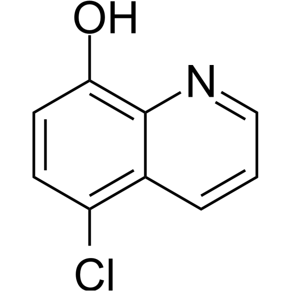
Cloxiquine (5-Chloro-8-quinolinol) is a PPARγ agonist with antibacterial, antifungal, antiamoebic, antiaging and antituberculosis activity. May be used for tuberculosis and dermatoses.
Physicochemical Properties
| Molecular Formula | C9H6CLNO |
| Molecular Weight | 179.6030 |
| Exact Mass | 179.013 |
| CAS # | 130-16-5 |
| Related CAS # | 25395-13-5 (hydrochloride) |
| PubChem CID | 2817 |
| Appearance | Light yellow to yellow solid powder |
| Density | 1.4±0.1 g/cm3 |
| Boiling Point | 348.7±22.0 °C at 760 mmHg |
| Melting Point | 122-124 °C(lit.) |
| Flash Point | 164.7±22.3 °C |
| Vapour Pressure | 0.0±0.8 mmHg at 25°C |
| Index of Refraction | 1.697 |
| LogP | 3.02 |
| Hydrogen Bond Donor Count | 1 |
| Hydrogen Bond Acceptor Count | 2 |
| Rotatable Bond Count | 0 |
| Heavy Atom Count | 12 |
| Complexity | 165 |
| Defined Atom Stereocenter Count | 0 |
| InChi Key | CTQMJYWDVABFRZ-UHFFFAOYSA-N |
| InChi Code | InChI=1S/C9H6ClNO/c10-7-3-4-8(12)9-6(7)2-1-5-11-9/h1-5,12H |
| Chemical Name | 5-chloroquinolin-8-ol |
| HS Tariff Code | 2934.99.9001 |
| Storage |
Powder-20°C 3 years 4°C 2 years In solvent -80°C 6 months -20°C 1 month |
| Shipping Condition | Room temperature (This product is stable at ambient temperature for a few days during ordinary shipping and time spent in Customs) |
Biological Activity
| Targets |
Peroxisome proliferator-activated receptor gamma (PPARγ) was identified as a potential molecular target through bioinformatic analyses (PharmMapper and DRAR-CPI). [2] |
| ln Vitro |
Chloroxiquin (cloxyquin) possesses anti-TB action, with a MIC range of 0.062 to 0.25 μg/mL against 9 standard strains and 150 species of Mycobacterium tuberculosis [3]. Cloxiquine (0.5-10 μM; 24 hours) suppresses B16F10 and A375 cell proliferation in a dose-dependent manner [2]. Cloxiquine (0.5-10 μM; 24 hours) inhibits the migration of B16F10 and A375 cells [2]. Cloxiquine (0.5-2.5 μM; 24 hours) inhibits glycolysis in B16F10 cells [2]. In a high-content screening of an FDA-approved drug library (1430 compounds at 10 µM), Cloxiquine (CLQ) was identified as a hit that strongly inhibited the viability of mouse B16F10 melanoma cells (inhibition ratio >75%) and human A375 melanoma cells. [2] Cloxiquine suppressed the proliferation of B16F10 and A375 cells in a dose-dependent manner, as shown by CCK-8 assay. It did not affect the viability of normal Melan-A and PIG1 melanocytes at tested doses. [2] EdU incorporation assay showed that Cloxiquine (2.5 µM and 10 µM) significantly reduced cell proliferation in B16F10 cells (by ~73%) and A375 cells (by ~64%) after 24-hour treatment. [2] Western blot analysis revealed that Cloxiquine treatment decreased protein levels of proliferation markers (PCNA) and cell cycle promoters (Cyclin E, Cyclin D1, CDK4, CDK2), while increasing levels of cell cycle inhibitors (p27, p21) in a concentration-dependent manner. [2] Cloxiquine inhibited the migration of B16F10 and A375 cells in transwell chamber and wound-healing assays. [2] Western blot analysis showed that Cloxiquine treatment decreased protein expression of migration-associated molecules (ICAM-1, VCAM-1, MMP-9, MMP-2) in a dose-dependent manner. [2] Cloxiquine suppressed glycolysis in B16F10 cells: it increased glucose levels in the culture medium, decreased lactate production and ATP generation, and reduced the extracellular acidification rate (ECAR). [2] RT-qPCR and Western blot analyses showed that Cloxiquine reduced mRNA and protein expression levels of glycolytic genes (GLUT1, HK2, PKM2, LDHA). [2] Treatment with Cloxiquine (1.5 µM) increased both mRNA and total protein expression levels of PPARγ in B16F10 cells in a time-dependent manner (up to 12 hours). [2] Subcellular fractionation and immunofluorescence confirmed that Cloxiquine treatment increased PPARγ protein levels in both the cytoplasm and nucleus of B16F10 cells. [2] The anti-proliferative and anti-migration effects of Cloxiquine, as well as its suppressive effects on glycolysis and related protein expression, were partially attenuated by co-treatment with the specific PPARγ inhibitor GW9662 (10 µM) or by transfection with PPARγ shRNA. [2] Combination of Cloxiquine and the glycolysis inhibitor 2-deoxyglucose (2-DG) did not exhibit a synergistic inhibitory effect on B16F10 cell proliferation and migration compared to each agent alone. [2] |
| ln Vivo |
In a mouse B16F10 melanoma xenograft model, coxiquine (5–25 mg/kg; daily intraperitoneal injection for 8 days) reduces tumor growth [2]. The mouse B16F10 melanoma lung metastasis model is used to study how well chloroxiquine (5–25 mg/kg; intraperitoneal injection every day for 14 days) inhibits tumor metastasis [2]. In a mouse B16F10 melanoma xenograft model, intraperitoneal (i.p.) administration of Cloxiquine (5 mg/kg and 25 mg/kg, daily for 8 days) starting from day 7 post-cell inoculation significantly suppressed tumor growth. Compared to the control group (average tumor volume ~5212.63 mm³), Cloxiquine at 5 mg/kg and 25 mg/kg reduced tumor volume by approximately 66.37% and 54.79%, respectively, and reduced tumor weights by approximately 75.91% and 63.41%, respectively. [2] Histological (H&E) and immunohistochemical (Ki-67) analysis of tumor sections showed that Cloxiquine treatment decreased tumor cell density and reduced the number of proliferating cells. [2] In a mouse B16F10 melanoma lung metastatic model, intravenous injection of B16F10 cells followed by i.p. administration of Cloxiquine (5 mg/kg and 25 mg/kg, daily for 14 days) starting from day 7 post-injection significantly decreased the number of macroscopic black lung metastases compared to the control group. H&E staining of lungs confirmed the reduction in metastatic nodules. [2] Combination treatment with Cloxiquine (25 mg/kg, i.p., daily) and 2-DG (500 mg/kg, i.p., every other day) did not show a synergistic effect on suppressing tumor growth and metastasis in vivo compared to Cloxiquine or 2-DG treatment alone. [2] |
| Cell Assay |
Drug Screening Assay: For initial screening, cells were seeded in 96-well plates and cultured overnight. After synchronization with serum-free medium, cells were treated with compounds from the library (10 µM final concentration) or vehicle (0.1% DMSO) in serum-free medium for 24 hours. Cell viability was then assessed by adding a water-soluble tetrazolium salt (WST-8) reagent, incubating for 2 hours, and measuring absorbance at 450 nm. Inhibition efficiency >75% was considered significant. [2] Cell Proliferation Assay (EdU): Cell proliferation was analyzed using an EdU (5-ethynyl-2’-deoxyuridine) incorporation assay. Cells were treated with Cloxiquine for 24 hours, then incubated with EdU to label newly synthesized DNA. Incorporated EdU was detected via a click chemistry reaction with a fluorescent azide, and proliferating cells were visualized and quantified. [2] Cell Migration Assays: Two methods were used. For the transwell chamber assay, cells were seeded into the upper chamber of a transwell insert with a porous membrane and allowed to migrate towards a chemoattractant in the lower chamber for a specified time. Migrated cells on the lower surface were fixed, stained, and counted. For the wound-healing assay, a scratch was made in a confluent cell monolayer. Cells were then treated with Cliquine, and the migration of cells into the scratch area was monitored and measured over time to assess wound closure. [2] Glycolysis Assays: For measuring glucose consumption, the concentration of glucose remaining in the cell culture medium after Cloxiquine treatment was quantified using a commercial glucose test kit. For lactate production, the intracellular lactate concentration was determined using a commercial kit. For ATP generation, intracellular ATP levels were measured using a commercial luciferase-based assay. [2] Extracellular Acidification Rate (ECAR) Assay: ECAR, an indicator of glycolytic flux, was measured using a extracellular flux analyzer. Cells were seeded in a specialized microplate. After treatment with Cloxiquine, cells were switched to a glucose-free medium. ECAR was measured in real-time following sequential injection of glucose, oligomycin, and 2-DG according to a glycolysis stress test protocol. [2] Gene Expression Analysis (RT-qPCR): Total RNA was extracted from treated cells using a reagent. RNA was reverse-transcribed into cDNA. Real-time PCR amplification was performed using a SYBR Green-based master mix and a thermal cycler. Expression levels of target genes (e.g., GLUT1, HK2, PKM2, LDHA, PPARγ) were normalized to a housekeeping gene (36B4). [2] Protein Expression Analysis (Western Blot): Cells were lysed, and protein concentration was determined. Equal amounts of protein were separated by SDS-PAGE, transferred to a membrane, and blocked. Membranes were incubated overnight with primary antibodies against target proteins (e.g., cell cycle regulators, migration markers, glycolytic enzymes, PPARγ), followed by incubation with HRP-conjugated secondary antibodies. Protein bands were visualized using chemiluminescence and quantified by densitometry. For nuclear fraction analysis, nuclear proteins were extracted using a commercial kit, and Histone H3 was used as a loading control. [2] Immunofluorescence (ICC) for PPARγ Localization: Cells were fixed, permeabilized, and blocked. They were then incubated overnight with an anti-PPARγ primary antibody at 4°C, followed by incubation with a fluorophore-conjugated secondary antibody. Nuclei were counterstained with DAPI. Cellular localization of PPARγ was visualized and analyzed using a fluorescence microscope. [2] Gene Knockdown (shRNA Transfection): To knockdown PPARγ expression, cells were transfected with specific PPARγ shRNA or a non-specific scramble shRNA using a lipid-based transfection reagent, following the manufacturer's instructions. Transfection was performed 24 hours prior to Cloxiquine treatment. Knockdown efficiency was verified by Western blot. [2] |
| Animal Protocol |
B16F10 Xenograft Tumor Growth Model: Mouse B16F10 melanoma cells were harvested and resuspended. A suspension containing 1 x 10⁶ cells was injected subcutaneously into the right flank of nude mice. Tumor growth was allowed for seven days until palpable. Mice were then randomly divided into groups and administered either vehicle (olive oil) or Cloxiquine (5 mg/kg or 25 mg/kg) via intraperitoneal (i.p.) injection daily for eight consecutive days. Tumor volumes were measured periodically. [2] B16F10 Lung Metastasis Model: Mouse B16F10 cells were harvested and resuspended. A suspension containing 1.5 x 10⁵ cells was injected intravenously into mice via the tail vein. Seven days post-injection, mice were randomly divided into groups and administered either vehicle (olive oil) or Cloxiquine (5 mg/kg or 25 mg/kg) via i.p. injection daily for fourteen consecutive days. Mice were then euthanized, lungs were harvested, and the number of surface metastatic nodules was counted. [2] Combination Therapy Model: In some experiments, to assess the role of glycolysis, mice bearing B16F10 tumors were treated with Cloxiquine (25 mg/kg, i.p., daily) alone or in combination with the glycolysis inhibitor 2-deoxyglucose (2-DG, 500 mg/kg, i.p., injected every other day). [2] |
| Toxicity/Toxicokinetics |
Cloxiquine is described as a common cause of epigastric discomfort, contact dermatitis, and neuropathy. It is also considered a mutagen. [1] Specific toxicokinetic parameters (e.g., LD50, organ toxicity, protein binding) are not provided. [1] In the in vivo studies, Cloxiquine administration at 5 mg/kg and 25 mg/kg (i.p., daily) did not cause apparent hepatotoxicity in mice. This was evidenced by only modest alterations in serum levels of alanine transaminase (ALT) and aspartate transaminase (AST) compared to controls. [2] Histological examination (H&E staining) of liver tissues from treated mice showed clear morphology of liver cells with discernible intracellular margins, comparable to controls. [2] Terminal deoxynucleotidyl transferase dUTP nick end labeling (TUNEL) assay on liver sections showed minimal DNA fragmentation, indicating a lack of significant hepatocyte apoptosis. [2] The study cites a previous maximum tolerated dose study in mice indicating that Cloxiquine dosages below 80 mg/kg did not lead to overt signs of toxicity. [2] |
| References |
[1]. Supramolecular synthon pattern in solid clioquinol and cloxiquine (APIs of antibacterial, antifungal, antiaging and antituberculosis drugs) studied by 35Cl NQR, 1H-17O and 1H-14N NQDR and DFT/QTAIM. J Mol Model. 2011 Jul;17(7):1781-800. [2]. Cloxiquine, a traditional antituberculosis agent, suppresses the growth and metastasis of melanoma cells through activation of PPARγ. Cell Death Dis. 2019 May 28;10(6):404. [3]. In vitro activities of cloxyquin (5-chloroquinolin-8-ol) against Mycobacterium tuberculosis. Antimicrob Agents Chemother. 2007 Mar;51(3):1105-6. |
| Additional Infomation |
Cloxiquine is a member of quinolines and an organochlorine compound. Cloxyquin is a monohalogenated 8-hydroxyquinoline with activity against bacteria, fungi, and protozoa. Cloxyquin is reported to have activity against Mycobacterium tuberculosis, including strains that show resistance to common first-line antibacterial agents. Cloxiquine (5-chloro-8-quinolinol) is an active pharmaceutical ingredient (API) with a broad spectrum of potent antibacterial, antifungal, and antiamebic activity, used in the treatment of dermatoses and in antiseptic/disinfectant formulations. [1] It is a member of the 8-quinolinol group of drugs which inhibit DNA replication and are active against both viral and protozoal infections. [1] The study primarily focuses on analyzing the supramolecular synthon patterns, hydrogen bonding (O-H...N), and π-π stacking interactions in the solid state of Cloxiquine and Clioquinol using ³⁵Cl NQR, ¹H-¹⁷O/¹H-¹⁴N NQDR spectroscopies, and DFT/QTAIM calculations. [1] Cloxiquine (CLQ) is a traditional antituberculosis drug with known anti-infection and antibacterial properties. [2] This study repositioned Cloxiquine as a potential anti-melanoma agent through high-content screening. [2] The proposed mechanism of action for its anti-melanoma effects involves the activation of PPARγ, leading to transcriptional changes. Activation of PPARγ contributes to the inhibition of melanoma cell proliferation and migration, and the suppression of glycolysis (Warburg effect). [2] The anti-melanoma effects of Cloxiquine were shown to be partially dependent on PPARγ activation, as they were attenuated by a PPARγ inhibitor (GW9662) or PPARγ shRNA. [2] The study suggests that Cloxiquine may represent a new non-thiazolidinedione (non-TZD) selective PPARγ agonist with potential applications beyond tuberculosis treatment. [2] |
Solubility Data
| Solubility (In Vitro) | DMSO : ~100 mg/mL (~556.79 mM) |
| Solubility (In Vivo) |
Solubility in Formulation 1: ≥ 2.5 mg/mL (13.92 mM) (saturation unknown) in 10% DMSO + 40% PEG300 + 5% Tween80 + 45% Saline (add these co-solvents sequentially from left to right, and one by one), clear solution. For example, if 1 mL of working solution is to be prepared, you can add 100 μL of 25.0 mg/mL clear DMSO stock solution to 400 μL PEG300 and mix evenly; then add 50 μL Tween-80 to the above solution and mix evenly; then add 450 μL normal saline to adjust the volume to 1 mL. Preparation of saline: Dissolve 0.9 g of sodium chloride in 100 mL ddH₂ O to obtain a clear solution. Solubility in Formulation 2: ≥ 2.5 mg/mL (13.92 mM) (saturation unknown) in 10% DMSO + 90% Corn Oil (add these co-solvents sequentially from left to right, and one by one), clear solution. For example, if 1 mL of working solution is to be prepared, you can add 100 μL of 25.0 mg/mL clear DMSO stock solution to 900 μL of corn oil and mix evenly. (Please use freshly prepared in vivo formulations for optimal results.) |
| Preparing Stock Solutions | 1 mg | 5 mg | 10 mg | |
| 1 mM | 5.5679 mL | 27.8396 mL | 55.6793 mL | |
| 5 mM | 1.1136 mL | 5.5679 mL | 11.1359 mL | |
| 10 mM | 0.5568 mL | 2.7840 mL | 5.5679 mL |