Physicochemical Properties
| Molecular Formula | C14H12O11 |
| Molecular Weight | 356.2385 |
| Exact Mass | 356.038 |
| CAS # | 23725-05-5 |
| PubChem CID | 71308174 |
| Appearance | White to off-white solid powder |
| Density | 1.882 |
| Boiling Point | 755.9±60.0 °C(Predicted) |
| LogP | -0.8 |
| Hydrogen Bond Donor Count | 6 |
| Hydrogen Bond Acceptor Count | 11 |
| Rotatable Bond Count | 5 |
| Heavy Atom Count | 25 |
| Complexity | 587 |
| Defined Atom Stereocenter Count | 3 |
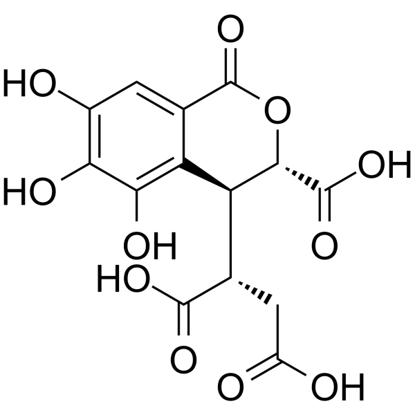
| SMILES | C1=C2C(=C(C(=C1O)O)O)[C@@H]([C@H](OC2=O)C(=O)O)[C@H](CC(=O)O)C(=O)O |
| InChi Key | COZMWVAACFYLBI-XJEVXTIOSA-N |
| InChi Code | InChI=1S/C14H12O11/c15-5-1-4-7(10(19)9(5)18)8(3(12(20)21)2-6(16)17)11(13(22)23)25-14(4)24/h1,3,8,11,15,18-19H,2H2,(H,16,17)(H,20,21)(H,22,23)/t3-,8-,11-/m0/s1 |
| Chemical Name | (2S)-2-[(3S,4S)-3-carboxy-5,6,7-trihydroxy-1-oxo-3,4-dihydroisochromen-4-yl]butanedioic acid |
| HS Tariff Code | 2934.99.9001 |
| Storage |
Powder-20°C 3 years 4°C 2 years In solvent -80°C 6 months -20°C 1 month |
| Shipping Condition | Room temperature (This product is stable at ambient temperature for a few days during ordinary shipping and time spent in Customs) |
Biological Activity
| Targets |
Chebulic acid targets oxidative stress-related enzymes (SOD, CAT, GPx) - enhances enzyme [4] Chebulic acid acts on advanced glycation endproduct (AGE) formation pathway - inhibits AGE-induced RAGE (receptor for AGEs) activation [3] Chebulic acid modulates inflammatory signaling pathways (NF-κB, MAPK) - suppresses pathway activation [1][3][5] |
| ln Vitro |
1. Alveolar epithelial cell protection: - Chebulic acid (10, 20, 40 μM) significantly increased viability of urban particulate matter (UPM)-exposed alveolar epithelial cells (A549) in a concentration-dependent manner (MTT assay) [1] - Chebulic acid reduced UPM-induced intracellular ROS generation and MDA levels, while increasing SOD and CAT activities in A549 cells [1] - Chebulic acid downregulated UPM-induced pro-inflammatory cytokines (TNF-α, IL-6, IL-1β) mRNA and protein expression via inhibiting NF-κB and MAPK pathways [1] 2. Endothelial cell dysfunction improvement: - Chebulic acid (5, 10, 20 μM) inhibited AGE-induced endothelial cell (HUVECs) apoptosis, as evidenced by reduced caspase-3 activity and TUNEL-positive cells [3] - Chebulic acid attenuated AGE-induced ROS production and NADPH oxidase activation in HUVECs, and restored NO production and eNOS activity [3] - Chebulic acid suppressed AGE-induced RAGE expression and downstream NF-κB p65 phosphorylation in HUVECs [3] 3. Hepatocyte antioxidant protection: - Chebulic acid (10, 25, 50 μM) protected isolated rat hepatocytes against H2O2-induced oxidative damage, increasing cell viability and reducing LDH leakage [4] - Chebulic acid enhanced SOD, CAT, and GPx activities, and decreased MDA and GSSG levels in H2O2-treated hepatocytes [4] - Chebulic acid inhibited H2O2-induced hepatocyte apoptosis by reducing Bax/Bcl-2 ratio and caspase-3 activation [4] |
| ln Vivo |
1. Ischemia-reperfusion (I/R) injury attenuation in diabetic rats: - Chebulic acid (25, 50 mg/kg, p.o.) administered for 14 days prior to I/R surgery significantly reduced serum AST, ALT, LDH, and CK-MB levels in streptozotocin-induced diabetic rats [2] - Chebulic acid restored antioxidant status in diabetic I/R rats by increasing SOD, CAT, GPx activities and GSH levels, while decreasing MDA and NO levels in liver and heart tissues [2] - Chebulic acid attenuated I/R-induced inflammatory response in diabetic rats by reducing TNF-α, IL-1β, and ICAM-1 expression in tissues [2] 2. Hepatoprotective activity: - Chebulic acid (10, 20, 40 mg/kg, p.o.) administered for 7 days protected mice against CCl4-induced liver injury, as shown by reduced serum ALT, AST, and ALP levels [5] - Chebulic acid alleviated CCl4-induced hepatic oxidative stress by increasing SOD, CAT, GPx activities and decreasing MDA content in liver tissue [5] - Chebulic acid suppressed CCl4-induced hepatic inflammation via inhibiting NF-κB p65 nuclear translocation and reducing TNF-α, IL-6, and COX-2 expression [5] - Chebulic acid attenuated CCl4-induced hepatocyte apoptosis by downregulating Bax, caspase-3, caspase-9 expression and upregulating Bcl-2 expression [5] |
| Enzyme Assay |
1. Antioxidant enzyme activity assay: - Tissue/cell homogenates are prepared and centrifuged to obtain supernatant [2][4][5] - Supernatant is incubated with specific substrates for SOD, CAT, and GPx respectively [2][4][5] - SOD activity is measured by monitoring the inhibition of nitrite formation from hydroxylamine hydrochloride [2][4][5] - CAT activity is determined by measuring the decomposition of H2O2 at 240 nm [2][4][5] - GPx activity is assayed by detecting the oxidation of NADPH at 340 nm [2][4][5] 2. AGE formation inhibition assay: - Bovine serum albumin (BSA) is incubated with glucose in the presence or absence of Chebulic acid at 37°C for 4 weeks [3] - Fluorescence intensity of AGE-BSA is measured at excitation 370 nm and emission 440 nm to evaluate AGE formation [3] - The inhibition rate of Chebulic acid on AGE formation is calculated compared to the control group [3] 3. Caspase-3 activity assay: - Cell/tissue lysates are prepared and incubated with caspase-3 substrate (Ac-DEVD-pNA) at 37°C for 2 hours [3][4][5] - The absorbance of released p-nitroaniline is measured at 405 nm to determine caspase-3 activity [3][4][5] |
| Cell Assay |
1. Alveolar epithelial cell (A549) protection assay: - A549 cells are cultured in DMEM medium supplemented with fetal bovine serum and antibiotics [1] - Cells are pre-treated with Chebulic acid (10, 20, 40 μM) for 2 hours, then exposed to UPM (100 μg/ml) for 24 hours [1] - Cell viability is detected by MTT assay, ROS generation by DCFH-DA staining, and MDA level by thiobarbituric acid reaction method [1] - TNF-α, IL-6, IL-1β mRNA expression is analyzed by RT-PCR, and protein levels by Western blot [1] 2. Endothelial cell (HUVECs) dysfunction assay: - HUVECs are cultured in EBM-2 medium and pre-treated with Chebulic acid (5, 10, 20 μM) for 1 hour, then incubated with AGE-BSA (100 μg/ml) for 24 hours [3] - Cell apoptosis is evaluated by TUNEL staining and caspase-3 activity assay [3] - ROS production is measured by DCFH-DA staining, NO level by Griess reagent, and eNOS activity by colorimetric assay [3] - RAGE and NF-κB p65 protein expression is detected by Western blot [3] 3. Hepatocyte oxidative damage assay: - Isolated rat hepatocytes are suspended in Williams' medium E and pre-treated with Chebulic acid (10, 25, 50 μM) for 30 minutes, then exposed to H2O2 (0.5 mM) for 1 hour [4] - Cell viability is determined by trypan blue exclusion test, and LDH leakage by colorimetric assay [4] - SOD, CAT, GPx activities and MDA, GSSG levels are measured using respective assay kits [4] - Bax and Bcl-2 protein expression is analyzed by Western blot [4] |
| Animal Protocol |
1. Diabetic rat I/R injury model: - Streptozotocin (60 mg/kg, i.p.) is injected into Wistar rats to induce diabetes (blood glucose >250 mg/dl) [2] - Chebulic acid is dissolved in 0.5% carboxymethyl cellulose (CMC) and administered orally at doses of 25, 50 mg/kg once daily for 14 days [2] - On day 14, rats are anesthetized, and hepatic/renal I/R injury is induced by clamping the hepatic portal vein/renal pedicle for 30 minutes followed by reperfusion for 24 hours [2] - Blood samples and liver/heart tissues are collected for biochemical and histological analysis [2] 2. Mouse CCl4-induced liver injury model: - BALB/c mice are divided into control, CCl4, and Chebulic acid treatment groups (10, 20, 40 mg/kg) [5] - Chebulic acid is dissolved in 0.5% CMC and administered orally once daily for 7 days [5] - On day 7, mice are injected with CCl4 (1 ml/kg, i.p., 10% in olive oil) 2 hours after the last Chebulic acid administration [5] - After 24 hours, blood samples and liver tissues are collected for serum biochemical, histological, and molecular biological analysis [5] |
| References |
[1]. Protective effects of chebulic acid on alveolar epithelial damage induced by urban particulate matter. BMC Complement Altern Med. 2017 Jul 19;17(1):373. [2]. Chebulic acid attenuates ischemia reperfusion induced biochemical alteration in diabetic rats. Pharm Biol. 2013 Jan;51(1):23-9. [3]. Preventive effects of chebulic acid isolated from Terminalia chebula on advanced glycation endproduct-induced endothelial cell dysfunction. Journal of Ethnopharmacology, 2010, 131(3): 567-574. [4]. Isolation of chebulic acid from Terminalia chebula Retz. and its antioxidant effect in isolated rat hepatocytes. Archives of Toxicology, 2007, 81: 211-218. [5]. In vivo hepatoprotective activity and the underlying mechanism of chebulinic acid from Terminalia chebula fruit. Phytomedicine, 2021, 83: 153479. |
| Additional Infomation |
Chebulic acid has been reported in Terminalia citrina, Phyllanthus emblica, and Terminalia chebula with data available. - Chebulic acid is a natural phenolic acid isolated from the fruits of Terminalia chebula Retz. [3][4][5] - Chebulic acid exerts biological activities mainly through antioxidant, anti-inflammatory, and anti-apoptotic mechanisms [1][2][3][4][5] - Chebulic acid shows potential protective effects against oxidative stress-related diseases, including respiratory, cardiovascular, and hepatic disorders [1][2][3][4][5] |
Solubility Data
| Solubility (In Vitro) | DMSO : ~250 mg/mL (~701.77 mM) |
| Solubility (In Vivo) |
Solubility in Formulation 1: ≥ 2.17 mg/mL (6.09 mM) (saturation unknown) in 10% DMSO + 40% PEG300 + 5% Tween80 + 45% Saline (add these co-solvents sequentially from left to right, and one by one), clear solution. For example, if 1 mL of working solution is to be prepared, you can add 100 μL of 21.7 mg/mL clear DMSO stock solution to 400 μL PEG300 and mix evenly; then add 50 μL Tween-80 to the above solution and mix evenly; then add 450 μL normal saline to adjust the volume to 1 mL. Preparation of saline: Dissolve 0.9 g of sodium chloride in 100 mL ddH₂ O to obtain a clear solution. Solubility in Formulation 2: ≥ 2.17 mg/mL (6.09 mM) (saturation unknown) in 10% DMSO + 90% (20% SBE-β-CD in Saline) (add these co-solvents sequentially from left to right, and one by one), clear solution. For example, if 1 mL of working solution is to be prepared, you can add 100 μL of 21.7 mg/mL clear DMSO stock solution to 900 μL of 20% SBE-β-CD physiological saline solution and mix evenly. Preparation of 20% SBE-β-CD in Saline (4°C,1 week): Dissolve 2 g SBE-β-CD in 10 mL saline to obtain a clear solution. (Please use freshly prepared in vivo formulations for optimal results.) |
| Preparing Stock Solutions | 1 mg | 5 mg | 10 mg | |
| 1 mM | 2.8071 mL | 14.0355 mL | 28.0710 mL | |
| 5 mM | 0.5614 mL | 2.8071 mL | 5.6142 mL | |
| 10 mM | 0.2807 mL | 1.4035 mL | 2.8071 mL |