Physicochemical Properties
| Molecular Formula | C32H36N2O5 |
| Molecular Weight | 528.6386 |
| Exact Mass | 528.262 |
| CAS # | 50335-03-0 |
| PubChem CID | 6438437 |
| Appearance | White to off-white solid powder |
| Density | 1.3 g/cm3 |
| Boiling Point | 789.7ºC at 760 mmHg |
| Flash Point | 431.4ºC |
| Index of Refraction | 1.651 |
| LogP | 4.161 |
| Hydrogen Bond Donor Count | 3 |
| Hydrogen Bond Acceptor Count | 5 |
| Rotatable Bond Count | 2 |
| Heavy Atom Count | 39 |
| Complexity | 1140 |
| Defined Atom Stereocenter Count | 9 |
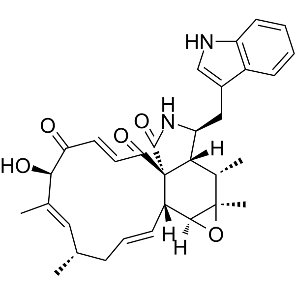
| SMILES | C[C@H]\1C/C=C/[C@H]2[C@H]3[C@](O3)([C@H]([C@@H]4[C@@]2(C(=O)/C=C/C(=O)[C@@H](/C(=C1)/C)O)C(=O)N[C@H]4CC5=CNC6=CC=CC=C65)C)C |
| InChi Key | OUMWCYMRLMEZJH-VOXRAUTJSA-N |
| InChi Code | InChI=1S/C32H36N2O5/c1-17-8-7-10-22-29-31(4,39-29)19(3)27-24(15-20-16-33-23-11-6-5-9-21(20)23)34-30(38)32(22,27)26(36)13-12-25(35)28(37)18(2)14-17/h5-7,9-14,16-17,19,22,24,27-29,33,37H,8,15H2,1-4H3,(H,34,38)/b10-7+,13-12+,18-14+/t17-,19-,22-,24-,27-,28+,29-,31+,32+/m0/s1 |
| Chemical Name | (1R,3E,6R,7E,9S,11E,13R,14S,16R,17S,18R,19S)-6-hydroxy-19-(1H-indol-3-ylmethyl)-7,9,16,17-tetramethyl-15-oxa-20-azatetracyclo[11.8.0.01,18.014,16]henicosa-3,7,11-triene-2,5,21-trione |
| HS Tariff Code | 2934.99.9001 |
| Storage |
Powder-20°C 3 years 4°C 2 years In solvent -80°C 6 months -20°C 1 month |
| Shipping Condition | Room temperature (This product is stable at ambient temperature for a few days during ordinary shipping and time spent in Customs) |
Biological Activity
| Targets |
- The primary target of Chaetoglobosin A is the cytoskeleton, specifically targeting actin filaments (F-actin) involved in maintaining cell structure and function [1] |
| ln Vitro |
- Chaetoglobosin A preferentially induces apoptosis in chronic lymphocytic leukemia (CLL) cells. In vitro culture of primary CLL cells (from patients) with Chaetoglobosin A (0.1–10 μM) for 24–48 hours results in dose-dependent apoptosis, with an IC₅₀ of 0.8 μM (assessed by Annexin V-FITC/PI flow cytometry). In contrast, it shows minimal apoptotic effects on normal peripheral blood mononuclear cells (PBMCs) from healthy donors (apoptosis rate <10% at 5 μM, vs. >60% in CLL cells at the same concentration) [1] - Chaetoglobosin A disrupts the cytoskeleton by inhibiting actin filament polymerization and inducing F-actin depolymerization. Immunofluorescence staining of CLL cells treated with 1 μM Chaetoglobosin A for 6 hours shows fragmented and disorganized F-actin networks, compared to intact, fibrous F-actin in untreated cells. This cytoskeletal disruption leads to loss of cell adhesion, abnormal cell morphology (shrinkage, rounding), and impaired cell migration (reduced migration distance by 70% in Transwell assays) [1] - Chaetoglobosin A modulates apoptosis-related signaling pathways in CLL cells. Western blot analysis reveals that treatment with 0.5–2 μM Chaetoglobosin A increases the expression of cleaved caspase-3, cleaved caspase-9, and Bax, while decreasing the expression of anti-apoptotic protein Bcl-2. It also downregulates the phosphatidylinositol 3-kinase (PI3K)/Akt pathway, as evidenced by reduced phosphorylation of Akt (p-Akt) [1] - Chaetoglobosin A inhibits CLL cell proliferation. MTT assays show that treatment with 0.1–5 μM Chaetoglobosin A for 72 hours reduces CLL cell viability by 50–90% in a dose-dependent manner, with no significant effect on the viability of normal B cells (viability >80% at 5 μM) [1] |
| ln Vivo |
- In a murine xenograft model of human CLL (NOD/SCID mice injected with primary CLL cells via tail vein), intraperitoneal injection of Chaetoglobosin A (2 mg/kg body weight, once every 2 days for 3 weeks) significantly inhibits tumor growth. The mean tumor burden (assessed by human CD5+/CD19+ CLL cell count in mouse peripheral blood and spleen) is reduced by 65% compared to the vehicle control group (DMSO + normal saline). Additionally, the treatment prolongs the median survival of mice by 21 days (from 42 days in controls to 63 days in treated group) [1] - Chaetoglobosin A shows no obvious systemic toxicity in vivo. Mice treated with 2 mg/kg Chaetoglobosin A maintain stable body weight (no >5% weight loss) throughout the treatment period. Histopathological analysis of major organs (liver, kidney, heart, lung, spleen) reveals no significant tissue damage or inflammation, compared to the control group [1] |
| Enzyme Assay |
1. Prepare purified rabbit muscle G-actin (globular actin) in G-actin buffer (containing Tris-HCl, ATP, CaCl₂, DTT). Adjust the G-actin concentration to 2 μM. 2. Add serial dilutions of Chaetoglobosin A (0.01–10 μM) to the G-actin solution; the vehicle control group receives an equal volume of DMSO. Incubate the mixture at room temperature for 10 minutes. 3. Initiate actin polymerization by adding polymerization buffer (containing KCl, MgCl₂) to the mixture, and transfer the solution to a 96-well black microplate. 4. Monitor the fluorescence intensity of pyrene-labeled G-actin (pre-mixed with unlabeled G-actin at a 1:10 ratio) every 2 minutes for 60 minutes using a microplate reader (excitation wavelength 365 nm, emission wavelength 407 nm). 5. Calculate the polymerization rate as the slope of the fluorescence increase curve in the initial 15 minutes. Determine the concentration of Chaetoglobosin A that inhibits 50% of actin polymerization (IC₅₀) by plotting the inhibition rate against drug concentration [1] |
| Cell Assay |
- CLL Cell Viability Assay: 1. Isolate primary CLL cells from patients’ peripheral blood using density gradient centrifugation (Ficoll-Paque). Culture the cells in RPMI 1640 medium supplemented with 10% fetal bovine serum, 100 U/mL penicillin, and 100 μg/mL streptomycin at 37°C with 5% CO₂. 2. Seed the cells in 96-well plates at a density of 5×10⁴ cells/well. Add serial dilutions of Chaetoglobosin A (0.1–10 μM) to the wells; the control group receives DMSO (final concentration <0.1%). 3. Incubate the cells for 72 hours, then add MTT reagent (5 mg/mL) to each well (10 μL/well) and continue incubating for 4 hours. 4. Add 100 μL of DMSO to each well to dissolve the formazan crystals. Measure the absorbance at 570 nm using a microplate reader. Calculate cell viability as (absorbance of treated group / absorbance of control group) × 100% [1] - Apoptosis Detection Assay: 1. Culture CLL cells (1×10⁶ cells/mL) with 0.5–2 μM Chaetoglobosin A for 24 hours. 2. Harvest the cells by centrifugation (1000 × g for 5 minutes), wash twice with cold PBS, and resuspend in binding buffer. 3. Add Annexin V-FITC (5 μL) and propidium iodide (PI, 10 μL) to the cell suspension, incubate in the dark at room temperature for 15 minutes. 4. Analyze the cells using flow cytometry to quantify the percentage of apoptotic cells (Annexin V-positive/PI-negative and Annexin V-positive/PI-positive) [1] - Actin Filament Localization Assay: 1. Seed CLL cells on glass coverslips in 24-well plates, culture overnight, then treat with 1 μM Chaetoglobosin A for 6 hours. 2. Fix the cells with 4% paraformaldehyde for 15 minutes, permeabilize with 0.1% Triton X-100 for 5 minutes, and block with 1% BSA for 30 minutes. 3. Incubate the cells with Alexa Fluor 488-conjugated phalloidin (specific for F-actin) for 1 hour at room temperature, then stain nuclei with DAPI for 5 minutes. 4. Mount the coverslips with antifade mounting medium and observe under a confocal laser scanning microscope to visualize F-actin distribution [1] |
| Animal Protocol |
- CLL Murine Xenograft Model Protocol: 1. Use 6–8 week-old female NOD/SCID mice. Irradiate the mice with 200 cGy 24 hours before cell injection to immunosuppress. 2. Prepare primary CLL cells from patients (1×10⁷ cells/mouse) in sterile PBS. Inject the cells into the mice via tail vein to establish the CLL xenograft model. 3. Seven days after cell injection, randomly divide the mice into two groups (n=6 per group): - Treatment group: Administer Chaetoglobosin A (dissolved in DMSO and diluted with normal saline to a final DMSO concentration of 5%) via intraperitoneal injection at a dose of 2 mg/kg body weight, once every 2 days for 3 weeks. - Control group: Administer an equal volume of vehicle (5% DMSO in normal saline) via intraperitoneal injection on the same schedule. 4. During the treatment period, measure the mice’s body weight once a week. Collect 50 μL of peripheral blood from the tail vein every 2 weeks to detect human CD5+/CD19+ CLL cells by flow cytometry (to assess tumor burden). 5. At the end of treatment, sacrifice the mice, harvest the spleen and liver, weigh the organs, and prepare tissue sections for hematoxylin-eosin (HE) staining and immunohistochemistry (to detect CLL cell infiltration). Record the mice’s survival time for survival curve analysis [1] |
| Toxicity/Toxicokinetics |
- In vitro, Chaetoglobosin A exhibits selective toxicity toward CLL cells. At a concentration of 5 μM (5-fold higher than the IC₅₀ for CLL cells), it induces apoptosis in only <10% of normal PBMCs and <15% of normal B cells from healthy donors, indicating low off-target toxicity [1] - In vivo, administration of Chaetoglobosin A (2 mg/kg, intraperitoneal, once every 2 days for 3 weeks) to NOD/SCID mice causes no significant systemic toxicity: - Body weight: No >5% reduction compared to baseline or the control group. - Major organs: HE staining of liver, kidney, heart, lung, and spleen shows no obvious necrosis, inflammation, or structural damage. - Hematology: No significant changes in white blood cell count, red blood cell count, or platelet count compared to the control group [1] |
| References |
[1]. Chaetoglobosin A preferentially induces apoptosis in chronic lymphocytic leukemia cells by targeting the cytoskeleton. Leukemia. 2014 Jun;28(6):1289-98. |
| Additional Infomation |
Chaetoglobosin A has been reported in Chaetomium subaffine, Cunila, and other organisms with data available. - Chaetoglobosin A is a secondary metabolite isolated from fungi of the genus Chaetomium (e.g., Chaetomium globosum). It belongs to the cytochalasin family of natural products, which are known for their ability to modulate the cytoskeleton [1] - The selective apoptosis induction of Chaetoglobosin A in CLL cells is attributed to the unique dependency of CLL cells on intact cytoskeletal function for survival and proliferation. Normal lymphocytes have more robust cytoskeletal repair mechanisms, making them less sensitive to the compound [1] - Chaetoglobosin A represents a potential lead compound for the development of novel CLL therapeutics, as it overcomes limitations of existing CLL drugs (e.g., high toxicity to normal cells, development of drug resistance) by targeting the cytoskeleton, a non-traditional but critical pathway in CLL cells [1] |
Solubility Data
| Solubility (In Vitro) | May dissolve in DMSO (in most cases), if not, try other solvents such as H2O, Ethanol, or DMF with a minute amount of products to avoid loss of samples |
| Solubility (In Vivo) |
Note: Listed below are some common formulations that may be used to formulate products with low water solubility (e.g. < 1 mg/mL), you may test these formulations using a minute amount of products to avoid loss of samples. Injection Formulations (e.g. IP/IV/IM/SC) Injection Formulation 1: DMSO : Tween 80: Saline = 10 : 5 : 85 (i.e. 100 μL DMSO stock solution → 50 μL Tween 80 → 850 μL Saline) *Preparation of saline: Dissolve 0.9 g of sodium chloride in 100 mL ddH ₂ O to obtain a clear solution. Injection Formulation 2: DMSO : PEG300 :Tween 80 : Saline = 10 : 40 : 5 : 45 (i.e. 100 μL DMSO → 400 μLPEG300 → 50 μL Tween 80 → 450 μL Saline) Injection Formulation 3: DMSO : Corn oil = 10 : 90 (i.e. 100 μL DMSO → 900 μL Corn oil) Example: Take the Injection Formulation 3 (DMSO : Corn oil = 10 : 90) as an example, if 1 mL of 2.5 mg/mL working solution is to be prepared, you can take 100 μL 25 mg/mL DMSO stock solution and add to 900 μL corn oil, mix well to obtain a clear or suspension solution (2.5 mg/mL, ready for use in animals). Injection Formulation 4: DMSO : 20% SBE-β-CD in saline = 10 : 90 [i.e. 100 μL DMSO → 900 μL (20% SBE-β-CD in saline)] *Preparation of 20% SBE-β-CD in Saline (4°C,1 week): Dissolve 2 g SBE-β-CD in 10 mL saline to obtain a clear solution. Injection Formulation 5: 2-Hydroxypropyl-β-cyclodextrin : Saline = 50 : 50 (i.e. 500 μL 2-Hydroxypropyl-β-cyclodextrin → 500 μL Saline) Injection Formulation 6: DMSO : PEG300 : castor oil : Saline = 5 : 10 : 20 : 65 (i.e. 50 μL DMSO → 100 μLPEG300 → 200 μL castor oil → 650 μL Saline) Injection Formulation 7: Ethanol : Cremophor : Saline = 10: 10 : 80 (i.e. 100 μL Ethanol → 100 μL Cremophor → 800 μL Saline) Injection Formulation 8: Dissolve in Cremophor/Ethanol (50 : 50), then diluted by Saline Injection Formulation 9: EtOH : Corn oil = 10 : 90 (i.e. 100 μL EtOH → 900 μL Corn oil) Injection Formulation 10: EtOH : PEG300:Tween 80 : Saline = 10 : 40 : 5 : 45 (i.e. 100 μL EtOH → 400 μLPEG300 → 50 μL Tween 80 → 450 μL Saline) Oral Formulations Oral Formulation 1: Suspend in 0.5% CMC Na (carboxymethylcellulose sodium) Oral Formulation 2: Suspend in 0.5% Carboxymethyl cellulose Example: Take the Oral Formulation 1 (Suspend in 0.5% CMC Na) as an example, if 100 mL of 2.5 mg/mL working solution is to be prepared, you can first prepare 0.5% CMC Na solution by measuring 0.5 g CMC Na and dissolve it in 100 mL ddH2O to obtain a clear solution; then add 250 mg of the product to 100 mL 0.5% CMC Na solution, to make the suspension solution (2.5 mg/mL, ready for use in animals). Oral Formulation 3: Dissolved in PEG400 Oral Formulation 4: Suspend in 0.2% Carboxymethyl cellulose Oral Formulation 5: Dissolve in 0.25% Tween 80 and 0.5% Carboxymethyl cellulose Oral Formulation 6: Mixing with food powders Note: Please be aware that the above formulations are for reference only. InvivoChem strongly recommends customers to read literature methods/protocols carefully before determining which formulation you should use for in vivo studies, as different compounds have different solubility properties and have to be formulated differently. (Please use freshly prepared in vivo formulations for optimal results.) |
| Preparing Stock Solutions | 1 mg | 5 mg | 10 mg | |
| 1 mM | 1.8916 mL | 9.4582 mL | 18.9165 mL | |
| 5 mM | 0.3783 mL | 1.8916 mL | 3.7833 mL | |
| 10 mM | 0.1892 mL | 0.9458 mL | 1.8916 mL |