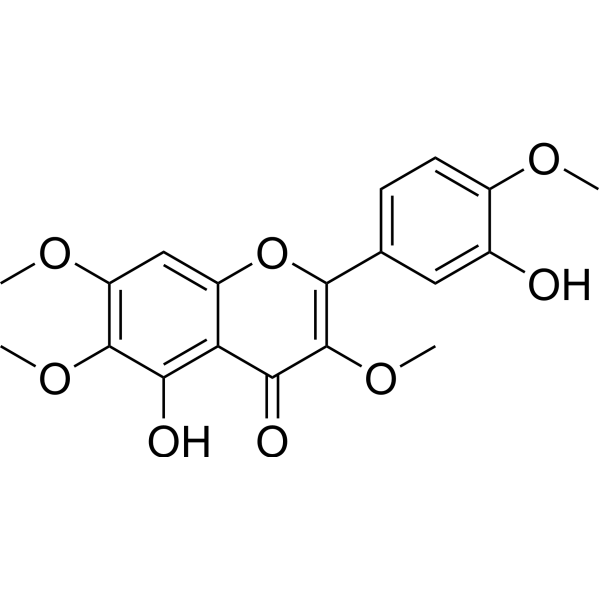
Physicochemical Properties
| Molecular Formula | C19H18O8 |
| Molecular Weight | 374.3414 |
| Exact Mass | 374.1 |
| CAS # | 479-91-4 |
| PubChem CID | 5315263 |
| Appearance | Light yellow to yellow solid powder |
| Density | 1.4±0.1 g/cm3 |
| Boiling Point | 617.7±55.0 °C at 760 mmHg |
| Melting Point | 186 - 187 °C |
| Flash Point | 223.5±25.0 °C |
| Vapour Pressure | 0.0±1.9 mmHg at 25°C |
| Index of Refraction | 1.640 |
| LogP | 2.13 |
| Hydrogen Bond Donor Count | 2 |
| Hydrogen Bond Acceptor Count | 8 |
| Rotatable Bond Count | 5 |
| Heavy Atom Count | 27 |
| Complexity | 576 |
| Defined Atom Stereocenter Count | 0 |
| InChi Key | PJQLSMYMOKWUJG-UHFFFAOYSA-N |
| InChi Code | InChI=1S/C19H18O8/c1-23-11-6-5-9(7-10(11)20)17-19(26-4)16(22)14-12(27-17)8-13(24-2)18(25-3)15(14)21/h5-8,20-21H,1-4H3 |
| Chemical Name | 5-hydroxy-2-(3-hydroxy-4-methoxyphenyl)-3,6,7-trimethoxychromen-4-one |
| HS Tariff Code | 2934.99.9001 |
| Storage |
Powder-20°C 3 years 4°C 2 years In solvent -80°C 6 months -20°C 1 month Note: This product requires protection from light (avoid light exposure) during transportation and storage. |
| Shipping Condition | Room temperature (This product is stable at ambient temperature for a few days during ordinary shipping and time spent in Customs) |
Biological Activity
| Targets |
- Casticin targets microtubule dynamics (mediates antimitotic activity and G2-M arrest), with IC50 values of ~2.5 μM (HeLa cells), ~3.1 μM (A549 cells), and ~3.5 μM (MCF-7 cells) for antiproliferative activity. [1] - Casticin targets TGF-β/Smad signaling pathway (inhibits Smad2/3 phosphorylation and nuclear translocation), effective at concentrations of 10–40 μM in hepatic stellate cells (HSCs). [2] - Casticin targets STAT3 signaling cascade (suppresses STAT3 phosphorylation at Tyr705), with an IC50 of ~4 μM for inhibiting p-STAT3 in A549 cells. [3] |
| ln Vitro |
Casticin (0.2-1.0 μM) had no discernible inhibitory impact on 3T3 Swiss Albino and TIG-103 cells, but it dose-dependently reduced the proliferation of KB cells, with an IC50 of 0.23 μM on day 3. Spindle morphology is altered by ricin (0.6 μM), which can result in spindle disorder or partial mitotic spindle disruption [1]. LX2 cell proliferation is dose-dependently inhibited by caucin (0–40 μM). Apoptosis is induced and L02 cell growth is inhibited by caucin (40 μM). Casticin assesses the mRNA levels of TGF-β, collagen α1(I), MMP-2, MMP-9, TIMP-1, and TIMP-2 and inhibits the fibrotic action of TGF-β1 on ECM deposition in LX2 cells [2]. The vitality of 786-O, YD-8, and HN-9 cells is decreased by cacticin (0-8 μM), whereas normal HEL 299 cells are not significantly affected. Casticin (5 μM) decreases stroma levels, vascular endothelial growth factor (VEGF), B-cell lymphoma-extra large (Bcl-xl), Bcl-2, IAP-1/-2, MMP-9, and COX-2 proteins in O, YD-8, and HN-9 cells, and promotes caspase-3 and PPAR cleavage. Additionally, calcin (5 μM) can cause apoptosis, prevent IL-6-induced STAT3 activation, block continuously active STAT3 in tumor cells, and regulate STAT3 activation by altering the activity of upstream STAT3 regulators. Furthermore, Casticin (2.5 μM) improves the therapeutic effect of radiation therapy and the effect of ionizing radiation in 786-O cells [3]. - Antimitotic & G2-M Arrest Activity: 1. Antiproliferation: Casticin (1–10 μM) dose-dependently inhibited viability of HeLa, A549, and MCF-7 cells; 72-hour treatment with 5 μM reduced cell viability by ~60%–75% (MTT assay). [1] 2. G2-M arrest: Flow cytometry showed Casticin (2.5 μM, 24 hours) increased HeLa cells in G2-M phase from ~12% (control) to ~45%; immunofluorescence revealed abnormal spindle formation (disorganized microtubules). [1] 3. Antimitotic effect: Casticin (5 μM) reduced mitotic index by ~70% vs. control, with most cells arrested at prometaphase. [1] - Hepatic Stellate Cell (HSC) Activation Inhibition: 1. HSC deactivation: Casticin (10–40 μM) dose-dependently reduced α-SMA (a HSC activation marker) and collagen I expression in activated HSCs; 40 μM reduced α-SMA protein levels by ~80% (western blot) vs. TGF-β-induced group. [2] 2. TGF-β/Smad blocking: Casticin (20–40 μM) inhibited TGF-β-induced Smad2/3 phosphorylation by ~50%–70% and nuclear translocation by ~65% (immunofluorescence). [2] - STAT3 Inhibition & Radiosensitization: 1. STAT3 suppression: Casticin (2–8 μM) dose-dependently reduced p-STAT3 (Tyr705) levels in A549 cells; 4 μM reduced p-STAT3 by ~60% (western blot) vs. control. [3] 2. Radiosensitization: Casticin (4 μM) + ionizing radiation (IR, 4 Gy) increased A549 cell apoptosis by ~2.5-fold (Annexin V-PI staining) vs. IR alone; enhanced cleavage of caspase-3 and PARP (western blot). [3] 3. Clone formation inhibition: Casticin (2–4 μM) + IR reduced A549 clone formation efficiency by ~70% vs. IR alone. [3] |
| ln Vivo |
Mice with liver impairment induced by BDL and CCl4 exhibit toxic effects on their livers when exposed to 20 mg/kg of cacticin po. In vivo, CCl4 or BDL-induced liver fibrosis is attenuated by cacticin. Casticin blocks TGF-β/Smad signaling in vivo to prevent HSC activation and collagen matrix expression [2]. - Liver Fibrosis Attenuation: 1. Animal model: C57BL/6 mice induced with liver fibrosis via CCl4 (0.5 mL/kg, ip, twice weekly for 4 weeks). [2] 2. Treatment: Casticin (20, 40 mg/kg, ig, daily) for 4 weeks during CCl4 induction. [2] 3. Efficacy: 40 mg/kg Casticin reduced liver collagen deposition by ~65% (Masson staining), decreased serum ALT/AST levels by ~50%–60%, and downregulated hepatic α-SMA/collagen I expression by ~70% (western blot) vs. CCl4 group. [2] - Tumor Growth Inhibition & Radiosensitization: 1. Animal model: A549 xenograft nude mice (5×10⁶ cells, subcutaneous injection). [3] 2. Treatment: Casticin (10 mg/kg, ip, every 2 days) + IR (2 Gy, weekly for 3 weeks); control groups: vehicle, Casticin alone, IR alone. [3] 3. Efficacy: Casticin + IR reduced tumor volume by ~75% and tumor weight by ~70% vs. IR alone; tumor tissues showed reduced p-STAT3 (~65%) and increased cleaved caspase-3 (~2.0-fold) vs. IR alone. [3] |
| Cell Assay |
- Cell Cycle & Antimitotic Assay: 1. Cell culture: HeLa/A549 cells seeded in 6-well plates (2×10⁵ cells/well) in RPMI 1640 (10% FBS) at 37°C (5% CO₂) for 24 hours. [1] 2. Treatment: Casticin (1–5 μM) added for 24–48 hours; vehicle (DMSO, ≤0.1% v/v) as control. [1] 3. Cell cycle detection: Cells fixed with 70% ethanol, stained with PI (50 μg/mL) for 30 minutes, analyzed via flow cytometry (excitation 488 nm) to quantify G2-M phase cells. [1] 4. Antimitotic observation: Cells fixed with 4% paraformaldehyde, stained with α-tubulin antibody (FITC-conjugated) and DAPI, imaged via confocal microscopy to observe spindle morphology. [1] - HSC Activation Assay: 1. HSC isolation: Primary HSCs isolated from rat liver via collagenase perfusion, cultured in DMEM (10% FBS) for 7 days to induce activation. [2] 2. Treatment: Casticin (10–40 μM) added with/without TGF-β (5 ng/mL) for 24–48 hours. [2] 3. Protein detection: Cells lysed, western blot performed with anti-α-SMA, anti-collagen I, anti-p-Smad2/3, anti-total Smad2/3 antibodies; β-actin as internal control. [2] - STAT3 & Apoptosis Assay: 1. Cell culture: A549 cells seeded in 6-well plates (1.5×10⁵ cells/well) in DMEM (10% FBS) for 24 hours. [3] 2. Treatment: Casticin (2–8 μM) added for 24 hours, then IR (4 Gy) applied; incubation continued for 48 hours. [3] 3. Apoptosis detection: Cells stained with Annexin V-FITC/PI for 15 minutes, analyzed via flow cytometry to count apoptotic cells. [3] 4. STAT3 detection: Western blot with anti-p-STAT3 (Tyr705), anti-total STAT3 antibodies; β-actin as internal control. [3] |
| Animal Protocol |
- CCl4-Induced Liver Fibrosis Mouse Model: 1. Animal preparation: Male C57BL/6 mice (6–8 weeks old, 20–22 g) acclimated for 1 week (free food/water, 25°C, 12h light/dark). [2] 2. Model induction: CCl4 (0.5 mL/kg, mixed with olive oil 1:1, ip) twice weekly for 4 weeks. [2] 3. Drug preparation: Casticin dissolved in DMSO (5% v/v) + 0.5% CMC-Na to 20/40 mg/kg. [2] 4. Administration: Casticin (20/40 mg/kg, ig, daily) for 4 weeks; control groups: vehicle (ig) + CCl4, normal mice (no CCl4, no drug). [2] 5. Sample collection: Mice euthanized, serum collected for ALT/AST detection; liver tissues fixed in 4% paraformaldehyde (Masson staining) or stored at -80°C (western blot). [2] - A549 Xenograft Nude Mouse Model: 1. Animal preparation: BALB/c nude mice (4–6 weeks old, female) acclimated for 1 week. [3] 2. Tumor induction: A549 cells (5×10⁶ cells in 100 μL PBS/Matrigel 1:1) subcutaneously injected into right flank. [3] 3. Drug preparation: Casticin dissolved in DMSO (5% v/v) + saline to 10 mg/kg. [3] 4. Administration: When tumors reached ~100 mm³, Casticin (10 mg/kg, ip, every 2 days) + IR (2 Gy, weekly for 3 weeks); control groups: vehicle (ip) + IR, Casticin alone, IR alone. [3] 5. Sample collection: Mice euthanized, tumors weighed/measured; tumor tissues fixed (IHC for p-STAT3) or stored at -80°C (western blot). [3] |
| Toxicity/Toxicokinetics |
- In Vitro Safety: Casticin (1–10 μM) had no significant cytotoxicity on normal cells (e.g., primary hepatocytes, normal lung fibroblasts), with cell viability >80% after 48-hour treatment. [2][3] - In Vivo Safety: In liver fibrosis and xenograft models, Casticin (up to 40 mg/kg, ig; 10 mg/kg, ip) did not affect mouse body weight, and serum creatinine/BUN remained normal vs. control groups. [2][3] |
| References |
[1]. G2-M arrest and antimitotic activity mediated by casticin, a flavonoid isolated from Viticis Fructus (Vitex rotundifolia Linne fil.). Cancer Lett. 2004 May 10;208(1):59-64. [2]. Casticin attenuates liver fibrosis and hepatic stellate cell activation by blocking TGF-β/Smad signaling pathway. Oncotarget. 2017 Apr 27;8(34):56267-56280. [3]. Casticin inhibits growth and enhances ionizing radiation-induced apoptosis through the suppression of STAT3 signaling cascade. J Cell Biochem. 2018 Dec 5. |
| Additional Infomation |
Casticin is a tetramethoxyflavone that consists of quercetagetin in which the hydroxy groups at positions 3, 6, 7 and 4' have been replaced by methoxy groups. It has been isolated from Eremophila mitchellii. It has a role as an apoptosis inducer and a plant metabolite. It is a tetramethoxyflavone and a dihydroxyflavone. It is functionally related to a quercetagetin. Casticin has been reported in Artemisia incanescens, Vitex quinata, and other organisms with data available. See also: Chaste tree fruit (part of). - Natural Source & Chemical Class: Casticin is a polymethoxylated flavonoid isolated from the fruits of Vitex rotundifolia Linne fil. (Viticis Fructus), a traditional Chinese medicinal herb. [1][2][3] - Mechanisms of Action: 1. Anticancer (Literature [1][3]): Interferes with microtubule polymerization to induce G2-M arrest; inhibits STAT3 phosphorylation to suppress tumor cell proliferation and enhance radiosensitivity. [1][3] 2. Antifibrotic: Blocks TGF-β/Smad signaling to inhibit hepatic stellate cell activation and collagen deposition, thereby attenuating liver fibrosis. [2] |
Solubility Data
| Solubility (In Vitro) | DMSO : ~100 mg/mL (~267.14 mM) |
| Solubility (In Vivo) |
Solubility in Formulation 1: ≥ 6.25 mg/mL (16.70 mM) (saturation unknown) in 10% DMSO + 90% (20% SBE-β-CD in Saline) (add these co-solvents sequentially from left to right, and one by one), clear solution. For example, if 1 mL of working solution is to be prepared, you can add 100 μL of 62.5 mg/mL clear DMSO stock solution to 900 μL of 20% SBE-β-CD physiological saline solution and mix evenly. Preparation of 20% SBE-β-CD in Saline (4°C,1 week): Dissolve 2 g SBE-β-CD in 10 mL saline to obtain a clear solution. (Please use freshly prepared in vivo formulations for optimal results.) |
| Preparing Stock Solutions | 1 mg | 5 mg | 10 mg | |
| 1 mM | 2.6714 mL | 13.3568 mL | 26.7137 mL | |
| 5 mM | 0.5343 mL | 2.6714 mL | 5.3427 mL | |
| 10 mM | 0.2671 mL | 1.3357 mL | 2.6714 mL |