Physicochemical Properties
| Molecular Formula | C20H37N3O14 |
| Molecular Weight | 4000-6000 |
| Exact Mass | 543.227 |
| CAS # | 83512-85-0 |
| PubChem CID | 71306969 |
| Appearance | Typically exists as White to off-white solid at room temperature |
| Density | 1.6±0.1 g/cm3 |
| Boiling Point | 971.8±65.0 °C at 760 mmHg |
| Flash Point | 541.5±34.3 °C |
| Vapour Pressure | 0.0±0.6 mmHg at 25°C |
| Index of Refraction | 1.648 |
| LogP | -1.66 |
| Hydrogen Bond Donor Count | 11 |
| Hydrogen Bond Acceptor Count | 16 |
| Rotatable Bond Count | 8 |
| Heavy Atom Count | 37 |
| Complexity | 753 |
| Defined Atom Stereocenter Count | 13 |
| SMILES | OC[C@H]1O[C@@H](OC2[C@@H](CO)O[C@@H](OC3[C@@H](CO)O[C@@H](O)[C@H](N)[C@H]3O)[C@H](NC(=O)C)[C@H]2O)[C@H](N)[C@@H](O)[C@@H]1O |
| InChi Key | HGNQXZLWHDQLRE-XADFZESNSA-N |
| InChi Code | InChI=1S/C20H37N3O14/c1-5(27)23-11-15(31)17(36-19-10(22)13(29)12(28)6(2-24)34-19)8(4-26)35-20(11)37-16-7(3-25)33-18(32)9(21)14(16)30/h6-20,24-26,28-32H,2-4,21-22H2,1H3,(H,23,27)/t6-,7-,8-,9-,10-,11-,12-,13-,14-,15-,16?,17?,18-,19+,20+/m1/s1 |
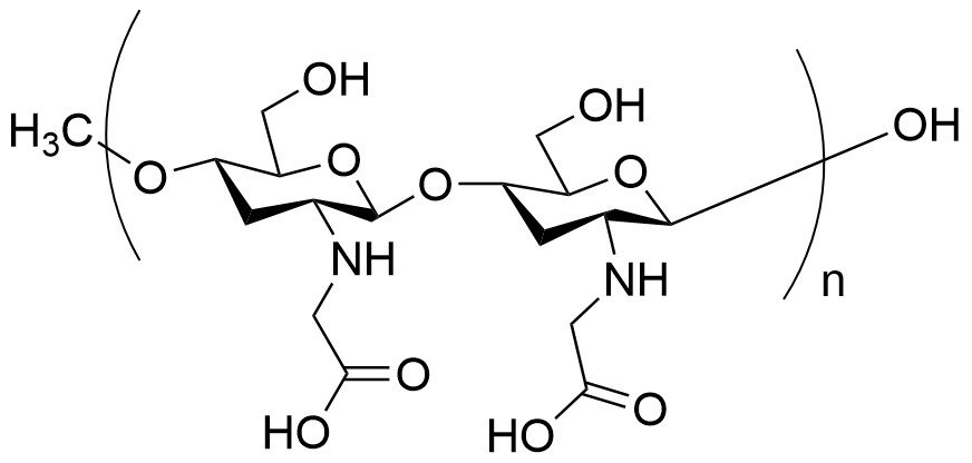
| Chemical Name | N-[(2S,3R,4R,6R)-5-[(2S,3R,4R,5S,6R)-3-amino-4,5-dihydroxy-6-(hydroxymethyl)oxan-2-yl]oxy-2-[(2R,4R,5R,6R)-5-amino-4,6-dihydroxy-2-(hydroxymethyl)oxan-3-yl]oxy-4-hydroxy-6-(hydroxymethyl)oxan-3-yl]acetamide |
| Synonyms | Carboxymethyl chitosan; 83512-85-0; N-[(2S,3R,4R,6R)-5-[(2S,3R,4R,5S,6R)-3-amino-4,5-dihydroxy-6-(hydroxymethyl)oxan-2-yl]oxy-2-[(2R,4R,5R,6R)-5-amino-4,6-dihydroxy-2-(hydroxymethyl)oxan-3-yl]oxy-4-hydroxy-6-(hydroxymethyl)oxan-3-yl]acetamide; MFCD00677440; N-Acetylchitosan; YC29683 |
| HS Tariff Code | 2934.99.9001 |
| Storage |
Powder-20°C 3 years 4°C 2 years In solvent -80°C 6 months -20°C 1 month Note: Please store this product in a sealed and protected environment, avoid exposure to moisture. |
| Shipping Condition | Room temperature (This product is stable at ambient temperature for a few days during ordinary shipping and time spent in Customs) |
Biological Activity
| Targets | Biochemical reagent |
| ln Vitro | The antibacterial carboxymethyl chitosan/ZnO nanocomposite hydrogels were successfully prepared via in situ formation of ZnO nanorods in the crosslinked carboxymethyl chitosan (CMCh) matrix, by treating the CMCh hydrogel matrix with zinc nitrate solution followed by the oxidation of zinc ions with alkaline solution. The resulting CMCh/ZnO hydrogels were characterized by using FTIR spectroscopy, X-ray diffractormetry and scanning electron microscopy (SEM). SEM micrographs revealed the formation of ZnO nanorods in the hydrogel matrix with the size ranging from 190nm to 600nm. The swelling behavior of the prepared nanocomposite hydrogels was also investigated in different pH solutions. The CMCh/ZnO nanocomposite hydrogel showed rather higher swelling behavior in different pH solutions in comparison with neat CMCh hydrogel. Furthermore, the antibacterial activity of CMCh/ZnO hydrogel was studied against Escherichia coli and Staphylococcus aureus by CFU assay[2]. |
| ln Vivo | The volume phase transition of a hydrogel initiated by shrinking may result in complex patterns on its surface. Based on this unique property of hydrogel, we have developed a novel solvent precipitation method to prepare a kind of novel superabsorbent polymers with excellent hemostatic properties. A porous carboxymethyl chitosan grafted poly (acrylic acid) (CMCTS-g-PAA) superabsorbent polymer was prepared by precipitating CMCTS-g-PAA hydrogel with ethanol. Its potential application in hemostatic wound dressing was investigated. The results indicate that the modified superabsorbent polymer is non-cytotoxic. It showed a high swelling capacity and better hemostatic performance in the treatments of hemorrhage model of ear artery, arteria cruralis and spleen of the New Zealand white rabbit than the unmodified polymer and other commonly used clinic wound dressings. The hemostatic mechanism of the porous CMCTS-g-PAA polymer was also discussed[1]. |
| Enzyme Assay |
Preparation and characterization of carboxymethyl chitosan Chitosan powder was alkalized using sodium hydroxide 40% (b/v) for 15 min. The mixture was added to monochloroacetic acid at a ratio of 1:7 and stirred for 4 h at 80 °C. The mixture was then neutralized with acetate acid 10% (v/v) and excess 70% methanol. Afterward, the mixture was filtered and washed using methanol. The remaining sediment in the filter was the modified chitosan product. Finally, the precipitate was dried at 55 °C. Characterization of carboxymethyl chitosan includes analysis with FTIR and a solubility test in 0.5% acetic acid. The effect of GSH concentration on the self-assembly behavior of PCL-SS-carboxymethyl chitosan-GA polymer was analyzed by the fluorene fluorescence probe method. As shown in the figure, the fluorescence intensity (I373) and I373/I383 did not change obviously in the GSH concentration range of 0.1 ∼ 100 μM, indicating that PCL-SS-carboxymethyl chitosan-GA micelles could exist in a stable micelle state. When the concentration of GSH increased to 1 ∼ 10 mM, I373 and I373/I383 changed drastically, indicating that the micelles were unstable at this concentration and transformed into irregular aggregates. As the concentration kept going up, PCL-SS-carboxymethyl chitosan-GA existed as random aggregates. This was confirmed by DLS, PCL-SS-carboxymethyl chitosan-GA possessed a narrower particle size distribution in the low-concentration GSH (< 100 μM) solution. However, the size distribution became scattered when the GSH concentration increased to 10 mM. [Healing effect of carboxymethyl chitosan-plantamajoside hydrogel on burn wound skin. Burns : journal of the International Society for Burn Injuries vol. 48,4 (2022): 902-914; Carboxymethyl chitosan based redox-responsive micelle for near-infrared fluorescence image-guided photo-chemotherapy of liver cancer. Carbohydrate polymers vol. 253 (2021): 117284.] |
| Cell Assay |
Cell viability assay The human skin fibroblasts (L929 cells) were cultured in DMEM medium containing 10% fetal bovine serum (FBS). The control, carboxymethyl chitosan-0.05% plantamajoside, carboxymethyl chitosan-0.1% plantamajoside, and carboxymethyl chitosan-0.25% plantamajoside hydrogels were dissolved in PBS, and then added to the 96-well plates. The hydrogels were cut to be able to cover all the L929 cells on the culture plate. 20 μL of MTT solution (5 mg/mL) was added, and incubated at 37 °C for 4 h, then 100 μL of dimethyl sulfoxide (DMSO) was added, shaking for 10 min to dissolve the MTT formazan crystals. After 12 h of carboxymethyl chitosan/plantamajoside hydrogel treatment, significant cell migration was observed at the edge of scratch, and the widest gap was seen in the control group. After 24 h, carboxymethyl chitosan/plantamajoside hydrogel further narrowed the gap. After 36 h, the gap was completely disappeared in the carboxymethyl chitosan-0.25% plantamajoside-hydrogel group. The cell migration rate of carboxymethyl chitosan/plantamajoside-0.05% plantamajoside (P = 0.045), chitosan/plantamajoside-0.1% plantamajoside(P = 0.038) and carboxymethyl chitosan-0.25% plantamajoside(P = 0.021) groups was significantly increased compared with the control group. The highest migration rate occurred in the carboxymethyl chitosan-0.25% plantamajoside group. The plantamajoside significantly improved gap closure with respect to plantamajoside-free conditions. These data indicate that carboxymethyl chitosan/plantamajoside hydrogel promotes the migration of L929 cells.[Healing effect of carboxymethyl chitosan-plantamajoside hydrogel on burn wound skin. Burns : journal of the International Society for Burn Injuries vol. 48,4 (2022): 902-914; Carboxymethyl chitosan based redox-responsive micelle for near-infrared fluorescence image-guided photo-chemotherapy of liver cancer. Carbohydrate polymers vol. 253 (2021): 117284.] |
| References |
[1].Preparation of porous carboxymethyl chitosan grafted poly (acrylic acid) superabsorbent by solvent precipitation and its application as a hemostatic wound dressing. Mater Sci Eng C Mater Biol Appl. 2016 Jun 1;63:18-29. [2]. Synthesis and characterization of antibacterial carboxymethyl Chitosan/ZnO nanocomposite hydrogels. Int J Biol Macromol. 2016 Mar 23;88:273-279. |
| Additional Infomation |
A novel interpenetrating network hydrogel for drug controlled release, composed of modified poly(aspartic acid) (KPAsp) and carboxymethyl chitosan (CMCTS), was prepared in aqueous system. The surface morphology and composition of hydrogels were characterized by SEM and FTIR. The swelling properties of KPAsp, KPAsp/CMCTS semi-IPN and KPAsp/CMCTS IPN hydrogels were investigated and the swelling dynamics of the hydrogels was analyzed based on the Fickian equation. The pH, temperature and salt sensitivities of hydrogels were further studied, and the prepared hydrogels showed extremely sensitive properties to pH, temperature, the ionic salts kinds and concentration. The results of controlled drug release behaviors of the hydrogels revealed that the introduction of IPN observably improved the drug release properties of hydrogels, the release rate of drug from hydrogels can be controlled by the structure of the hydrogels and pH value of the external environment, a relative large amount of drug released was preferred under simulated intestinal fluid.[One-step synthesis of interpenetrating network hydrogels: Environment sensitivities and drug delivery properties. Saudi J Biol Sci. 2016 Jan;23(1):S22-31.] Here we describe a novel bioinspired hydrogel material that can be hardened with calcium ions to yield a scaffold material with viscoelastic properties matching those of cartilage. This material consists of a negatively charged biopolymer triplet, composed of morphogenetically active natural inorganic polyphosphate (polyP), along with the likewise biocompatible natural polymers N,O-carboxymethyl chitosan (N,O-CMC) and alginate. The porosity of the hardened scaffold material obtained after calcium exposure can be adjusted by varying the pre-processing conditions. Various compression tests were applied to determine the local (nanoindentation) and bulk mechanical properties (tensile/compression test system for force measurements) of the N,O-CMC-polyP-alginate material. Determinations of the Young's modulus revealed that the stiffness of this comparably water rich (and mouldable) material increases during successive compression cycles to values measured for native cartilage.[Morphogenetically active scaffold for osteochondral repair (polyphosphate/alginate/N,O-carboxymethyl chitosan). Eur Cell Mater. 2016 Feb 22;31:174-90.] Carboxymethyl chitosan (carboxymethyl chitosanS) is a derivative of chitosan prepared by carboxymethylation and has a wide range of applications. Hemostasis and wound healing: Due to its biocompatibility and biodegradability, carboxymethyl chitosanS are widely used in the preparation of hemostatic materials and wound healing promoters. carboxymethyl chitosanS can form a gel-like substance that promotes the platelet aggregation and clotting process, speeding up wound healing. Drug delivery carrier: carboxymethyl chitosanS can be used as a drug carrier for the delivery of bioactive substances, such as drugs, growth factors and genes. Its cationic properties make it biocompatible and biodegradable in vivo, which is suitable for controlled release and targeted delivery of drugs. Tissue engineering: carboxymethyl chitosanS can be used as a substrate for tissue engineering materials to support cell attachment, proliferation and differentiation. By regulating the structural and physical properties of carboxymethyl chitosanS, precise control of tissue regeneration and repair, such as regeneration of bone, cartilage and skin tissue, can be achieved. Bioimaging: carboxymethyl chitosan can be modified into nanoparticles with bioimaging capabilities for applications such as biomarkers and fluorescence imaging. These functionalized carboxymethyl chitosanS nanoparticles have potential applications in biomedical imaging and diagnostics. Antibacterial and anti-inflammatory: carboxymethyl chitosanS has antibacterial and anti-inflammatory effects, and can be used to prepare antibacterial covering materials, antibacterial drug carriers, etc., for the prevention and treatment of infectious diseases, such as trauma infection and skin infection. |
Solubility Data
| Solubility (In Vitro) | H2O: ~10 mg/mL |
| Solubility (In Vivo) |
Note: Listed below are some common formulations that may be used to formulate products with low water solubility (e.g. < 1 mg/mL), you may test these formulations using a minute amount of products to avoid loss of samples. Injection Formulations (e.g. IP/IV/IM/SC) Injection Formulation 1: DMSO : Tween 80: Saline = 10 : 5 : 85 (i.e. 100 μL DMSO stock solution → 50 μL Tween 80 → 850 μL Saline) *Preparation of saline: Dissolve 0.9 g of sodium chloride in 100 mL ddH ₂ O to obtain a clear solution. Injection Formulation 2: DMSO : PEG300 :Tween 80 : Saline = 10 : 40 : 5 : 45 (i.e. 100 μL DMSO → 400 μLPEG300 → 50 μL Tween 80 → 450 μL Saline) Injection Formulation 3: DMSO : Corn oil = 10 : 90 (i.e. 100 μL DMSO → 900 μL Corn oil) Example: Take the Injection Formulation 3 (DMSO : Corn oil = 10 : 90) as an example, if 1 mL of 2.5 mg/mL working solution is to be prepared, you can take 100 μL 25 mg/mL DMSO stock solution and add to 900 μL corn oil, mix well to obtain a clear or suspension solution (2.5 mg/mL, ready for use in animals). Injection Formulation 4: DMSO : 20% SBE-β-CD in saline = 10 : 90 [i.e. 100 μL DMSO → 900 μL (20% SBE-β-CD in saline)] *Preparation of 20% SBE-β-CD in Saline (4°C,1 week): Dissolve 2 g SBE-β-CD in 10 mL saline to obtain a clear solution. Injection Formulation 5: 2-Hydroxypropyl-β-cyclodextrin : Saline = 50 : 50 (i.e. 500 μL 2-Hydroxypropyl-β-cyclodextrin → 500 μL Saline) Injection Formulation 6: DMSO : PEG300 : castor oil : Saline = 5 : 10 : 20 : 65 (i.e. 50 μL DMSO → 100 μLPEG300 → 200 μL castor oil → 650 μL Saline) Injection Formulation 7: Ethanol : Cremophor : Saline = 10: 10 : 80 (i.e. 100 μL Ethanol → 100 μL Cremophor → 800 μL Saline) Injection Formulation 8: Dissolve in Cremophor/Ethanol (50 : 50), then diluted by Saline Injection Formulation 9: EtOH : Corn oil = 10 : 90 (i.e. 100 μL EtOH → 900 μL Corn oil) Injection Formulation 10: EtOH : PEG300:Tween 80 : Saline = 10 : 40 : 5 : 45 (i.e. 100 μL EtOH → 400 μLPEG300 → 50 μL Tween 80 → 450 μL Saline) Oral Formulations Oral Formulation 1: Suspend in 0.5% CMC Na (carboxymethylcellulose sodium) Oral Formulation 2: Suspend in 0.5% Carboxymethyl cellulose Example: Take the Oral Formulation 1 (Suspend in 0.5% CMC Na) as an example, if 100 mL of 2.5 mg/mL working solution is to be prepared, you can first prepare 0.5% CMC Na solution by measuring 0.5 g CMC Na and dissolve it in 100 mL ddH2O to obtain a clear solution; then add 250 mg of the product to 100 mL 0.5% CMC Na solution, to make the suspension solution (2.5 mg/mL, ready for use in animals). Oral Formulation 3: Dissolved in PEG400 Oral Formulation 4: Suspend in 0.2% Carboxymethyl cellulose Oral Formulation 5: Dissolve in 0.25% Tween 80 and 0.5% Carboxymethyl cellulose Oral Formulation 6: Mixing with food powders Note: Please be aware that the above formulations are for reference only. InvivoChem strongly recommends customers to read literature methods/protocols carefully before determining which formulation you should use for in vivo studies, as different compounds have different solubility properties and have to be formulated differently. (Please use freshly prepared in vivo formulations for optimal results.) |