CaCCinh-A01 (also known as TMEM16 Blocker I) is an inhibitor of TMEM16A and calcium-activated chloride channel (CaCC) with IC50s of 2.1 and 10 μM, respectively. TMEM16A (ANO1) functions as a calcium-activated chloride channel (CaCC). CaCCs are widely expressed in mammalian tissues, including intestinal epithelia, where they facilitate fluid secretion. CaCCinh-A01 is a non-selective inhibitor of CaCCs that blocks ATP-stimulated chloride conductance in human salivary gland, intestinal, and bronchial epithelium (mean IC50 = 10 µM). As a TMEM16A/CaCC inhibitor, CaCCinh-A01 is a potential development candidate for drug therapy of hypertension, pain, diarrhea, and excessive mucus production.
Physicochemical Properties
| Molecular Formula | C18H21NO4S | |
| Molecular Weight | 347.43 | |
| Exact Mass | 347.119 | |
| CAS # | 407587-33-1 | |
| Related CAS # |
|
|
| PubChem CID | 2898877 | |
| Appearance | White to off-white solid powder | |
| LogP | 4.515 | |
| Hydrogen Bond Donor Count | 2 | |
| Hydrogen Bond Acceptor Count | 5 | |
| Rotatable Bond Count | 4 | |
| Heavy Atom Count | 24 | |
| Complexity | 504 | |
| Defined Atom Stereocenter Count | 0 | |
| InChi Key | ACLUEOBQFRYTQS-UHFFFAOYSA-N | |
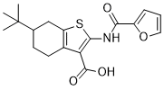
| InChi Code | InChI=1S/C18H21NO4S/c1-18(2,3)10-6-7-11-13(9-10)24-16(14(11)17(21)22)19-15(20)12-5-4-8-23-12/h4-5,8,10H,6-7,9H2,1-3H3,(H,19,20)(H,21,22) | |
| Chemical Name | 6-tert-butyl-2-(furan-2-carbonylamino)-4,5,6,7-tetrahydro-1-benzothiophene-3-carboxylic acid | |
| Synonyms |
|
|
| HS Tariff Code | 2934.99.9001 | |
| Storage |
Powder-20°C 3 years 4°C 2 years In solvent -80°C 6 months -20°C 1 month |
|
| Shipping Condition | Room temperature (This product is stable at ambient temperature for a few days during ordinary shipping and time spent in Customs) |
Biological Activity
| Targets |
CaCCinh-A01 targets TMEM16A (calcium-activated chloride channel, CaCC) (IC50 = 1.1 μM in T84 cells using forskolin-induced chloride secretion assay) [2] CaCCinh-A01 targets TMEM16A (CaCC) [1] CaCCinh-A01 targets TMEM16A (CaCC) [3] |
| ln Vitro |
After ATP stimulation, CaCC current is strongly inhibited by 30 μM CaCCinh-A01 and 100 μM tannic acid[1]. 0.1, 1 and 10 μM CaCCinh-A01 reduces calcium-dependent chloride current by 38±14, 66±10 and 91±1%, respectively. When ATP-induced short-circuit currents are present at 10 and 30 μM CaCCinh-A01, respectively, they are decreased by 38±7 and 78±3%[2]. In human bronchial epithelial cells (HBECs) and mouse intestinal epithelial cells, CaCCinh-A01 (10 μM) inhibited calcium-activated chloride channel (CaCC) currents by ~70-80% as measured by whole-cell patch clamp; the inhibition was reversible and concentration-dependent, with minimal effect on other ion channels (e.g., CFTR, K+ channels) [1] - In T84 human colonic epithelial cells, CaCCinh-A01 dose-dependently suppressed forskolin-induced chloride secretion (IC50 = 1.1 μM) and carbachol-induced CaCC currents, as detected by Ussing chamber short-circuit current (Isc) measurements; the drug did not affect cAMP levels or CFTR channel activity, indicating specific inhibition of TMEM16A-mediated CaCC [2] - In bEnd.3 mouse brain microvascular endothelial cells subjected to oxygen-glucose deprivation (OGD) for 4 hours, CaCCinh-A01 (1-10 μM) dose-dependently reduced endothelial cell paracellular permeability (measured by FITC-dextran flux assay), with 10 μM treatment decreasing permeability by ~65% compared to OGD alone; Western blot analysis showed that CaCCinh-A01 preserved the expression of tight junction proteins (ZO-1, occludin, claudin-5) and reduced phosphorylation of myosin light chain (MLC), a regulator of cytoskeletal rearrangement [3] - In OGD-treated bEnd.3 cells, CaCCinh-A01 (5 μM) inhibited TMEM16A channel activity (detected by patch clamp) and attenuated intracellular calcium overload, as evidenced by reduced Fluo-4 AM fluorescence intensity [3] |
| ln Vivo |
In a middle cerebral artery occlusion model in mice, CaCCinh-A01 (vein injection; 5 mg/kg; caudal vein injection within 15 min after the onset of reperfusion) significantly reduces infarction when compared with MCAO-saline treatment at 24 h or 72 h[3]. In a rat model of transient middle cerebral artery occlusion (tMCAO)-induced ischemic stroke, intraperitoneal administration of CaCCinh-A01 (10 mg/kg, 30 minutes before ischemia and 2 hours after reperfusion) significantly reduced blood-brain barrier (BBB) leakage (measured by Evans blue extravasation, reduced by ~58% compared to vehicle control) and brain edema (brain water content decreased by ~4.2%) [3] - In the tMCAO rat model, CaCCinh-A01 treatment (10 mg/kg, i.p.) reduced cerebral infarct volume by ~40% (assessed by TTC staining) and improved neurological function scores (from 3.2 ± 0.5 to 1.8 ± 0.4 on a 5-point scale) at 24 hours after reperfusion; immunohistochemical staining of brain sections showed preserved tight junction protein (ZO-1, occludin) expression in cerebral microvessels [3] |
| Enzyme Assay |
Whole-cell patch clamp assay for CaCC currents: Epithelial cells (HBECs, mouse intestinal epithelial cells) were seeded on glass coverslips and cultured to confluence. Cells were enzymatically dissociated into single cells and placed in a recording chamber. Patch pipettes were filled with intracellular solution, and the bath was perfused with extracellular solution. Calcium-activated chloride currents were induced by adding ionomycin (a calcium ionophore) to the bath solution. Serial dilutions of CaCCinh-A01 were applied, and currents were recorded using a patch clamp amplifier. Current amplitude was measured at steady state, and inhibition rates were calculated relative to vehicle control [1] - Fluorometric chloride flux assay: T84 cells were loaded with a chloride-sensitive fluorescent dye and seeded in 96-well plates. After incubation with CaCCinh-A01 at various concentrations for 30 minutes, forskolin (a cAMP activator) was added to induce chloride secretion. Changes in fluorescence intensity were monitored using a microplate reader, and IC50 values were determined by nonlinear regression analysis of dose-response curves [2] - Patch clamp assay for TMEM16A currents in bEnd.3 cells: bEnd.3 cells were cultured on coverslips and subjected to OGD for 4 hours. Whole-cell patch clamp recordings were performed with intracellular solution containing calcium and extracellular solution with chloride as the main anion. TMEM16A currents were activated by intracellular calcium release, and CaCCinh-A01 was added to the bath to assess current inhibition [3] |
| Cell Assay |
Ussing chamber short-circuit current (Isc) assay: T84 cells were cultured on permeable filters until forming a confluent monolayer. The filters were mounted in Ussing chambers, with apical and basolateral sides perfused with physiological solutions. CaCCinh-A01 was added to the apical side 30 minutes before stimulation with forskolin or carbachol. Isc was recorded continuously, and changes in chloride secretion were calculated as the difference in Isc before and after agonist stimulation [2] - FITC-dextran permeability assay: bEnd.3 cells were seeded on transwell inserts and cultured to confluence. Cells were pretreated with CaCCinh-A01 (1-10 μM) for 1 hour, then subjected to OGD for 4 hours. FITC-dextran (4 kDa) was added to the apical chamber, and the fluorescence intensity in the basolateral chamber was measured after 2 hours. Permeability was calculated as the percentage of FITC-dextran transported across the cell monolayer [3] - Western blot assay for tight junction proteins: OGD-treated bEnd.3 cells were lysed in RIPA buffer after CaCCinh-A01 treatment. Proteins were separated by SDS-PAGE, transferred to PVDF membranes, and probed with antibodies against ZO-1, occludin, claudin-5, phosphorylated MLC (p-MLC), and GAPDH (loading control). Chemiluminescent detection was used to visualize bands, and densitometric analysis was performed to quantify protein expression levels [3] - Calcium imaging assay: bEnd.3 cells were loaded with Fluo-4 AM (a calcium indicator) for 30 minutes, pretreated with CaCCinh-A01 (5 μM) for 1 hour, then subjected to OGD. Fluorescence intensity was monitored using a confocal microscope, and intracellular calcium concentration was calculated based on fluorescence ratios [3] |
| Animal Protocol |
Animal/Disease Models: Two-month-old male C57/BL6J mice[3] Doses: 5 mg/kg Route of Administration: Vein injection; 5 mg/kg; caudal vein injection within 15 min after the onset of reperfusion Experimental Results: Attenuated brain infarct size, improved neurological outcomes and lowered BBB permeability after ischemic stroke in mice. Rat transient middle cerebral artery occlusion (tMCAO) model: Male Sprague-Dawley rats (250-300 g) were anesthetized, and the middle cerebral artery (MCA) was occluded using a nylon suture for 90 minutes, followed by reperfusion. CaCCinh-A01 was dissolved in DMSO and diluted with saline (final DMSO concentration ≤5%) to a concentration of 2 mg/mL. The drug was administered via intraperitoneal injection at a dose of 10 mg/kg, with the first injection 30 minutes before MCA occlusion and a second injection 2 hours after reperfusion. Vehicle control rats received the same volume of DMSO/saline solution. At 24 hours after reperfusion, rats were euthanized to measure brain water content, Evans blue extravasation, and infarct volume; brain tissues were collected for immunohistochemical staining [3] |
| Toxicity/Toxicokinetics |
In vitro cytotoxicity: CaCCinh-A01 (up to 30 μM) did not affect the viability of T84 cells, HBECs, or bEnd.3 cells after 24-hour treatment, as measured by MTT assay [1][2][3] - Acute in vivo toxicity: Single intraperitoneal administration of CaCCinh-A01 (10 mg/kg) to rats did not cause mortality or significant clinical signs of toxicity (e.g., weight loss, lethargy, abnormal behavior) within 24 hours [3] |
| References |
[1]. TMEM16A inhibitors reveal TMEM16A as a minor component of calcium-activated chloridechannel conductance in airway and intestinal epithelial cells. J Biol Chem. 2011 Jan 21;286(3):2365-74. [2]. Small-molecule screen identifies inhibitors of a human intestinal calcium-activated chloridechannel. Mol Pharmacol. 2008 Mar;73(3):758-68. [3]. TMEM16A Inhibition Preserves Blood-Brain Barrier Integrity After Ischemic Stroke.Front Cell Neurosci. 2019 Aug 6;13:360. |
| Additional Infomation |
CaCCinh-A01 is a selective small-molecule inhibitor of the TMEM16A calcium-activated chloride channel (CaCC), identified through high-throughput screening for inhibitors of intestinal epithelial chloride secretion [2] - The inhibitory effect of CaCCinh-A01 on TMEM16A is specific, with no significant activity against other ion channels (e.g., CFTR, voltage-gated chloride channels, potassium channels) at concentrations up to 30 μM [1][2] - CaCCinh-A01 preserves blood-brain barrier integrity after ischemic stroke by inhibiting TMEM16A-mediated endothelial cell permeability and maintaining tight junction protein expression, which is associated with reduced cerebral edema and infarct volume [3] - TMEM16A is expressed in airway and intestinal epithelial cells, where it mediates calcium-dependent chloride secretion; CaCCinh-A01 has been used as a tool compound to study the physiological role of TMEM16A in epithelial transport [1][2] |
Solubility Data
| Solubility (In Vitro) |
|
|||
| Solubility (In Vivo) |
Solubility in Formulation 1: ≥ 2.5 mg/mL (7.20 mM) (saturation unknown) in 10% DMSO + 40% PEG300 + 5% Tween80 + 45% Saline (add these co-solvents sequentially from left to right, and one by one), clear solution. For example, if 1 mL of working solution is to be prepared, you can add 100 μL of 25.0 mg/mL clear DMSO stock solution to 400 μL PEG300 and mix evenly; then add 50 μL Tween-80 to the above solution and mix evenly; then add 450 μL normal saline to adjust the volume to 1 mL. Preparation of saline: Dissolve 0.9 g of sodium chloride in 100 mL ddH₂ O to obtain a clear solution. Solubility in Formulation 2: ≥ 2.5 mg/mL (7.20 mM) (saturation unknown) in 10% DMSO + 90% (20% SBE-β-CD in Saline) (add these co-solvents sequentially from left to right, and one by one), clear solution. For example, if 1 mL of working solution is to be prepared, you can add 100 μL of 25.0 mg/mL clear DMSO stock solution to 900 μL of 20% SBE-β-CD physiological saline solution and mix evenly. Preparation of 20% SBE-β-CD in Saline (4°C,1 week): Dissolve 2 g SBE-β-CD in 10 mL saline to obtain a clear solution. Solubility in Formulation 3: ≥ 2.5 mg/mL (7.20 mM) (saturation unknown) in 10% DMSO + 90% Corn Oil (add these co-solvents sequentially from left to right, and one by one), clear solution. For example, if 1 mL of working solution is to be prepared, you can add 100 μL of 25.0 mg/mL clear DMSO stock solution to 900 μL of corn oil and mix evenly. (Please use freshly prepared in vivo formulations for optimal results.) |
| Preparing Stock Solutions | 1 mg | 5 mg | 10 mg | |
| 1 mM | 2.8783 mL | 14.3914 mL | 28.7828 mL | |
| 5 mM | 0.5757 mL | 2.8783 mL | 5.7566 mL | |
| 10 mM | 0.2878 mL | 1.4391 mL | 2.8783 mL |