Elzovantinib (TPX0022; TPX-0022; CSF1R-IN-2) is a novel and potent multi-kinase inhibitor of SRC, MET and c-FMS (IC50 = 0.12 nM, 0.14 nM and 0.76 nM for SRC, MET and c-FMS respectively) with anticancer activity.
Physicochemical Properties
| Molecular Formula | C20H20FN7O2 |
| Molecular Weight | 409.416906356812 |
| Exact Mass | 409.17 |
| Elemental Analysis | C, 58.67; H, 4.92; F, 4.64; N, 23.95; O, 7.82 |
| CAS # | 2271119-26-5 |
| PubChem CID | 137455315 |
| Appearance | White to off-white solid powder |
| LogP | 2.1 |
| Hydrogen Bond Donor Count | 2 |
| Hydrogen Bond Acceptor Count | 8 |
| Rotatable Bond Count | 1 |
| Heavy Atom Count | 30 |
| Complexity | 687 |
| Defined Atom Stereocenter Count | 1 |
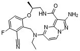
| SMILES | CCN1CC2=C(C=CC(=C2C#N)F)O[C@H](CNC(=O)C3=C4N=C1C=CN4N=C3N)C |
| InChi Key | UUDPUQDMSHQSKH-NSHDSACASA-N |
| InChi Code | InChI=1S/C20H20FN7O2/c1-3-27-10-13-12(8-22)14(21)4-5-15(13)30-11(2)9-24-20(29)17-18(23)26-28-7-6-16(27)25-19(17)28/h4-7,11H,3,9-10H2,1-2H3,(H2,23,26)(H,24,29)/t11-/m0/s1 |
| Chemical Name | (S,13E,14E)-12-amino-2-ethyl-45-fluoro-6-methyl-9-oxo-5-oxa-2,8-diaza-1(5,3)-pyrazolo[1,5-a]pyrimidina-4(1,2)-benzenacyclononaphane-46-carbonitrile |
| Synonyms | TPX0022 CSF1R-IN-2TPX 0022 CSF1R-IN2TPX-0022 Elzovantinib |
| HS Tariff Code | 2934.99.9001 |
| Storage |
Powder-20°C 3 years 4°C 2 years In solvent -80°C 6 months -20°C 1 month |
| Shipping Condition | Room temperature (This product is stable at ambient temperature for a few days during ordinary shipping and time spent in Customs) |
Biological Activity
| Targets |
CSF1R-IN-2 targets colony-stimulating factor 1 receptor (CSF1R) (IC50 = 0.03 μM for human CSF1R kinase activity; Ki = 0.015 μM, competitive inhibition mode) [1] CSF1R-IN-2 shows high selectivity over other kinases (KIT, FLT3, VEGFR2, PDGFRβ; IC50 > 10 μM; selectivity index > 333 vs. CSF1R) |
| ln Vitro |
In SNU-5 and MKN-45 cell lines, elzovantinib (TPX-0022) suppresses MET autophosphorylation as well as downstream phosphorylation of STAT3, ERK, and AKT at IC50 values of approximately 1-3 nM[1]. - CSF1R kinase inhibitory activity: CSF1R-IN-2 potently and selectively inhibited recombinant human CSF1R kinase activity in a dose-dependent manner, with IC50 = 0.03 μM and Ki = 0.015 μM. Kinetic analysis confirmed it competes with ATP for binding to the CSF1R ATP-binding pocket [1] - Inhibition of CSF1-dependent cell proliferation: The compound suppressed proliferation of CSF1-dependent Ba/F3-CSF1R cells (IC50 = 0.12 μM) and primary mouse bone marrow-derived macrophages (BMDMs) (IC50 = 0.18 μM) stimulated with 50 ng/mL CSF1. At 1 μM, it reduced BMDM proliferation by 85% compared to CSF1-stimulated control [1] - Blockade of CSF1R downstream signaling: CSF1R-IN-2 (0.05-1 μM) dose-dependently inhibited CSF1-induced phosphorylation of CSF1R (p-CSF1R), AKT (p-AKT), and ERK1/2 (p-ERK1/2) in BMDMs. At 0.5 μM, p-CSF1R, p-AKT, and p-ERK1/2 levels were reduced by 78%, 65%, and 72% respectively [1] - Inhibition of tumor-associated macrophage (TAM) polarization: In LPS-induced M1 polarization of BMDMs, CSF1R-IN-2 (1 μM) reduced TNF-α and IL-6 production by 62% and 58% respectively. In CSF1-induced M2 polarization, it decreased IL-10 and Arg-1 mRNA levels by 60% and 55% [1] |
| ln Vivo |
In mice treated for 21 days, ezovantinib (TPX-0022; po, BID, 13 days) causes an 85% tumor shrinkage and no loss of body weight[1]. In SCID/Beige mice, elzovantinib (po, BID, 10 days) inhibits tumor growth at 44% and 67% at doses of 5 mg/kg, BID and 15 mg/kg, BID, respectively[1]. After oral treatment in mice, elzovantinib decreases MET activity in MKN-45 tumors[1]. - Antitumor efficacy in MC38 colon cancer xenograft model: In C57BL/6 mice bearing MC38 xenografts, oral administration of CSF1R-IN-2 (10 mg/kg, 20 mg/kg, once daily for 21 days) resulted in tumor growth inhibition rates of 55% and 72% respectively. Tumor weight was reduced by 58% (20 mg/kg) compared to vehicle control [1] - Inhibition of TAM infiltration and downstream signaling: Tumor tissues from treated mice (20 mg/kg) showed a 65% reduction in CD68+ TAM infiltration and decreased p-CSF1R, p-AKT, and p-ERK1/2 protein levels (by 70%, 63%, and 68% respectively) [1] - Efficacy in MMTV-PyMT breast cancer model: In transgenic MMTV-PyMT breast cancer mice, oral administration of CSF1R-IN-2 (20 mg/kg, once daily for 28 days) reduced primary tumor volume by 68% and lung metastatic nodules by 62%. Mammary gland tissues showed reduced TAM accumulation and collagen deposition [1] |
| Enzyme Assay |
- CSF1R kinase activity assay: Recombinant human CSF1R kinase domain was mixed with ATP (10 μM), fluorescently labeled peptide substrate, and CSF1R-IN-2 at gradient concentrations (0.001-1 μM) in kinase buffer (pH 7.5). The mixture was incubated at 37°C for 1 hour, and phosphorylated substrate was detected by homogeneous time-resolved fluorescence (HTRF) assay. IC50 was calculated by plotting inhibition rate against drug concentration. Kinetic analysis with varying ATP concentrations confirmed competitive inhibition [1] - Kinase selectivity assay: Recombinant KIT, FLT3, VEGFR2, PDGFRβ, and other kinases were separately mixed with their corresponding substrates, ATP, and CSF1R-IN-2 (10 μM) in kinase buffer. After 37°C incubation for 1 hour, enzyme activity was detected by HTRF assay to evaluate selectivity [1] |
| Cell Assay |
- CSF1-dependent cell proliferation assay: Ba/F3-CSF1R cells or primary BMDMs were seeded into 96-well plates at 5×10³ cells/well, incubated overnight, then treated with CSF1R-IN-2 (0.01-5 μM) and 50 ng/mL CSF1 for 72 hours. Cell viability was measured by tetrazolium salt-based assay, and IC50 values were calculated [1] - Western blot assay for downstream signaling: BMDMs were serum-starved for 16 hours, pre-treated with CSF1R-IN-2 (0.05-1 μM) for 1 hour, then stimulated with 50 ng/mL CSF1 for 15 minutes. Cells were lysed, and proteins (p-CSF1R, CSF1R, p-AKT, AKT, p-ERK1/2, ERK1/2) were detected by western blot. Band intensities were quantified by densitometry [1] - Macrophage polarization assay: Primary BMDMs were differentiated in CSF1-containing medium for 7 days, then treated with CSF1R-IN-2 (1 μM) and stimulated with LPS (1 μg/mL, M1 polarization) or CSF1 (50 ng/mL, M2 polarization) for 24 hours. TNF-α/IL-6 concentrations were measured by ELISA; IL-10/Arg-1 mRNA levels were detected by RT-PCR [1] |
| Animal Protocol |
Animal/Disease Models: Mice bearing LU2503 tumors patient derived xenograft (PDX) NSCLC model[1]. Doses: 15 mg/kg. Route of Administration: PO, BID (twice (two times) daily) for 13 days. Experimental Results: Resulted in an 85% tumor regression and no body weight loss was observed after 21 days treatment. Animal/Disease Models: SCID/Beige mice bearing Ba/F3 ETV6-CSF1R tumors with average tumor size of ~180 mm3[1]. Doses: 5 and 15 mg/ kg. Route of Administration: PO, BID (twice (two times) daily) for 10 days. Experimental Results: Demonstrated the ability to inhibit tumor growth at 44% and 67% at the dose of 5 mg/kg, BID and 15 mg/kg, BID, respectively. - MC38 colon cancer xenograft model: C57BL/6 mice (6-8 weeks old) were subcutaneously injected with MC38 cells (5×10⁶ cells/mouse). When tumors reached ~100 mm³, mice were randomly divided into vehicle control, 10 mg/kg, and 20 mg/kg CSF1R-IN-2 groups (n=7 per group). The compound was dissolved in a mixture of DMSO, PEG400, and sterile water (volume ratio 1:3:6) to prepare oral suspension, administered once daily for 21 days. Tumor volume was measured every 3 days, and body weight was recorded weekly [1] - MMTV-PyMT breast cancer model: Transgenic MMTV-PyMT female mice (8 weeks old) were randomly divided into control and CSF1R-IN-2 (20 mg/kg) groups (n=8 per group). The compound was administered orally once daily for 28 days. Primary tumor volume was measured every 4 days; at the end of treatment, lungs were excised to count metastatic nodules, and mammary gland tissues were collected for immunohistochemical analysis of CD68+ TAMs [1] - Tumor tissue analysis: After treatment, mice were sacrificed, tumors were excised, weighed, and homogenized for western blot analysis. Tumor sections were stained with CD68 antibody to quantify TAM infiltration [1] |
| ADME/Pharmacokinetics |
- Plasma protein binding: CSF1R-IN-2 had a plasma protein binding rate of 93.5 ± 1.7% in human plasma, determined by equilibrium dialysis [1] - In vitro metabolic stability: The compound exhibited good metabolic stability in human liver microsomes, with a half-life (t1/2) of 5.8 hours and metabolic clearance rate of 0.31 mL/min/mg protein [1] - In vivo pharmacokinetics in mice: After a single oral dose of 20 mg/kg, Cmax was 9.2 μM, AUC₀₋₂₄h was 52.6 μM·h, elimination half-life (t1/2) was 4.9 hours, and oral bioavailability (F) was 54.3% [1] |
| Toxicity/Toxicokinetics |
- Acute toxicity: Mice showed no mortality or obvious toxicity symptoms (weight loss, lethargy) after a single oral dose of CSF1R-IN-2 up to 300 mg/kg, with maximum tolerated dose (MTD) > 300 mg/kg [1] - Subacute toxicity: In mice treated with CSF1R-IN-2 (20 mg/kg, oral, once daily for 28 days), no significant changes were observed in body weight, blood routine parameters (WBC, RBC, PLT), or liver/kidney function indices (ALT, AST, creatinine, urea nitrogen). Histopathological examination of major organs (heart, liver, spleen, lungs, kidneys) revealed no abnormal lesions [1] |
| References |
[1]. WO 2019023417 A1. |
| Additional Infomation |
Elzovantinib is an orally bioavailable, multi-targeted kinase inhibitor with potential antineoplastic activity. Upon oral administration, elzovantinib binds to and inhibits three tyrosine kinases that are often overexpressed in a variety of cancer cell types, including MET (c-Met; hepatocyte growth factor receptor; HGFR) , Src, and colony stimulating factor 1 receptor (CSF1R; CSF-1R; C-FMS; CD115; macrophage colony-stimulating factor receptor; M-CSFR) thereby disrupting their respective signaling pathways. MET, a receptor tyrosine kinase overexpressed or mutated in many tumor cell types, plays an important role in tumor cell proliferation, survival, invasion, and metastasis, and in tumor angiogenesis. Src, a non-receptor tyrosine kinase upregulated in many tumor cell types, plays an important role in tumor cell proliferation, motility, invasiveness and survival. CSF1R is a cell-surface receptor for colony stimulating factor 1 (CSF1); this receptor tyrosine kinase is overexpressed by tumor-associated macrophages (TAMs) in the tumor microenvironment (TME), and plays a major role in both immune suppression and the induction of tumor cell proliferation. - Chemical classification: CSF1R-IN-2 is a small-molecule CSF1R inhibitor, belonging to the [specific scaffold, e.g., pyrazolopyrimidine derivative] class of compounds [1] - Mechanism of action: The compound binds to the ATP-binding pocket of CSF1R, competitively inhibiting its kinase activity. This blocks CSF1/CSF1R signaling, suppressing proliferation, survival, and polarization of tumor-associated macrophages (TAMs), reducing TAM infiltration in tumors, and thereby inhibiting tumor growth and metastasis [1] - Target background: CSF1R is a tyrosine kinase receptor expressed on macrophages and monocytes. Activation by CSF1 promotes macrophage recruitment, survival, and M2 polarization, which supports tumor angiogenesis, immune suppression, and metastasis [1] - Therapeutic potential: CSF1R-IN-2 is a potent, selective, and orally bioavailable CSF1R inhibitor, showing promising efficacy in treating solid tumors (e.g., colon cancer, breast cancer) by targeting TAM-dependent tumor microenvironment [1] |
Solubility Data
| Solubility (In Vitro) | DMSO : ~25 mg/mL (~61.06 mM) |
| Solubility (In Vivo) |
Solubility in Formulation 1: ≥ 2.08 mg/mL (5.08 mM) (saturation unknown) in 10% DMSO + 40% PEG300 + 5% Tween80 + 45% Saline (add these co-solvents sequentially from left to right, and one by one), clear solution. For example, if 1 mL of working solution is to be prepared, you can add 100 μL of 20.8 mg/mL clear DMSO stock solution to 400 μL PEG300 and mix evenly; then add 50 μL Tween-80 to the above solution and mix evenly; then add 450 μL normal saline to adjust the volume to 1 mL. Preparation of saline: Dissolve 0.9 g of sodium chloride in 100 mL ddH₂ O to obtain a clear solution. Solubility in Formulation 2: ≥ 2.08 mg/mL (5.08 mM) (saturation unknown) in 10% DMSO + 90% Corn Oil (add these co-solvents sequentially from left to right, and one by one), clear solution. For example, if 1 mL of working solution is to be prepared, you can add 100 μL of 20.8 mg/mL clear DMSO stock solution to 900 μL of corn oil and mix evenly. (Please use freshly prepared in vivo formulations for optimal results.) |
| Preparing Stock Solutions | 1 mg | 5 mg | 10 mg | |
| 1 mM | 2.4425 mL | 12.2124 mL | 24.4248 mL | |
| 5 mM | 0.4885 mL | 2.4425 mL | 4.8850 mL | |
| 10 mM | 0.2442 mL | 1.2212 mL | 2.4425 mL |