Physicochemical Properties
| Molecular Formula | C25H20F3N5O2 |
| Molecular Weight | 479.453815460205 |
| Exact Mass | 479.16 |
| Elemental Analysis | C, 62.63; H, 4.20; F, 11.89; N, 14.61; O, 6.67 |
| CAS # | 2095849-04-8 |
| Related CAS # | 2095849-04-8 |
| PubChem CID | 137333440 |
| Appearance | White to off-white solid powder |
| LogP | 3.7 |
| Hydrogen Bond Donor Count | 2 |
| Hydrogen Bond Acceptor Count | 7 |
| Rotatable Bond Count | 5 |
| Heavy Atom Count | 35 |
| Complexity | 751 |
| Defined Atom Stereocenter Count | 0 |
| InChi Key | BMEXIUCHJUGPRB-UHFFFAOYSA-N |
| InChi Code | InChI=1S/C25H20F3N5O2/c1-15-6-7-21(31-23(34)16-4-3-5-20(9-16)25(26,27)28)10-22(15)32-24(35)18-8-17(11-29-12-18)19-13-30-33(2)14-19/h3-14H,1-2H3,(H,31,34)(H,32,35) |
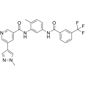
| Chemical Name | 5-(1-methylpyrazol-4-yl)-N-[2-methyl-5-[[3-(trifluoromethyl)benzoyl]amino]phenyl]pyridine-3-carboxamide |
| Synonyms | BUN49048; BUN 49048; BUN-49048; CSF1R-IN-1 |
| HS Tariff Code | 2934.99.9001 |
| Storage |
Powder-20°C 3 years 4°C 2 years In solvent -80°C 6 months -20°C 1 month |
| Shipping Condition | Room temperature (This product is stable at ambient temperature for a few days during ordinary shipping and time spent in Customs) |
Biological Activity
| Targets |
CSF1R (IC50 = 0.5 nM); EGFR T790M (IC50 = 0.18 nM); EGFR (WT) (IC50 = 7.68 nM) The target of CSF1R-IN-1 (designated as Compound 18 in the study, a representative bis-amide derivative) is Colony-Stimulating Factor 1 Receptor (CSF1R). Key activity data include: - CSF1R (enzyme level): IC₅₀ = 0.045 μM [1] - c-Kit: IC₅₀ = 3.8 μM (selectivity > 84-fold vs. CSF1R) [1] - VEGFR2: IC₅₀ = 5.2 μM (selectivity > 115-fold vs. CSF1R) [1] |
| ln Vitro |
CSF1R is believed to be crucial for the development and recruitment of tumor-associated macrophages (TAMs). In the Caco2 assay, CSF1R-IN-1 (compound 22) exhibits good intestinal permeability[1]. 1. CSF1R enzyme inhibitory activity: CSF1R-IN-1 exhibited potent and selective inhibition of CSF1R kinase activity with an IC₅₀ of 0.045 μM. It showed weak inhibitory effects on off-target kinases (c-Kit, VEGFR2), demonstrating high target selectivity [1] 2. Inhibition of CSF1-induced cell proliferation: RAW264.7 macrophages were treated with serial concentrations of CSF1R-IN-1 in the presence of CSF1 (50 ng/mL). After 72 hours of incubation, cell viability was measured, and the compound inhibited cell proliferation with an IC₅₀ of 0.32 μM [1] 3. Suppression of CSF1R downstream signaling: RAW264.7 cells were pretreated with CSF1R-IN-1 (1 μM) for 1 hour, then stimulated with CSF1 (50 ng/mL) for 15 minutes. Western blot analysis showed reduced phosphorylation of CSF1R downstream molecules (p-STAT5, p-AKT, p-ERK1/2) compared to the CSF1-stimulated control group [1] |
| ln Vivo | CSF1R-IN-1 exhibits favorable pharmacokinetics when dosed orally to mice. It seems appropriate for proof of concept in vivo pharmacology testing in the relevant preclinical tumor model.[1]. |
| Enzyme Assay |
A homogeneous time-resolved fluorescence (HTRF) assay was used to evaluate the inhibitory activity of CSF1R-IN-1 against CSF1R. Recombinant CSF1R kinase domain was mixed with a specific peptide substrate, ATP (at a concentration near its Km value), and serial dilutions of CSF1R-IN-1. The reaction mixture was incubated at room temperature for 120 minutes to allow kinase-catalyzed phosphorylation of the substrate. HTRF detection reagents were added to bind phosphorylated and non-phosphorylated substrates, and the fluorescence signal was measured. The inhibition rate at each compound concentration was calculated relative to the vehicle control, and the IC₅₀ value (0.045 μM) was derived by fitting the dose-response curve. Parallel assays were performed with c-Kit and VEGFR2 kinases to assess selectivity [1] |
| Cell Assay |
1. CSF1-induced cell proliferation assay: RAW264.7 macrophage cells were seeded in 96-well plates at a density of 5×10³ cells per well and cultured overnight. Serial concentrations of CSF1R-IN-1 were added to the wells, followed by the addition of CSF1 (50 ng/mL) to stimulate cell proliferation. After 72 hours of incubation at 37°C with 5% CO₂, a cell viability assay reagent was added, and the absorbance was measured at the appropriate wavelength. The IC₅₀ value (0.32 μM) was calculated based on the dose-response relationship [1] 2. Downstream signaling pathway inhibition assay: RAW264.7 cells were seeded in 6-well plates and cultured to 80% confluence. The cells were pretreated with CSF1R-IN-1 (1 μM) for 1 hour, then stimulated with CSF1 (50 ng/mL) for 15 minutes. The cells were lysed with RIPA buffer containing protease and phosphatase inhibitors, and total proteins were extracted. Equal amounts of protein were separated by SDS-PAGE, transferred to PVDF membranes, and blocked with non-fat milk. The membranes were probed with primary antibodies against p-STAT5, p-AKT, p-ERK1/2, and total STAT5/AKT/ERK1/2 (as loading controls) overnight at 4°C, followed by incubation with peroxidase-conjugated secondary antibodies. The protein bands were visualized with chemiluminescent reagents, and the intensity of phosphorylated protein bands was quantified relative to the control group [1] |
| Animal Protocol |
Male CD-1 mice, 25-35 grams (8-11 weeks old) 2 mg/kg IV or 10 mg/kg orally (Per Os) i.v. or oral |
| ADME/Pharmacokinetics |
1. Metabolic stability: CSF1R-IN-1 was incubated with human and mouse liver microsomes in the presence of NADPH-regenerating system. The remaining compound concentration was measured at different time points (0, 15, 30, 60, 120 minutes) by LC-MS/MS. The half-life (t₁/₂) in human liver microsomes was 3.2 hours, and in mouse liver microsomes was 4.5 hours [1] 2. Caco-2 permeability: Caco-2 cells were cultured on transwell inserts until forming a confluent monolayer. CSF1R-IN-1 solution (10 μM) was added to either the apical (A) or basolateral (B) compartment, and samples were collected from the opposite compartment at 30, 60, 90, and 120 minutes. The apparent permeability coefficient (Papp) was calculated as 1.8×10⁻⁶ cm/s (A→B direction), indicating moderate intestinal absorption potential [1] 3. Oral bioavailability: In mice, CSF1R-IN-1 was administered via oral gavage (10 mg/kg) and intravenous injection (5 mg/kg). Plasma samples were collected at different time points, and the concentration of CSF1R-IN-1 was determined by LC-MS/MS. The oral bioavailability (F) was calculated as 35% [1] |
| References |
[1]. Design, synthesis and optimization of bis-amide derivatives as CSF1R inhibitors. Bioorg Med Chem Lett. 2017 May 15;27(10):2153-2160. |
| Additional Infomation |
1. Structural and optimization background: CSF1R-IN-1 is a bis-amide derivative optimized from initial lead compounds through structural modification (e.g., adjustment of the linker length and substitution of aromatic rings). The optimization aimed to improve CSF1R inhibitory activity, metabolic stability, and intestinal permeability [1] 2. Mechanism of action: CSF1R-IN-1 binds to the ATP-binding pocket of CSF1R, inhibiting its kinase activity and blocking downstream STAT5/AKT/ERK signaling pathways. This suppresses the proliferation, survival, and activation of macrophages dependent on CSF1-CSF1R signaling [1] 3. Therapeutic potential: Due to its selective inhibition of CSF1R and favorable pharmacokinetic properties, CSF1R-IN-1 is a potential candidate for the treatment of diseases associated with abnormal activation of the CSF1-CSF1R pathway, such as inflammatory disorders and tumors with high tumor-associated macrophage infiltration [1] |
Solubility Data
| Solubility (In Vitro) | DMSO: 83.3~96 mg/mL (173.8~200.2 mM) |
| Solubility (In Vivo) |
Solubility in Formulation 1: ≥ 2.08 mg/mL (4.34 mM) (saturation unknown) in 10% DMSO + 40% PEG300 + 5% Tween80 + 45% Saline (add these co-solvents sequentially from left to right, and one by one), clear solution. For example, if 1 mL of working solution is to be prepared, you can add 100 μL of 20.8 mg/mL clear DMSO stock solution to 400 μL PEG300 and mix evenly; then add 50 μL Tween-80 to the above solution and mix evenly; then add 450 μL normal saline to adjust the volume to 1 mL. Preparation of saline: Dissolve 0.9 g of sodium chloride in 100 mL ddH₂ O to obtain a clear solution. Solubility in Formulation 2: ≥ 2.08 mg/mL (4.34 mM) (saturation unknown) in 10% DMSO + 90% Corn Oil (add these co-solvents sequentially from left to right, and one by one), clear solution. For example, if 1 mL of working solution is to be prepared, you can add 100 μL of 20.8 mg/mL clear DMSO stock solution to 900 μL of corn oil and mix evenly. (Please use freshly prepared in vivo formulations for optimal results.) |
| Preparing Stock Solutions | 1 mg | 5 mg | 10 mg | |
| 1 mM | 2.0857 mL | 10.4286 mL | 20.8572 mL | |
| 5 mM | 0.4171 mL | 2.0857 mL | 4.1714 mL | |
| 10 mM | 0.2086 mL | 1.0429 mL | 2.0857 mL |