CP-673451 (CP673451) is a novel, potent, selective and ATP-competitive inhibitor of PDGFR(α/β) with potential antitumor activity. It exhibits >450-fold selectivity for PDGFR(α/β) over other angiogenic receptors and inhibits PDGFR(α/β) with IC50s of 10 nM/1 nM in cell-free assays. There are antiangiogenic and antitumor properties to CP-673451. and is being looked into as a possible cancer treatment. While high-dose CP-673451 (40 mg/kg) strongly inhibits tumor growth in mice without significantly reducing weight, CP-673451 exhibits excellent anti-proliferative activity in vitro and high in vivo antitumor efficacy (at 20 mg/kg) with a medium suppression of tumor growth.
Physicochemical Properties
| Molecular Formula | C24H27N5O2 | |
| Molecular Weight | 417.5 | |
| Exact Mass | 417.216 | |
| Elemental Analysis | C, 69.04; H, 6.52; N, 16.77; O, 7.66 | |
| CAS # | 343787-29-1 | |
| Related CAS # |
|
|
| PubChem CID | 10158940 | |
| Appearance | White to off-white solid powder | |
| Density | 1.3±0.1 g/cm3 | |
| Boiling Point | 656.3±65.0 °C at 760 mmHg | |
| Flash Point | 350.7±34.3 °C | |
| Vapour Pressure | 0.0±2.0 mmHg at 25°C | |
| Index of Refraction | 1.677 | |
| LogP | 2.77 | |
| Hydrogen Bond Donor Count | 1 | |
| Hydrogen Bond Acceptor Count | 6 | |
| Rotatable Bond Count | 6 | |
| Heavy Atom Count | 31 | |
| Complexity | 572 | |
| Defined Atom Stereocenter Count | 0 | |
| SMILES | O(C([H])([H])C([H])([H])OC([H])([H])[H])C1C([H])=C([H])C2=C(C=1[H])N=C([H])N2C1C([H])=C([H])C2C([H])=C([H])C([H])=C(C=2N=1)N1C([H])([H])C([H])([H])C([H])(C([H])([H])C1([H])[H])N([H])[H] |
|
| InChi Key | DEEOXSOLTLIWMG-UHFFFAOYSA-N | |
| InChi Code | InChI=1S/C24H27N5O2/c1-30-13-14-31-19-6-7-21-20(15-19)26-16-29(21)23-8-5-17-3-2-4-22(24(17)27-23)28-11-9-18(25)10-12-28/h2-8,15-16,18H,9-14,25H2,1H3 | |
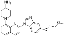
| Chemical Name | 1-[2-[5-(2-methoxyethoxy)benzimidazol-1-yl]quinolin-8-yl]piperidin-4-amine | |
| Synonyms | CP-673451; CP673451; CP 673451; CP-673,451; CP 673,451 | |
| HS Tariff Code | 2934.99.9001 | |
| Storage |
Powder-20°C 3 years 4°C 2 years In solvent -80°C 6 months -20°C 1 month |
|
| Shipping Condition | Room temperature (This product is stable at ambient temperature for a few days during ordinary shipping and time spent in Customs) |
Biological Activity
| Targets |
PDGFRα (IC50 = 10 nM); PDGFRβ (IC50 = 1 nM) CP-673451 is a potent and selective inhibitor of platelet-derived growth factor receptors (PDGFRs), with IC₅₀ values of 5 nM for PDGFRβ, 10 nM for PDGFRα, and 40 nM for vascular endothelial growth factor receptor 2 (VEGFR2) [1] It exhibits weak inhibitory activity against c-Kit (IC₅₀ = 120 nM) and no significant activity against epidermal growth factor receptor (EGFR), Abl, or Src kinases (IC₅₀ > 1000 nM) [1] In extended kinase profiling, the drug showed no off-target inhibition of 28 other kinases (IC₅₀ > 500 nM) [2] |
| ln Vitro |
CP 673451 demonstrates >450-fold selectivity over other angiogenic receptors and is a selective inhibitor of PDGFRα/β with an IC50 of 10 nM/1 nM. For four hours, CP-673451 (33 mg/kg) in glioblastoma tumors inhibits the PDGFR-β receptor by more than 50%, resulting in an EC50 of 120 ng/mL in plasma at Cmax. In an angiogenesis model involving sponges, CP-673451 at 3 mg/kg (q.d. ×5, p.o., or 5.5 ng/mL at Cmax) inhibits 70% of PDGF-BB-stimulated angiogenesis.[1] Through mechanisms involving reduced phosphorylation of GSK-3α and GSK-3β, CP-673451 reduces the rate of cell proliferation. CP-673451 inhibits rhabdosphere-forming ability and cell differentiation, leading to increased senescence in both RD and RUCH2 cultures.[2] CP-673451 dose-dependently inhibited proliferation of PDGFR-overexpressing tumor cell lines: C6 rat glioma cells (PDGFRβ-positive, IC₅₀ = 0.08 μM) and UM-UC-3 human bladder cancer cells (PDGFRα-positive, IC₅₀ = 0.1 μM). It blocked PDGFR phosphorylation (Tyr751 for β subtype, Tyr754 for α subtype) and downstream AKT/ERK1/2 signaling at concentrations ≥ 0.05 μM [1] In human umbilical vein endothelial cells (HUVECs), the drug (0.5-2 μM) suppressed vascular endothelial growth factor (VEGF)-induced tube formation by ~50% at 1 μM and reduced cell migration by ~60% at 1.5 μM, inhibiting angiogenesis [2] It had minimal effect on PDGFR-low tumor cells (e.g., A549 lung cancer cells), with cell viability remaining >85% even at 5 μM [1] |
| ln Vivo |
CP 673451 (once-daily p.o.?0 days dosing regularly) inhibits tumor growth (ED50 < 33 mg/kg) in several human tumor xenografts grown s.c. in athymic mice, such as U87MG human glioblastoma multiforme, Colo205 and LS174T human colon carcinomas, and H460 human lung carcinoma.[1] CP 673451 inhibits tumor growth and stromal cell infiltration in RUCH2 xenograft-bearing mice.[2] CP-673451 significantly inhibited tumor growth in nude mice bearing C6 glioma xenografts. Oral administration of 30 mg/kg/day for 21 days reduced tumor volume by ~60% compared to controls, with intratumoral PDGFRβ phosphorylation downregulated by ~80% [1] In a murine model of UM-UC-3 bladder cancer xenografts, the drug (50 mg/kg/day, oral for 28 days) achieved a 70% tumor growth inhibition rate and prolonged median survival by 35% [2] It attenuated tumor angiogenesis in C6 xenografts: CD31⁺ microvessel density was reduced by ~45% in the treatment group, confirming anti-angiogenic activity [1] |
| Enzyme Assay |
Sf-9 cells (baculovirus expression system) express a glutathione S-transferase-tagged kinase domain construct of the intracellular portion of PDGFR-β (amino acids 693-1401, accession no. J03278). The phosphorylation buffer [50 mmol/L HEPES (pH 7.3), 125 mmol/L NaCl, 24 mmol/L MgCl2 in Nunc Immuno MaxiSorp 96-well plates previously coated with 100 μL of 100 μg/mL poly-Glu-Tyr (4:1 ratio) diluted in PBS] is used to incubate the enzyme at increasing concentrations of ATP to determine the enzyme's kinetics. The plates are incubated with anti-phosphotyrosine-horseradish peroxidase antibody for 10 minutes, followed by a 30-minute room-temperature diluting step in PBS, 0.05% Tween 20, and 3% BSA. After the previously mentioned washing, the plates are incubated with 3,3',5,5'-tetramethylbenzidine. By adding an equal volume of 0.09 NaH2SO4, the reaction is terminated. Next, the phosphotyrosine-dependent signal is measured at 450 nm using a plate reader. For standard enzyme tests, the enzyme is incubated for 30 minutes at room temperature with 10 μM (final) ATP in the presence of a compound diluted in DMSO (1.6% v/v DMSO assay final) in the previously mentioned plates coated with 100 μL of 6.25 μg/mL poly-Glu-Tyr. The assay is completed as previously described, and IC50 values are computed as a percentage of control's inhibition. Recombinant human PDGFRα, PDGFRβ, and VEGFR2 kinase domains were individually incubated with serial dilutions of CP-673451 (0.001-100 nM) in kinase buffer containing 10 μM ATP and a synthetic peptide substrate (PDGFR/VEGFR-specific sequence). Reactions were conducted at 37°C for 60 minutes, and phosphorylated substrates were detected using a radiometric assay (³²P-ATP incorporation). Inhibition rates were calculated by comparing radioactivity with vehicle controls, and IC₅₀ values were derived from sigmoidal dose-response curves [1] To assess selectivity, recombinant EGFR, Abl, and Src kinase domains were tested using the same protocol. Reaction conditions (buffer composition, temperature, ATP concentration) were identical, and <10% inhibition was observed at 100 nM CP-673451 [1] |
| Cell Assay |
It is possible to generate PAE cells that express full-length PDGFR and VEGFR consistently. Full-length human PDGFR-a, PDGFR-h, or VEGFR-2 are transfected into PAE cells in order to perform cell-based selectivity assays. 50 μL of growth medium (Ham's F-12 media supplemented with 10% fetal bovine serum, 50,000 units of penicillin and streptomycin, and 500 μg/mL gentamicin) is used per well in 96-well plates to seed cells at a density of 4×10 3 cells/mL. After 6 to 8 hours, the cells are incubated overnight in 50 μL of serum-depleted medium (as above, but with 0.1% fetal bovine serum) in place of the growth medium. 95 μL of serum-depleted medium was added to the medium just before the compound was added. The compounds are added to the cells at a final DMSO concentration of 0.25% v/v after being diluted in 100% DMSO, and they are then incubated for 10 minutes at 37°C. The proper ligand is used to stimulate the cells, and they are then incubated for a further eight minutes. The medium is taken out, and the cells are rinsed once with PBS before being lysed for five minutes at room temperature in 50 μL HNTG buffer (20 mmol/L HEPES (pH 7.5), 150 mmol/L NaCl, 2% Triton X-100, 10% glycerol, 5 μmol/L EDTA, 2 mmol/L NaVO4, and 1 EDTA-free complete protease inhibitor tablet per 25 mL). 50 μL of HG buffer (20 mmol/L HEPES (pH 7.5), 10% glycerol)] is then added to the lysates to dilute them. Once the diluted cell lysates are well combined, 50 μL of the supernatant is added to the ELISA capture plate and it is incubated for two hours at room temperature while being stirred. Coating 96-well ReactiBind goat-antirabbit plates with 100 μL/well of rabbit anti-human PDGFR-h, anti-PDGFR-a, or anti-VEGFR-2 antibody for 60 to 90 minutes is the process used to prepare ELISA capture plates. Following a 2-hour incubation period, the plates are cleaned in PBS with 0.1% Tween 20 and then incubated for 30 minutes at room temperature with a diluted anti-phosphotyrosine-horseradish peroxidase antibody (PBS with 0.05% Tween 20). The plates are once more cleaned, tetramethylbenzidine is added, and they are assessed as previously mentioned. Percentage inhibition of control is used to compute IC50 values. C6 and UM-UC-3 cells were seeded in 96-well plates at 5×10³ cells/well and treated with CP-673451 (0.01-10 μM) for 72 hours. Cell viability was measured via tetrazolium-based (MTT) assay, and IC₅₀ values were calculated from dose-response curves [1,2] For Western blot analysis: C6 cells were serum-starved for 16 hours, treated with 0.02-0.2 μM CP-673451 for 1 hour, then stimulated with PDGF-BB (50 ng/mL) for 10 minutes. Cells were lysed, and lysates were probed with antibodies against p-PDGFRβ (Tyr751), p-AKT (Ser473), p-ERK1/2 (Thr202/Tyr204), and GAPDH (loading control) [1] HUVEC tube formation assay: Matrigel-coated 24-well plates were seeded with HUVECs (2×10⁴ cells/well) pre-treated with 0.5-2 μM CP-673451 for 1 hour. VEGF (20 ng/mL) was added, and after 24 hours, tube formation was visualized and quantified under a microscope [2] |
| Animal Protocol |
The anticancer efficacy of CP-673451 is assessed in nude mice using a subcutaneous A549 xenograft model. In short, mice are given 2×10 6 cells/mouse of A549 cells injected into their axillary regions. Upon reaching 70 mm 3 , the mice are randomized into two groups: one for control and the other for CP-673451 (n = 6 per group) at low and high doses (20 mg/kg and 40 mg/kg, respectively) (vehicle 10% 1-methyl-2-pyrrolidinone and 90% polyethylene glycol 300). CP-673451 (20 or 40 mg/kg/day) or a vehicle is injected intraperitoneally into these animals. The implanted tumors are measured blindly once a day with a calliper during the course of treatment. Simultaneously, the animal body weights are measured. Tumor volume is computed. Following therapy, the tumors are removed and examined, and the mice are euthanized. Nude mice (6-8 weeks old) were subcutaneously implanted with C6 glioma cells (5×10⁶ cells/mouse) to establish xenografts. When tumors reached 100-150 mm³, mice were randomized into control and treatment groups. CP-673451 was suspended in 0.5% carboxymethylcellulose (CMC) and administered orally at 30 mg/kg/day for 21 days. Tumor volume was measured every 3 days using calipers (volume = length × width² / 2) [1] For UM-UC-3 xenograft studies: nude mice were implanted with UM-UC-3 cells (4×10⁶ cells/mouse), and CP-673451 was given orally at 50 mg/kg/day for 28 days. Survival time was recorded daily, and tumors were harvested at sacrifice for CD31 immunohistochemistry (microvessel density analysis) [2] |
| ADME/Pharmacokinetics |
CP-673451 had an oral bioavailability of ~40% in mice after a single 30 mg/kg dose. Maximum plasma concentration (Cmax) was 2.5 μg/mL achieved at 1.5 hours post-administration, and plasma half-life (t₁/₂) was ~6 hours [1] In rats, oral administration of 50 mg/kg resulted in an AUC₀-24h of 38 μg·h/mL. The drug distributed preferentially to tumor tissues, with a tumor-to-plasma concentration ratio of 2.3 at 4 hours post-dose [2] It was metabolized primarily by cytochrome P450 3A4 (CYP3A4) in human liver microsomes, with a metabolic clearance rate of 1.2 mL/min/mg protein [1] |
| Toxicity/Toxicokinetics |
Mice treated with CP-673451 at 30 mg/kg/day for 21 days showed mild weight loss (~5%) but no significant liver or kidney toxicity. Serum ALT, AST, creatinine, and BUN levels were within normal ranges [1] The plasma protein binding rate of CP-673451 was ~90% in human plasma, determined via equilibrium dialysis [1] In vitro cytotoxicity assays: the drug showed no significant damage to normal human astrocytes (NHAs) or bladder epithelial cells (SV-HUC-1) at concentrations up to 2 μM [2] |
| References |
[1]. Cancer Res . 2005 Feb 1;65(3):957-66. [2]. Cancer Res . 2013 Apr 1;73(7):2139-49. |
| Additional Infomation |
1-[2-[5-(2-methoxyethoxy)-1-benzimidazolyl]-8-quinolinyl]-4-piperidinamine is an aminoquinoline. CP-673451 is a small-molecule, orally active inhibitor designed to target PDGFR/VEGFR2 signaling axes, which are critical for tumor proliferation and angiogenesis [1] It was developed for the treatment of PDGFR-driven solid tumors, including glioma and bladder cancer—tumors where PDGFR overexpression correlates with poor prognosis [2] Preclinical data demonstrate that its dual inhibition of PDGFR and VEGFR2 provides synergistic antitumor effects by targeting both tumor cells and the tumor microenvironment (angiogenesis) [1] |
Solubility Data
| Solubility (In Vitro) |
|
|||
| Solubility (In Vivo) |
Solubility in Formulation 1: ≥ 2.75 mg/mL (6.59 mM) (saturation unknown) in 10% DMSO + 40% PEG300 + 5% Tween80 + 45% Saline (add these co-solvents sequentially from left to right, and one by one), clear solution. For example, if 1 mL of working solution is to be prepared, you can add 100 μL of 27.5 mg/mL clear DMSO stock solution to 400 μL PEG300 and mix evenly; then add 50 μL Tween-80 to the above solution and mix evenly; then add 450 μL normal saline to adjust the volume to 1 mL. Preparation of saline: Dissolve 0.9 g of sodium chloride in 100 mL ddH₂ O to obtain a clear solution. Solubility in Formulation 2: ≥ 2.75 mg/mL (6.59 mM) (saturation unknown) in 10% DMSO + 90% (20% SBE-β-CD in Saline) (add these co-solvents sequentially from left to right, and one by one), clear solution. For example, if 1 mL of working solution is to be prepared, you can add 100 μL of 27.5 mg/mL clear DMSO stock solution to 900 μL of 20% SBE-β-CD physiological saline solution and mix evenly. Preparation of 20% SBE-β-CD in Saline (4°C,1 week): Dissolve 2 g SBE-β-CD in 10 mL saline to obtain a clear solution. Solubility in Formulation 3: 30% PEG 400+5% propylene glycol+1% Tween 80+ddH2O: 30mg/mL (Please use freshly prepared in vivo formulations for optimal results.) |
| Preparing Stock Solutions | 1 mg | 5 mg | 10 mg | |
| 1 mM | 2.3952 mL | 11.9760 mL | 23.9521 mL | |
| 5 mM | 0.4790 mL | 2.3952 mL | 4.7904 mL | |
| 10 mM | 0.2395 mL | 1.1976 mL | 2.3952 mL |