CORM-3 is a water-soluble carbon monoxide (CO)-releasing molecule with potent anti-inflammatory and cardioprotective activity by attenuatingNF-κB p65 nuclear translocation, reducing ROS generation and enhancing intracellular glutathione and superoxide dismutase levels. CORM-3, a CO-releasing molecule, can activate the soluble guanylyl cyclase to mimic the anti-allergic and anti-anaphylactic effects of heme oxygenase. According to reports, CORM-3 has an impact on cardiovascular inflammation. It lessens the in vitro model's induction of CD54 expression on EC by PMN. Both in vitro and in vivo, CORM-3 has vasoactive qualities. By controlling blood pressure and vessel tone, it exhibits significant vasodilatation.
Physicochemical Properties
| Molecular Formula | C5H4CLNO5RU | |
| Molecular Weight | 294.61 | |
| Exact Mass | 296.898 | |
| CAS # | 475473-26-8 | |
| Related CAS # |
|
|
| Appearance | Light yellow to yellow solid | |
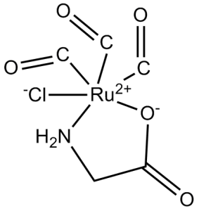
| SMILES | O=C[Ru+2]1(NCC(=O)[O-]1)([Cl-])(C=O)C=O |
|
| Synonyms |
|
|
| HS Tariff Code | 2934.99.9001 | |
| Storage |
Powder-20°C 3 years 4°C 2 years In solvent -80°C 6 months -20°C 1 month |
|
| Shipping Condition | Room temperature (This product is stable at ambient temperature for a few days during ordinary shipping and time spent in Customs) |
Biological Activity
| Targets |
NLRP3 Soluble guanylate cyclase (sGC) [1] - Inducible nitric oxide synthase (iNOS, inhibiting its expression) [2] - Nuclear factor κB (NF-κB) [4] |
| ln Vitro |
CORM-3 protects against hypoxia-reoxygenation and oxidative stress by promoting CO release in cardiac cells. [1] In RAW264.7 murine macrophages, CORM-3 reduces the inflammatory response brought on by LPS. [2] Through interaction with the phosphate carrier, CORM-3 also dissociates mitochondrial respiration. [4] CORM-3 could dilate rat aortic rings in a concentration-dependent manner, which was dependent on the activation of soluble guanylate cyclase (sGC) and not blocked by atropine or propranolol [1] - CORM-3 significantly inhibited human platelet aggregation, with a marked inhibitory effect on ADP-induced platelet aggregation [1] - In lipopolysaccharide (LPS)-induced mouse macrophages, CORM-3 dose-dependently reduced nitric oxide (NO) production and downregulated the mRNA and protein expression levels of inducible nitric oxide synthase (iNOS) [2] - Treatment of HeLa cells with CORM-3 inhibited cell proliferation and induced apoptosis, accompanied by decreased nuclear factor κB (NF-κB) activity and altered mitochondrial membrane potential [4] - CORM-3 suppressed the secretion of tumor necrosis factor α (TNF-α) and interleukin 6 (IL-6) in vitro-cultured macrophages [3] |
| ln Vivo |
CORM-3 (40 mg/kg i.p.) prolongs the survival of murine cardiac grafts and attenuates organ rejection in CBA mice transplanted with BALB/c hearts. [1] In a mouse model of collagen-induced arthritis, CORM-3 (20 mg/kg i.p.) reduces cellular infiltration, joint inflammation, and degeneration. [3] In rat in vivo experiments, CORM-3 regulated vasomotor function and improved endothelium-dependent vasodilation [1] - In carrageenan-induced paw edema model in rats, administration of CORM-3 significantly inhibited paw edema, exerting in vivo anti-inflammatory effects [2] - In collagen-induced arthritis (CIA) mouse model, CORM-3 alleviated joint swelling and inflammatory infiltration, reduced serum levels of TNF-α and IL-6, and minimized joint cartilage and bone tissue damage [3] |
| Enzyme Assay |
Soluble guanylate cyclase (sGC) activity assay: The activation of sGC by CORM-3 was evaluated by detecting cyclic guanosine monophosphate (cGMP) production in aortic ring tissues. Blank control group and enzyme inhibitor control group were set up to compare cGMP level changes after treatment with different concentrations of the drug [1] - Inducible nitric oxide synthase (iNOS) activity assay: After macrophages were induced by LPS and treated with CORM-3, the content of NO metabolite (nitrite) in cell culture supernatant was determined by colorimetric method to indirectly reflect iNOS activity [2] - NF-κB activity assay: Electrophoretic mobility shift assay (EMSA) was used to detect the binding activity of NF-κB in nuclear extracts of HeLa cells treated with CORM-3 to DNA probes, with untreated group and positive control group set for comparison [4] |
| Cell Assay |
Platelet aggregation assay: Human venous blood was collected to isolate platelets. The effect of different concentrations of CORM-3 on ADP-induced platelet aggregation rate was detected by a platelet aggregometer, and the aggregation curve was recorded to calculate the inhibition rate [1] - Aortic ring relaxation assay: Thoracic aortas were isolated from rats to prepare vascular rings, which were placed in an organ bath. Cumulative concentrations of CORM-3 were added, and the changes in vascular ring tension were recorded to calculate the relaxation percentage [1] - Macrophage inflammatory factor detection: Mouse macrophages were seeded, induced by LPS and treated with CORM-3. After culturing for a specific time, the supernatant was collected. The concentrations of TNF-α and IL-6 were detected by enzyme-linked immunosorbent assay (ELISA), and the protein and mRNA expressions of iNOS were detected by Western blot and RT-PCR [2] - Cell apoptosis and proliferation assay: HeLa cells were seeded and treated with CORM-3. Cell proliferation activity was detected by MTT assay, apoptosis rate was detected by flow cytometry (Annexin V/PI double staining), and the expression of apoptosis-related proteins (such as caspase-3) was detected by Western blot [4] |
| Animal Protocol |
WT mice (C57BL/6J male, 8e10 weeks old) 4 mg/kg. IP for 3 h before i.p. injection of E. coli LPS (10 mg/kg), and IP injection after 7-day-treatment of S0130. Vascular function evaluation experiment: Rats were adaptively fed and injected with different doses of CORM-3 via tail vein. At different time points after administration, the thoracic aorta was isolated for vascular ring relaxation experiment [1] - Paw edema anti-inflammatory experiment: Inflammation was induced by subcutaneous injection of carrageenan into the right hind paw of rats. CORM-3 was subcutaneously injected before or after modeling. The paw volume was measured at regular intervals to calculate the edema inhibition rate [2] - Arthritis model experiment: DBA/1 mice were induced to develop arthritis by subcutaneous injection of bovine type Ⅱ collagen. From a specific time point after modeling, CORM-3 was intraperitoneally injected several times a week for several weeks. During the period, the degree of joint swelling was monitored, and serum and joint tissues were collected for detection at the end of the experiment [3] |
| References |
[1]. Circ Res . 2003 Jul 25;93(2):e2-8. [2]. Br J Pharmacol . 2005 Jul;145(6):800-10. [3]. Ann Rheum Dis . 2008 Sep;67(9):1211-7. [4]. Biochim Biophys Acta . 2014 Jan;1837(1):201-9. |
| Additional Infomation |
CORM-3 is a water-soluble carbon monoxide-releasing molecule that can slowly release carbon monoxide (CO) under physiological conditions, and CO is the key mediator for its biological activities [1][2][3][4] - The vasodilatory effect of CORM-3 is independent of endothelial cells but can be blocked by soluble guanylate cyclase inhibitors, suggesting that its mechanism of action is related to the activation of the sGC-cGMP signaling pathway [1] - In inflammatory responses, CORM-3 can inhibit the activation of the NF-κB pathway, reduce the expression of inflammatory factors and iNOS, and exert anti-inflammatory effects [2][4] - The joint protective effect of CORM-3 in the collagen-induced arthritis model suggests its potential application value in the treatment of autoimmune arthritis [3] |
Solubility Data
| Solubility (In Vitro) |
|
|||
| Solubility (In Vivo) |
Solubility in Formulation 1: 3.75 mg/mL (12.73 mM) in 10% DMSO + 40% PEG300 + 5% Tween80 + 45% Saline (add these co-solvents sequentially from left to right, and one by one), clear solution; with sonication. For example, if 1 mL of working solution is to be prepared, you can add 100 μL of 37.5 mg/mL clear DMSO stock solution to 400 μL PEG300 and mix evenly; then add 50 μL Tween-80 to the above solution and mix evenly; then add 450 μL normal saline to adjust the volume to 1 mL. Preparation of saline: Dissolve 0.9 g of sodium chloride in 100 mL ddH₂ O to obtain a clear solution. Solubility in Formulation 2: 3.75 mg/mL (12.73 mM) in 10% DMSO + 90% (20% SBE-β-CD in Saline) (add these co-solvents sequentially from left to right, and one by one), clear solution; with ultrasonication. For example, if 1 mL of working solution is to be prepared, you can add 100 μL of 37.5 mg/mL clear DMSO stock solution to 900 μL of 20% SBE-β-CD physiological saline solution and mix evenly. Preparation of 20% SBE-β-CD in Saline (4°C,1 week): Dissolve 2 g SBE-β-CD in 10 mL saline to obtain a clear solution. Solubility in Formulation 3: 3.75 mg/mL (12.73 mM) in 10% DMSO + 90% Corn Oil (add these co-solvents sequentially from left to right, and one by one), clear solution; with ultrasonication. For example, if 1 mL of working solution is to be prepared, you can add 100 μL of 37.5 mg/mL clear DMSO stock solution to 900 μL of corn oil and mix evenly. (Please use freshly prepared in vivo formulations for optimal results.) |
| Preparing Stock Solutions | 1 mg | 5 mg | 10 mg | |
| 1 mM | 3.3943 mL | 16.9716 mL | 33.9432 mL | |
| 5 mM | 0.6789 mL | 3.3943 mL | 6.7886 mL | |
| 10 mM | 0.3394 mL | 1.6972 mL | 3.3943 mL |