Physicochemical Properties
| Molecular Formula | C21H30N2O |
| Molecular Weight | 326.4757 |
| Exact Mass | 326.236 |
| CAS # | 32421-46-8 |
| PubChem CID | 36131 |
| Appearance | Typically exists as solid at room temperature |
| Density | 1.026g/cm3 |
| Boiling Point | 476ºC at 760 mmHg |
| Flash Point | 192ºC |
| Index of Refraction | 1.562 |
| LogP | 4.423 |
| Hydrogen Bond Donor Count | 0 |
| Hydrogen Bond Acceptor Count | 2 |
| Rotatable Bond Count | 9 |
| Heavy Atom Count | 24 |
| Complexity | 367 |
| Defined Atom Stereocenter Count | 0 |
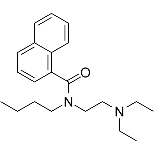
| SMILES | O=C(C1=C2C=CC=CC2=CC=C1)N(CCCC)CCN(CC)CC |
| InChi Key | WWGZXRYELYWJBD-UHFFFAOYSA-N |
| InChi Code | InChI=1S/C21H30N2O/c1-4-7-15-23(17-16-22(5-2)6-3)21(24)20-14-10-12-18-11-8-9-13-19(18)20/h8-14H,4-7,15-17H2,1-3H3 |
| Chemical Name | N-butyl-N-[2-(diethylamino)ethyl]naphthalene-1-carboxamide |
| HS Tariff Code | 2934.99.9001 |
| Storage |
Powder-20°C 3 years 4°C 2 years In solvent -80°C 6 months -20°C 1 month |
| Shipping Condition | Room temperature (This product is stable at ambient temperature for a few days during ordinary shipping and time spent in Customs) |
Biological Activity
| Targets |
Bunaftide involves myocardial cell ion channels [2] Bunaftide affects cellular microfilament cytoskeleton and mitosis-related pathways [1] |
| ln Vitro |
Bunaftide causes rounding and shrinking of the cells, as well as the loss of microvilli-like processes, at concentrations between 0.5 and 2.0 mM. Within the cytoplasm, dense, partially granular, partially fibrous aggregates form, causing the rough endoplasmic reticulum to become vesicular. Loops, coils, and granules are formed when actin filament bundles are broken. Bunaftide raises the proportion of mitotic cells by 0.4 mM, but it dramatically lowers the proportion of mitotic cells without cells accumulating at any particular stage of mitosis by 0.8–1.0 mM. The medication may have a critical concentration above which the arrest becomes irreversible, and it may stop the cell cycle before mitosis [1]. In guinea pig papillary muscle preparations, bunaftide at 1, 5, and 10 mg/L produced a concentration-dependent inhibition of the maximum rise rate of action potentials without altering the resting membrane potential and action potential amplitude. While treatment with 10 mg/L did not alter the action potential duration, treatment with 1 and 5 mg/L brinatilide significantly extended the action potential duration (APD50, APD90) [2]. - Cytotoxic and cytoskeletal effects: In cultured human fibroblasts and HeLa cells, Bunaftide (10-50 μg/mL) exhibited dose-dependent effects; at 10 μg/mL, it induced mild microfilament fragmentation without significant morphological changes; at 25 μg/mL, it severely disrupted microfilament integrity, leading to cell rounding and reduced adherence; at 50 μg/mL, it inhibited mitotic activity by 62% (HeLa cells) and 58% (fibroblasts) compared to control, with no obvious cell lysis [1] - Electrophysiological effects: In isolated rabbit ventricular muscle preparations, Bunaftide (1-10 μM) dose-dependently prolonged action potential duration (APD₅₀ and APD₉₉); 10 μM concentration extended APD₅₀ by 45% and APD₉₉ by 52% without altering maximum diastolic potential or the rate of depolarization (Vmax); it also reduced the amplitude of action potential by 15% at 10 μM, consistent with its antiarrhythmic activity [2] |
| Cell Assay |
- Fibroblast/HeLa cell culture assay: Human fibroblasts and HeLa cells were seeded in culture dishes and grown to 70% confluence; serial concentrations of Bunaftide (10-50 μg/mL) were added to the culture medium, and cells were incubated at 37°C in a 5% CO₂ atmosphere for 24-48 hours [1] - Morphological observation: Cells were observed under a phase-contrast microscope to record changes in shape, adherence, and cytoskeletal structure; microfilaments were visualized by fluorescent staining, and the degree of fragmentation was quantified [1] - Mitotic activity assay: After incubation, cells were fixed and stained with hematoxylin-eosin; the number of mitotic cells (metaphase/anaphase/telophase) was counted in 10 random high-power fields, and the mitotic index was calculated [1] |
| Toxicity/Toxicokinetics |
- In vitro cytotoxicity: Bunaftide (10-50 μg/mL) inhibited the proliferation of human fibroblasts and HeLa cells in a dose-dependent manner, with the mitotic index decreasing by 30-62% compared to control; it disrupted microfilament cytoskeleton but did not induce significant cell necrosis at concentrations up to 50 μg/mL [1] |
| References |
[1]. Effects of bunaftine on morphology, microfilament integrity, and mitotic activity in cultured human fibroblasts and HeLa cells. Cell Tissue Res. 1984;236(1):107-15. [2]. Electrophysiological effects of bunaftine, an antiarrhythmic drug, on action potential characteristics in ventricular muscle preparations. Nihon Yakurigaku Zasshi. 1986 Jul;88(1):1-7. |
| Additional Infomation |
Bunaftine is a naphthalenecarboxamide. N-Butyl-N-(2-(diethylamino)ethyl)-1-naphthamide. A proposed antiarrhythmic that prolongs myocardial refractory period and stabilizes cell membranes. - Bunaftide (originally named "bunaftine" in literatures) is a synthetic antiarrhythmic drug [2] - Its antiarrhythmic mechanism is associated with modifying ion channel activity in ventricular muscle cells, leading to prolonged action potential duration and stabilized cardiac electrical activity [2] - The compound exerts cytotoxic effects on cultured cells by damaging microfilament integrity, which is essential for cell shape maintenance, adherence, and mitosis [1] - The effects of Bunaftide on both cardiac tissue and cultured cells are dose-dependent, with higher concentrations causing more pronounced biological responses [1][2] |
Solubility Data
| Solubility (In Vitro) | May dissolve in DMSO (in most cases), if not, try other solvents such as H2O, Ethanol, or DMF with a minute amount of products to avoid loss of samples |
| Solubility (In Vivo) |
Note: Listed below are some common formulations that may be used to formulate products with low water solubility (e.g. < 1 mg/mL), you may test these formulations using a minute amount of products to avoid loss of samples. Injection Formulations (e.g. IP/IV/IM/SC) Injection Formulation 1: DMSO : Tween 80: Saline = 10 : 5 : 85 (i.e. 100 μL DMSO stock solution → 50 μL Tween 80 → 850 μL Saline) *Preparation of saline: Dissolve 0.9 g of sodium chloride in 100 mL ddH ₂ O to obtain a clear solution. Injection Formulation 2: DMSO : PEG300 :Tween 80 : Saline = 10 : 40 : 5 : 45 (i.e. 100 μL DMSO → 400 μLPEG300 → 50 μL Tween 80 → 450 μL Saline) Injection Formulation 3: DMSO : Corn oil = 10 : 90 (i.e. 100 μL DMSO → 900 μL Corn oil) Example: Take the Injection Formulation 3 (DMSO : Corn oil = 10 : 90) as an example, if 1 mL of 2.5 mg/mL working solution is to be prepared, you can take 100 μL 25 mg/mL DMSO stock solution and add to 900 μL corn oil, mix well to obtain a clear or suspension solution (2.5 mg/mL, ready for use in animals). Injection Formulation 4: DMSO : 20% SBE-β-CD in saline = 10 : 90 [i.e. 100 μL DMSO → 900 μL (20% SBE-β-CD in saline)] *Preparation of 20% SBE-β-CD in Saline (4°C,1 week): Dissolve 2 g SBE-β-CD in 10 mL saline to obtain a clear solution. Injection Formulation 5: 2-Hydroxypropyl-β-cyclodextrin : Saline = 50 : 50 (i.e. 500 μL 2-Hydroxypropyl-β-cyclodextrin → 500 μL Saline) Injection Formulation 6: DMSO : PEG300 : castor oil : Saline = 5 : 10 : 20 : 65 (i.e. 50 μL DMSO → 100 μLPEG300 → 200 μL castor oil → 650 μL Saline) Injection Formulation 7: Ethanol : Cremophor : Saline = 10: 10 : 80 (i.e. 100 μL Ethanol → 100 μL Cremophor → 800 μL Saline) Injection Formulation 8: Dissolve in Cremophor/Ethanol (50 : 50), then diluted by Saline Injection Formulation 9: EtOH : Corn oil = 10 : 90 (i.e. 100 μL EtOH → 900 μL Corn oil) Injection Formulation 10: EtOH : PEG300:Tween 80 : Saline = 10 : 40 : 5 : 45 (i.e. 100 μL EtOH → 400 μLPEG300 → 50 μL Tween 80 → 450 μL Saline) Oral Formulations Oral Formulation 1: Suspend in 0.5% CMC Na (carboxymethylcellulose sodium) Oral Formulation 2: Suspend in 0.5% Carboxymethyl cellulose Example: Take the Oral Formulation 1 (Suspend in 0.5% CMC Na) as an example, if 100 mL of 2.5 mg/mL working solution is to be prepared, you can first prepare 0.5% CMC Na solution by measuring 0.5 g CMC Na and dissolve it in 100 mL ddH2O to obtain a clear solution; then add 250 mg of the product to 100 mL 0.5% CMC Na solution, to make the suspension solution (2.5 mg/mL, ready for use in animals). Oral Formulation 3: Dissolved in PEG400 Oral Formulation 4: Suspend in 0.2% Carboxymethyl cellulose Oral Formulation 5: Dissolve in 0.25% Tween 80 and 0.5% Carboxymethyl cellulose Oral Formulation 6: Mixing with food powders Note: Please be aware that the above formulations are for reference only. InvivoChem strongly recommends customers to read literature methods/protocols carefully before determining which formulation you should use for in vivo studies, as different compounds have different solubility properties and have to be formulated differently. (Please use freshly prepared in vivo formulations for optimal results.) |
| Preparing Stock Solutions | 1 mg | 5 mg | 10 mg | |
| 1 mM | 3.0630 mL | 15.3149 mL | 30.6297 mL | |
| 5 mM | 0.6126 mL | 3.0630 mL | 6.1259 mL | |
| 10 mM | 0.3063 mL | 1.5315 mL | 3.0630 mL |