Physicochemical Properties
| Molecular Formula | C15H26O |
| Molecular Weight | 222.3663 |
| Exact Mass | 222.198 |
| CAS # | 473-15-4 |
| PubChem CID | 91457 |
| Appearance | White to off-white solid powder |
| Density | 1.0±0.1 g/cm3 |
| Boiling Point | 301.7±11.0 °C at 760 mmHg |
| Melting Point | 72-74ºC |
| Flash Point | 108.6±15.6 °C |
| Vapour Pressure | 0.0±1.4 mmHg at 25°C |
| Index of Refraction | 1.499 |
| LogP | 4.68 |
| Hydrogen Bond Donor Count | 1 |
| Hydrogen Bond Acceptor Count | 1 |
| Rotatable Bond Count | 1 |
| Heavy Atom Count | 16 |
| Complexity | 292 |
| Defined Atom Stereocenter Count | 3 |
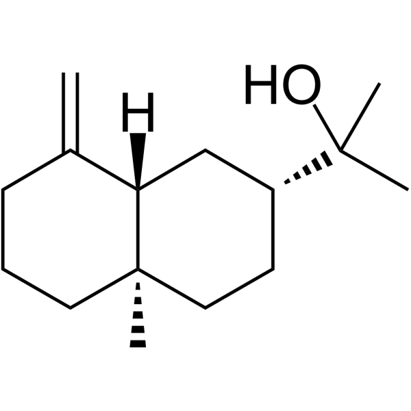
| SMILES | C[C@]12CCCC(=C)[C@@H]1C[C@@H](CC2)C(C)(C)O |
| InChi Key | BOPIMTNSYWYZOC-VNHYZAJKSA-N |
| InChi Code | InChI=1S/C15H26O/c1-11-6-5-8-15(4)9-7-12(10-13(11)15)14(2,3)16/h12-13,16H,1,5-10H2,2-4H3/t12-,13+,15-/m1/s1 |
| Chemical Name | 2-[(2R,4aR,8aS)-4a-methyl-8-methylidene-1,2,3,4,5,6,7,8a-octahydronaphthalen-2-yl]propan-2-ol |
| Synonyms | Beta-Eudesmol |
| HS Tariff Code | 2934.99.9001 |
| Storage |
Powder-20°C 3 years 4°C 2 years In solvent -80°C 6 months -20°C 1 month Note: Please store this product in a sealed and protected environment (e.g. under nitrogen), avoid exposure to moisture. |
| Shipping Condition | Room temperature (This product is stable at ambient temperature for a few days during ordinary shipping and time spent in Customs) |
Biological Activity
| Targets |
- `Beta-Eudesmol` inhibits the NF-κB signaling pathway to alleviate septic liver injury; it reduces NF-κB p65 nuclear translocation with an EC50 of ~15 μM in LPS-induced RAW264.7 cells [2] - `Beta-Eudesmol` antagonizes cholinergic receptors to reverse neostigmine-induced neuromuscular failure. [3] - `Beta-Eudesmol` induces apoptosis in HL-60 cells via promoting cytochrome c release from mitochondria; the IC50 for HL-60 cell viability inhibition is ~28 μM [4] |
| ln Vitro |
Beta-Eudesmol (30-100 μM; 6 days) inhibits the growth of HUVEC cells induced by vascular endothelial growth factor (VEGF) (30 ng/ml) and basic fibroblast growth factor (bFGF) (30 ng/ml). In a time- and dose-dependent manner, Beta-Eudesmol (10-100 μM; 72 hours) inhibits the proliferation of cells in tumors HeLa, SGC-7901, and BEL-7402 [1]. -Eudesmol (10–120 μM; 4 hours) can decrease HL-60 cell viability [4]. In HL-60 cells, beta-eudesmol (40-80 μM; 4 hours) can detect cytochrome c. - In human umbilical vein endothelial cells (HUVECs), `Beta-Eudesmol` (5-40 μM) for 12 hours inhibited tube formation dose-dependently: 5 μM reduced tube length by 21%, 20 μM by 58%, and 40 μM by 89% (Matrigel assay). In A549 lung cancer cells, 40 μM `Beta-Eudesmol` for 48 hours reduced cell viability by 72% (MTT assay) [1] - In LPS-induced RAW264.7 macrophages, `Beta-Eudesmol` (10-50 μM) for 24 hours reduced TNF-α and IL-6 secretion: 20 μM reduced TNF-α by 45% and IL-6 by 52% (ELISA). Western blot showed 20 μM `Beta-Eudesmol` downregulated NF-κB p65 (0.4-fold) and phospho-p65 (0.3-fold) [2] - In mouse diaphragm preparations, `Beta-Eudesmol` (10-100 μM) reversed neostigmine (1 μM)-induced neuromuscular failure: 50 μM restored muscle twitch amplitude to 82% of the control (electromyography) [3] - In HL-60 leukemia cells, `Beta-Eudesmol` (15-60 μM) for 48 hours increased apoptotic rate: 30 μM increased apoptosis to 41.3% (Annexin V/PI flow cytometry) and promoted cytochrome c release from mitochondria (Western blot, cytosolic cytochrome c increased by 3.2-fold) [4] |
| ln Vivo |
In MK mice, the intraperitoneal injection of beta-eudesmol (2.5–5 mg/kg; once daily for 7 days) greatly suppresses the growth of tumor cells and new blood vessels [1]. intraperitoneal; 50–100 mg/kg) beta-eudesmol - In nude mice bearing A549 lung cancer xenografts (n=6 per group), intraperitoneal injection of `Beta-Eudesmol` (10 mg/kg/day or 20 mg/kg/day) for 21 days reduced tumor volume by 38% (10 mg/kg) and 65% (20 mg/kg) vs. vehicle. Tumor weight in the 20 mg/kg group was 0.35 ± 0.06 g, significantly lower than vehicle’s 1.02 ± 0.11 g [1] - In mice with LPS-induced septic liver injury (n=8 per group), oral gavage of `Beta-Eudesmol` (20 mg/kg/day or 40 mg/kg/day) for 7 days reduced serum ALT (40 mg/kg: 68 ± 9 U/L vs. vehicle: 215 ± 18 U/L) and AST (40 mg/kg: 75 ± 10 U/L vs. vehicle: 242 ± 21 U/L). Liver tissue NF-κB p65 expression was downregulated by 58% (40 mg/kg, immunohistochemistry) [2] |
| Enzyme Assay |
- NF-κB p65 nuclear translocation assay (RAW264.7 cells): Cells were treated with `Beta-Eudesmol` (0-50 μM) for 1 hour, then stimulated with LPS (1 μg/mL) for 2 hours. Nuclear and cytoplasmic proteins were extracted, and NF-κB p65 was quantified by ELISA (detection at 450 nm). `Beta-Eudesmol` (20 μM) reduced nuclear p65 by 62% vs. LPS-only group [2] - Neuromuscular transmission assay (mouse diaphragm): Diaphragm strips were isolated and mounted in Krebs solution. Neostigmine (1 μM) was added to induce twitch depression, then `Beta-Eudesmol` (10-100 μM) was added. Twitch amplitude was recorded via a force transducer, and the reversal rate was calculated [3] |
| Cell Assay |
Western Blot Analysis[4] Cell Types: HL-60 Tested Concentrations: 40 μM, 80 μM Incubation Duration: 4 h Experimental Results: Increased expression of caspase-3 protein. 80 µM. Cell viability assay [4] Cell Types: HL-60 Tested Concentrations: 10 μM, 20 μM, 40 μM, 60 μM, 80 μM, 120 μM Incubation Duration: 4 h Experimental Results: Cell viability was not affected at 40 μM dose, but at 80 μM The cell activity diminished at the dose, and the cell viability diminished by 51.6%. Apoptosis analysis [4] Cell Types: HL-60 Tested Concentrations: 40 μM, 80 μM Incubation Duration: 4 h Experimental Results: 40 μM dose had no effect on cell apoptosis, 80 μM dose induced apoptosis, and the cell apoptosis rate is 31.7%. - HUVEC tube formation assay: Matrigel was coated onto 24-well plates and polymerized. HUVECs (2×10⁴ cells/well) suspended in medium containing `Beta-Eudesmol` (0-40 μM) were seeded. After 12 hours, tube length was measured via image analysis software [1] - A549 cell viability assay: Cells were seeded in 96-well plates (5×10³ cells/well) and treated with `Beta-Eudesmol` (0-40 μM) for 48 hours. MTT solution was added, incubated for 4 hours, DMSO dissolved formazan, and absorbance was measured at 570 nm [1] - HL-60 cell apoptosis assay: Cells (2×10⁵ cells/well) were treated with `Beta-Eudesmol` (0-60 μM) for 48 hours. Cells were stained with Annexin V-FITC/PI and analyzed by flow cytometry. Mitochondrial and cytosolic fractions were isolated for cytochrome c detection via Western blot [4] |
| Animal Protocol |
Animal/Disease Models: Male C57BL/6 mouse model [2] Doses: 50 mg/kg, 100 mg/kg Route of Administration: intraperitoneal (ip) injection; single dose. 2 hrs (hrs (hours)) before cecal ligation and puncture (CLP) surgery. Experimental Results: The expression of TNF-α, IL-1β and IL-6 increased in a dose-dependent manner. Increases levels of superoxide dismutase (SOD) and glutathione (GSH), and decreases levels of malondialdehyde (MDA) and myeloperoxidase (MPO). Inhibits the phosphorylation of p65. - A549 xenograft model (nude mice): 5×10⁶ A549 cells were subcutaneously injected into nude mice (6-8 weeks old). When tumors reached ~100 mm³, mice were grouped: vehicle (0.1% DMSO + saline), `Beta-Eudesmol` 10 mg/kg, 20 mg/kg. Drugs were administered intraperitoneally once daily for 21 days. Tumor volume (length×width²/2) and weight were measured [1] - Septic liver injury model (mice): C57BL/6 mice (20-25 g) were injected with LPS (10 mg/kg, ip) to induce sepsis. Mice were grouped: vehicle (0.5% carboxymethyl cellulose), `Beta-Eudesmol` 20 mg/kg, 40 mg/kg. Drugs were given by oral gavage once daily for 7 days. Serum ALT/AST and liver NF-κB p65 were detected [2] |
| Toxicity/Toxicokinetics |
- In nude mice treated with `Beta-Eudesmol` (up to 20 mg/kg/day, ip, 21 days), no significant weight loss (weight change <5%) or mortality was observed. Serum creatinine and urea nitrogen were normal [1] - In mice with septic liver injury, `Beta-Eudesmol` (up to 40 mg/kg/day, po, 7 days) did not cause additional liver or kidney damage; serum bilirubin and creatinine were within normal ranges [2] - In mouse diaphragm preparations, `Beta-Eudesmol` (up to 100 μM) showed no cytotoxicity; diaphragm cell viability remained >95% (trypan blue staining) [3] |
| References |
[1]. Beta-eudesmol suppresses tumour growth through inhibition of tumour neovascularisation and tumour cell proliferation. J Asian Nat Prod Res. 2008 Jan-Feb;10(1-2):159-67. [2]. Protective effects of β-eudesmol against septic liver injury via inhibition of NF-κB signaling. Tropical Journal of Pharmaceutical Research, 2022, 21(6): 1183-1188. [3]. Antagonism by beta-eudesmol of neostigmine-induced neuromuscular failure in mouse diaphragms. Eur J Pharmacol. 1992 Jun 5;216(2):199-206. [4]. Eudesmols induce apoptosis through release of cytochrome c in HL-60 cells. Natural Product Sciences, 2010, 16(2): 88-92. |
| Additional Infomation |
Beta-eudesmol is a carbobicyclic compound that is trans-decalin substituted at positions 2, 4a, and 8 by 2-hydroxypropan-2-yl, methyl and methylidene groups, respectively (the 2R,4aR,8aS-diastereoisomer). It has a role as a volatile oil component. It is a carbobicyclic compound, a tertiary alcohol and a eudesmane sesquiterpenoid. beta-Eudesmol has been reported in Rhododendron dauricum, Humulus lupulus, and other organisms with data available. See also: Arctium lappa Root (part of); Cannabis sativa subsp. indica top (part of); Pterocarpus marsupium wood (part of) ... View More ... - `Beta-Eudesmol` is a natural sesquiterpene alcohol isolated from plants such as ` Atractylodes lancea ` and ` Eucalyptus globulus ` [1][2] - Its anti-tumor mechanism involves dual effects: inhibiting tumor neovascularization (via blocking HUVEC tube formation) and suppressing tumor cell proliferation (via inducing G2/M cycle arrest in A549 cells) [1] - In septic liver injury, `Beta-Eudesmol` reduces inflammation by inhibiting NF-κB activation, thereby alleviating liver tissue necrosis and inflammatory cell infiltration [2] - The neuromuscular protective effect of `Beta-Eudesmol` is mediated by competitive antagonism of cholinergic receptors, preventing excessive acetylcholine accumulation-induced muscle paralysis [3] |
Solubility Data
| Solubility (In Vitro) |
DMSO : ~100 mg/mL (~449.70 mM) H2O : < 0.1 mg/mL |
| Solubility (In Vivo) |
Solubility in Formulation 1: 2.5 mg/mL (11.24 mM) in 10% DMSO + 40% PEG300 + 5% Tween80 + 45% Saline (add these co-solvents sequentially from left to right, and one by one), suspension solution; with sonication. For example, if 1 mL of working solution is to be prepared, you can add 100 μL of 25.0 mg/mL clear DMSO stock solution to 400 μL PEG300 and mix evenly; then add 50 μL Tween-80 to the above solution and mix evenly; then add 450 μL normal saline to adjust the volume to 1 mL. Preparation of saline: Dissolve 0.9 g of sodium chloride in 100 mL ddH₂ O to obtain a clear solution. Solubility in Formulation 2: ≥ 2.5 mg/mL (11.24 mM) (saturation unknown) in 10% DMSO + 90% (20% SBE-β-CD in Saline) (add these co-solvents sequentially from left to right, and one by one), clear solution. For example, if 1 mL of working solution is to be prepared, you can add 100 μL of 25.0 mg/mL clear DMSO stock solution to 900 μL of 20% SBE-β-CD physiological saline solution and mix evenly. Preparation of 20% SBE-β-CD in Saline (4°C,1 week): Dissolve 2 g SBE-β-CD in 10 mL saline to obtain a clear solution. Solubility in Formulation 3: ≥ 2.5 mg/mL (11.24 mM) (saturation unknown) in 10% DMSO + 90% Corn Oil (add these co-solvents sequentially from left to right, and one by one), clear solution. For example, if 1 mL of working solution is to be prepared, you can add 100 μL of 25.0 mg/mL clear DMSO stock solution to 900 μL of corn oil and mix evenly. (Please use freshly prepared in vivo formulations for optimal results.) |
| Preparing Stock Solutions | 1 mg | 5 mg | 10 mg | |
| 1 mM | 4.4970 mL | 22.4850 mL | 44.9701 mL | |
| 5 mM | 0.8994 mL | 4.4970 mL | 8.9940 mL | |
| 10 mM | 0.4497 mL | 2.2485 mL | 4.4970 mL |