Bay 65-1942 HCl is the hydrochloride salt of of BAY65-1942 which is a novel, potent and selective ATP-competitive inhibitor of IKKβ.It targets the IKKbeta kinase activity specifically. Following an acute ischemia-reperfusion injury, IKKbeta inhibition reduces myocardial damage and dysfunction. The CK-MB levels of the animals pretreated with Bay 65-1942 (n=3) were significantly lower than those of the untreated animals before IR (14,170 ±3,219 units, P<0.05 vs. vehicle).
Physicochemical Properties
| Molecular Formula | C22H25N3O4.HCL |
| Molecular Weight | 431.91254 |
| Exact Mass | 431.161 |
| Elemental Analysis | C, 61.18; H, 6.07; Cl, 8.21; N, 9.73; O, 14.82 |
| CAS # | 600734-06-3 |
| Related CAS # | 758683-21-5 (BAY65-1942 R-isomer); 600734-02-9 |
| PubChem CID | 135454903 |
| Appearance | White to off-white solid powder |
| LogP | 5.04 |
| Hydrogen Bond Donor Count | 4 |
| Hydrogen Bond Acceptor Count | 6 |
| Rotatable Bond Count | 5 |
| Heavy Atom Count | 30 |
| Complexity | 586 |
| Defined Atom Stereocenter Count | 1 |
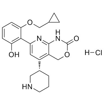
| SMILES | O=C1OCC2=C([C@H]3CNCCC3)C=C(C4=C(O)C=CC=C4OCC5CC5)N=C2N1.[H]Cl |
| InChi Key | XZTOAEZYOFWVHB-PFEQFJNWSA-N |
| InChi Code | InChI=1S/C22H25N3O4.ClH/c26-18-4-1-5-19(28-11-13-6-7-13)20(18)17-9-15(14-3-2-8-23-10-14)16-12-29-22(27)25-21(16)24-17;/h1,4-5,9,13-14,23,26H,2-3,6-8,10-12H2,(H,24,25,27);1H/t14-;/m1./s1 |
| Chemical Name | 7-[2-(cyclopropylmethoxy)-6-hydroxyphenyl]-5-[(3S)-piperidin-3-yl]-1,4-dihydropyrido[2,3-d][1,3]oxazin-2-one;hydrochloride |
| Synonyms | BAY 65-1942 HCl; BAY-65-1942 hydrochloride; BAY65-1942; BAY 651942; BAY-651942; BAY651942 |
| HS Tariff Code | 2934.99.03.00 |
| Storage |
Powder-20°C 3 years 4°C 2 years In solvent -80°C 6 months -20°C 1 month Note: Please store this product in a sealed and protected environment, avoid exposure to moisture. |
| Shipping Condition | Room temperature (This product is stable at ambient temperature for a few days during ordinary shipping and time spent in Customs) |
Biological Activity
| Targets |
IKKβ Compared to animals receiving a vehicle, the size of the left ventricular infarct is significantly reduced when Bay 65-1942 is administered before ischemia. Animals receiving vehicles have a significantly higher infarct-to-area at risk (AAR) ratio than sham animals (70.7±3.4 vs. 5.8±3.4%, P<0.05). Treatment with Bay 65-1942 at each time point significantly lowers this ratio (prior to ischemia 42.7±4.1%, at reperfusion 42.7±7.5%, 2 hours after reperfusion 29.4±5.2%; each group P<0.05 vs. vehicle). The CK-MB levels in the animals pretreated with Bay 65-1942 (n=3) were significantly lower than those in the control group before IR (14,170 ±3,219 units, P<0.05 vs. vehicle)[1]. |
| ln Vitro |
Compared to animals receiving a vehicle, the size of the left ventricular infarct is significantly reduced when Bay 65-1942 is administered before ischemia. Animals receiving vehicles have a significantly higher infarct-to-area at risk (AAR) ratio than sham animals (70.7±3.4 vs. 5.8±3.4%, P<0.05). Treatment with Bay 65-1942 at each time point significantly lowers this ratio (prior to ischemia 42.7±4.1%, at reperfusion 42.7±7.5%, 2 hours after reperfusion 29.4±5.2%; each group P<0.05 vs. vehicle). The CK-MB levels in the animals pretreated with Bay 65-1942 (n=3) were significantly lower than those in the control group before IR (14,170 ±3,219 units, P<0.05 vs. vehicle)[1]. In imatinib-resistant, Lyn-dependent chronic myeloid leukemia cells (MYL-R), treatment with Bay 65-1942 (10 µM) for 2 hours effectively blocked the phosphorylation of the NF-κB p65 subunit at Ser536, a marker of NF-κB activity. [2] Treatment of MYL-R cells with Bay 65-1942 (10 µM) for 12 hours resulted in an approximately 2-fold decrease in IκBα mRNA expression, but led to a 4.4-fold increase in IL-6 mRNA expression compared to vehicle-treated cells. [2] Treatment of MYL-R cells with Bay 65-1942 (10 µM) for 24 hours increased MEK/ERK pathway activation, as demonstrated by a significant increase in ERK phosphorylation. This activation was prevented by co-treatment with the MEK inhibitor AZD6244 (5 µM). [2] Co-treatment of MYL-R cells with Bay 65-1942 (10 µM) and AZD6244 (5 µM) for 12 hours prevented the Bay 65-1942-induced increase in IL-6 mRNA expression. [2] In cell viability assays (MTS), treatment of MYL-R cells with Bay 65-1942 (10 µM) for 48 hours reduced cell viability by approximately 37% compared to DMSO-treated controls. Combined treatment with Bay 65-1942 (10 µM) and AZD6244 (5 µM) showed synergistic effects, reducing viability by approximately 84%. The combination index (CI) at this dose was 0.48, indicating synergism. [2] In apoptosis assays, treatment of MYL-R cells with Bay 65-1942 (10 µM) for 24 hours induced a 1.3-fold increase in caspase 3/7 activity. Combined treatment with Bay 65-1942 and AZD6244 induced a 3.2-fold increase in caspase 3/7 activity and enhanced cleavage of poly(ADP-ribose) polymerase (PARP). [2] Multiplexed kinase inhibitor bead/mass spectrometry (MIB/MS) analysis of MYL-R cells treated with Bay 65-1942 (10 µM) for 24 hours revealed an increase in the capture of kinases in the MEK/ERK pathway (B-Raf, ERK, RSK1) by the beads, indicating increased pathway activity. [2] |
| ln Vivo | In order to sufficiently inhibit kinase activity, inhibitors of MEK (AZD6244) and IKK (BAY 65-1942) are used at their IC50 concentrations, as determined by a 48-hour MTS assay. AZD6244 (5 µM), BAY 65-1942 (10 µM), or a combination of these inhibitors at the same concentrations are applied to MYL-R cells for 24 hours. At the dose combination of (5 µM AZD6244+10 µM BAY 65-1942), which correlates with IC75 (CI = 0.48±0.01), AZD6244 and BAY 65-1942 show synergistic inhibition of cell viability. Additionally, the software reports that synergism is present at the IC50 (CI = 0.56±0.09) and IC90 (CI = 0.460.02) dose combinations (CI values are the mean of three independent experiments, ±standard deviation). In comparison to DMSO-treated cells, the effects of AZD6244 and BAY 65-1942 on caspase 3/7 activation are 1.3 and 2 times greater, respectively. Caspase 3/7 activity is 3.2 times higher after treatment with AZD6244 and BAY 65-1942[2]. |
| Cell Assay |
MYL-R cells are plated on a 96-well plate at a density of 4×10⁴ cells/well in a growth medium of 100 µL RPMI containing kinase inhibitors to test the viability of the cells. During the first 24 and second 48 hours, growth media and kinase inhibitors are replenished. Each well is filled with 20 L of the MTS assay reagent. The absorbance at 490 nm is measured after the plate has been in the incubator for roughly an hour. Cells are cultivated and tested for combination index (CI) experiments. Cells are treated with a series of three-fold dilutions of either AZD6244 alone or in combination with BAY 65-1942 (10 µM) while maintaining a constant ratio of 1:2, respectively, to ascertain the dose-effects of each drug. To calculate CI values, cell viability test results are analyzed. Three independent experiments' CI values are averaged[2]. For Western blot analysis of kinase phosphorylation, MYL-R cells were treated with Bay 65-1942 (10 µM) for specified durations (e.g., 2 or 24 hours). Cells were then lysed, proteins were separated by SDS-PAGE, transferred to PVDF membranes, and probed with specific primary and secondary antibodies. Signal was detected using enhanced chemiluminescence reagents. [2] For qRT-PCR analysis of gene expression, MYL-R cells were treated with Bay 65-1942 (10 µM) for 12 or 24 hours. Total RNA was isolated, reverse transcribed into cDNA, and analyzed by real-time PCR using specific TaqMan assays. [2] For cell viability assessment (MTS assay), MYL-R cells were seeded in 96-well plates and treated with Bay 65-1942 (10 µM) alone or in combination with AZD6244. Media and drugs were replenished at 24 hours, and cell viability was measured at 48 hours by adding MTS reagent and measuring absorbance at 490 nm. [2] For caspase 3/7 activity assays, MYL-R cells were treated with Bay 65-1942 (10 µM) for 24 hours, lysed, and the supernatant was incubated with a caspase 3/7 substrate (Ac-DEVD-AMC). The fluorescence of liberated AMC was measured to determine caspase activity. [2] For MIB/MS kinome profiling, MYL-R cells were treated with Bay 65-1942 (10 µM) for 24 hours. Cells were lysed, and kinases were enriched using multiplexed kinase inhibitor-conjugated beads (MIBs). Captured proteins were eluted, digested with trypsin, and analyzed by liquid chromatography coupled with MALDI-TOF/TOF mass spectrometry for identification and relative quantification. [2] |
| Animal Protocol | Mice: 8–10 week-old male C57BL/6 mice are used. Mice are given 30 minutes of cardiac ischemia followed by various amounts of reperfusion in order to study the effects of IKK inhibition on myocardial IR injury. At the proper dosing times, Bay 65-1942 (5 mg/kg) is injected intraperitoneally. A 10% cremaphor in water vehicle is given to non-treatment groups. In treatment groups, Bay 65-1942 is administered either before ischemia, during reperfusion, or two hours after reperfusion injury. The size of the infarct is determined 24 hours after the injury caused by the reperfusion in the vehicle, sham, and treatment groups. Serum creatine kinase-muscle-brain fraction (CK-MB) levels are assessed 1 hour after reperfusion in animals that have received Bay 65-1942 treatment in order to confirm myocardial injury. |
| References |
[1]. Inhibition of IκB kinase-β protects dopamine neurons against lipopolysaccharide-induced neurotoxicity By Zhang, Feng; Qian, Li; Flood, Patrick M.; Shi, Jing-Shan; Hong, Jau-Shyong; Gao, Hui-Ming J Pharmacol Exp Ther. 2010 June; 333(3): 822-833. [2]. Application of multiplexed kinase inhibitor beads to study kinome adaptations in drug-resistant leukemia. PLoS One. 2013 Jun 24;8(6):e66755. |
| Additional Infomation |
Bay 65-1942 hydrochloride is an ATP-competitive and selective IKKb inhibitor. Bay 65-1942 was used in this study as a pharmacological tool to investigate the role of the IKK/NF-κB pathway in imatinib-resistant leukemia. [2] The study found that inhibition of IKK with Bay 65-1942 led to a compensatory activation of the MEK/ERK pathway in resistant cells. This finding provided a rationale for the synergistic anti-leukemic effect observed when Bay 65-1942 was combined with a MEK inhibitor. [2] The research demonstrates that kinome profiling using MIB/MS can identify adaptive signaling responses (like MEK/ERK upregulation upon IKK inhibition) and guide effective combination therapy strategies. [2] |
Solubility Data
| Solubility (In Vitro) |
DMSO: ~50 mg/mL (~115.8 mM) H2O: ~2.2 mg/mL (~5.0 mM) |
| Solubility (In Vivo) |
Solubility in Formulation 1: ≥ 2.5 mg/mL (5.79 mM) (saturation unknown) in 10% DMSO + 40% PEG300 + 5% Tween80 + 45% Saline (add these co-solvents sequentially from left to right, and one by one), clear solution. For example, if 1 mL of working solution is to be prepared, you can add 100 μL of 25.0 mg/mL clear DMSO stock solution to 400 μL PEG300 and mix evenly; then add 50 μL Tween-80 to the above solution and mix evenly; then add 450 μL normal saline to adjust the volume to 1 mL. Preparation of saline: Dissolve 0.9 g of sodium chloride in 100 mL ddH₂ O to obtain a clear solution. Solubility in Formulation 2: ≥ 2.5 mg/mL (5.79 mM) (saturation unknown) in 10% DMSO + 90% (20% SBE-β-CD in Saline) (add these co-solvents sequentially from left to right, and one by one), clear solution. For example, if 1 mL of working solution is to be prepared, you can add 100 μL of 25.0 mg/mL clear DMSO stock solution to 900 μL of 20% SBE-β-CD physiological saline solution and mix evenly. Preparation of 20% SBE-β-CD in Saline (4°C,1 week): Dissolve 2 g SBE-β-CD in 10 mL saline to obtain a clear solution. Solubility in Formulation 3: ≥ 2.5 mg/mL (5.79 mM) (saturation unknown) in 10% DMSO + 90% Corn Oil (add these co-solvents sequentially from left to right, and one by one), clear solution. For example, if 1 mL of working solution is to be prepared, you can add 100 μL of 25.0 mg/mL clear DMSO stock solution to 900 μL of corn oil and mix evenly. (Please use freshly prepared in vivo formulations for optimal results.) |
| Preparing Stock Solutions | 1 mg | 5 mg | 10 mg | |
| 1 mM | 2.3153 mL | 11.5765 mL | 23.1530 mL | |
| 5 mM | 0.4631 mL | 2.3153 mL | 4.6306 mL | |
| 10 mM | 0.2315 mL | 1.1576 mL | 2.3153 mL |