Physicochemical Properties
| Molecular Formula | C47H76O18 |
| Molecular Weight | 929.0956 |
| Exact Mass | 928.503 |
| Elemental Analysis | C, 60.76; H, 8.25; O, 31.00 |
| CAS # | 382146-66-9 |
| PubChem CID | 9876264 |
| Appearance | White to off-white solid powder |
| Density | 1.43 |
| LogP | 2 |
| Hydrogen Bond Donor Count | 10 |
| Hydrogen Bond Acceptor Count | 18 |
| Rotatable Bond Count | 10 |
| Heavy Atom Count | 65 |
| Complexity | 1770 |
| Defined Atom Stereocenter Count | 25 |
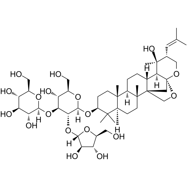
| SMILES | CC(=C[C@@H]1CO[C@]23C[C@]4(CO2)[C@@H]([C@H]3[C@@]1(C)O)CC[C@H]5[C@]4(CC[C@@H]6[C@@]5(CC[C@@H](C6(C)C)O[C@H]7[C@@H]([C@H]([C@@H]([C@H](O7)CO)O)O[C@H]8[C@@H]([C@H]([C@@H]([C@H](O8)CO)O)O)O)O[C@H]9[C@@H]([C@H]([C@@H](O9)CO)O)O)C)C)C |
| InChi Key | WZWPYJOPCULCLQ-UOXCDNDQSA-N |
| InChi Code | InChI=1S/C47H76O18/c1-21(2)14-22-18-58-47-19-46(20-59-47)23(38(47)45(22,7)57)8-9-28-43(5)12-11-29(42(3,4)27(43)10-13-44(28,46)6)63-41-37(65-39-34(55)31(52)25(16-49)61-39)36(32(53)26(17-50)62-41)64-40-35(56)33(54)30(51)24(15-48)60-40/h14,22-41,48-57H,8-13,15-20H2,1-7H3/t22-,23-,24-,25+,26-,27+,28-,29+,30-,31+,32-,33+,34-,35-,36+,37-,38+,39+,40+,41+,43+,44-,45+,46+,47-/m1/s1 |
| Chemical Name | (2S,3R,4S,5S,6R)-2-[(2R,3R,4S,5R,6R)-5-[(2S,3R,4R,5S)-3,4-dihydroxy-5-(hydroxymethyl)oxolan-2-yl]oxy-3-hydroxy-2-(hydroxymethyl)-6-[[(1S,2R,5R,7S,10R,11R,14R,15S,16S,17R,20R)-16-hydroxy-2,6,6,10,16-pentamethyl-17-(2-methylprop-1-enyl)-19,21-dioxahexacyclo[18.2.1.01,14.02,11.05,10.015,20]tricosan-7-yl]oxy]oxan-4-yl]oxy-6-(hydroxymethyl)oxane-3,4,5-triol |
| Synonyms | bacopaside II; 382146-66-9; BACOPASIDE II(P); Bacopaside ii, (-)-; ZO6L404Y9T; UNII-ZO6L404Y9T; Bacopaside II (constituent of bacopa) [DSC]; 3-O-alpha-L-arabinofuranosyl-(1-2)-(beta-D-glucopyranosyl (1-3))-beta-D-glucopyranosyl pseudo-jujubogenin; |
| HS Tariff Code | 2934.99.9001 |
| Storage |
Powder-20°C 3 years 4°C 2 years In solvent -80°C 6 months -20°C 1 month Note: This product requires protection from light (avoid light exposure) during transportation and storage. |
| Shipping Condition | Room temperature (This product is stable at ambient temperature for a few days during ordinary shipping and time spent in Customs) |
Biological Activity
| Targets |
Aquaporin-1 (AQP1) water channel The action target of Bacopaside II is Aquaporin 1 (AQP1) [1] |
| ln Vitro |
Researchers tested the AQP1 inhibitor, bacopaside II, derived from medicinal plant Bacopa monnieri, on endothelial cell migration and tube-formation in vitro using mouse endothelial cell lines (2H11 and 3B11) and human umbilical vein endothelial cells (HUVEC). The effect of bacopaside II on viability, apoptosis, migration and tubulogenesis was assessed by a proliferation assay, annexin-V/propidium iodide flow cytometry, the scratch wound assay and endothelial tube-formation, respectively. Cell viability was reduced significantly for 2H11 at 15 μM (p = 0.037), 3B11 at 12.5 μM (p = 0.017) and HUVEC at 10 μM (p < 0.0001). At 15 μM, the reduced viability was accompanied by an increase in apoptosis of 38%, 50% and 32% for 2H11, 3B11 and HUVEC, respectively. Bacopaside II at ≥10 μM significantly reduced migration of 2H11 (p = 0.0002) and 3B11 (p = 0.034). HUVECs were most sensitive with a significant reduction at ≥7.5 μM (p = 0.037). Tube-formation was reduced with a 15 μM dose for all cell lines and 10 μM for 3B11 (p < 0.0001). These results suggest that bacopaside II is a potential anti-angiogenic agent.[1] Aquaporin-1 (AQP1), a transmembrane pore-forming molecule, facilitates the rapid movement of water and small solutes across cell membranes. Researchers have previously shown that bacopaside II, an extract from the medicinal herb Bacopa monnieri, blocks the AQP1 water channel and impairs migration of cells that express AQP1. The aim of this study was to further elucidate the anti-tumour potential of bacopaside II in colon cancer cells. Expression of AQP1 in HT-29, SW480, SW620 and HCT116 was determined by quantitative PCR and western immunoblot. Cells were treated with bacopaside II, and morphology, growth, autophagy, cell cycle and apoptosis assessed by time-lapse microscopy, crystal violet, acridine orange, propidium iodide (PI) and annexin V/PI staining respectively. AQP1 expression was significantly higher in HT-29 than SW480, SW620 and HCT116. Bacopaside II significantly reduced growth at ≥20 µM for HT-29 and ≥15 µM for SW480, SW620 and HCT116. Inhibition of HT-29 at 20 µM was primarily mediated by G0/G1 cell cycle arrest, and at 30 µM by G2/M arrest and apoptosis. Inhibition of SW480, SW620 and HCT116 at ≥15 µM was mediated by G2/M arrest and apoptosis. These results are the first to show that bacopaside II inhibits colon cancer cell growth by inducing cell cycle arrest and apoptosis. [2] 1. Effects on human umbilical vein endothelial cells (HUVECs): HUVECs were treated with Bacopaside II at concentrations of 5 μM, 10 μM, and 20 μM for 24–48 hours: - Inhibition of cell migration: Scratch assay showed 20 μM Bacopaside II reduced migration rate by 68.3 ± 4.5% vs control; Transwell assay showed 20 μM reduced migrated cell number by 72.1 ± 5.2% [1] - Suppression of tubulogenesis: Matrigel assay showed 20 μM Bacopaside II decreased tube length by 65.7 ± 3.8% and branch number by 69.2 ± 4.1% vs control [1] - Induction of apoptosis: Annexin V-FITC/PI staining showed 20 μM Bacopaside II increased apoptosis rate to 28.5 ± 2.3% (vs control 3.2 ± 0.5%) [1] - Downregulation of AQP1: Western blot showed 20 μM Bacopaside II reduced AQP1 protein expression by 75.4 ± 5.8% vs control [1] 2. Effects on human colon cancer cells (HT-29, HCT116): Cells were treated with Bacopaside II (10 μM, 20 μM, 40 μM) for 24–72 hours: - Antiproliferative activity: MTT assay showed concentration- and time-dependent inhibition. IC50 values at 72 hours: HT-29 (18.6 ± 1.2 μM), HCT116 (15.3 ± 0.9 μM) [2] - Cell cycle arrest: Flow cytometry showed 20 μM Bacopaside II increased G2/M phase cell proportion: HT-29 (38.2 ± 2.5% vs control 12.1 ± 1.3%), HCT116 (42.5 ± 2.8% vs control 11.8 ± 1.1%) [2] - Induction of apoptosis: Annexin V-FITC/PI staining showed 20 μM Bacopaside II increased apoptosis rate: HT-29 (32.6 ± 2.4%), HCT116 (35.8 ± 2.7%) [2] - Modulation of apoptotic/cell cycle proteins: Western blot showed 20 μM Bacopaside II activated caspase-3 (cleaved caspase-3 increased by 3.2-fold in HT-29) and downregulated Cyclin B1 (reduced by 65.3% in HCT116) [2] |
| Cell Assay |
MTS Viability Assay [1] Endothelial cells were seeded in complete medium (2H11 and 3B11) or endothelial growth medium (HUVEC) at 1 × 10⁴ cells per well of a 96-well plate and were incubated overnight. Cells were treated with various concentrations of bacopaside II for 20 h. The cell viability was determined using the CellTiter 96® AQueous Non-Radioactive Cell Proliferation Assay according to the manufacturer’s instructions and absorbance read at 492 nm. Results were calculated as the mean absorbance normalized to the vehicle control. Apoptosis Assay [1] Cells (1 × 10⁵) were seeded in six-well plates and left for 48 h to reach confluence. Apoptosis controls were treated with paclitaxel (400 nM) for 24 h prior to the Time 0 collection. Necrosis controls were prepared by heating harvested cells to 63 °C for 30 min. Cells were treated either with medium (untreated), vehicle (1% methanol) or 10, 12.5 or 15 µM bacopaside II. Cells were collected at 0 and 6 h after treatment, stained with the Annexin-V-FLUOS staining kit as per the manufacturer’s instructions and run on the BD FACSCanto II cell analyser. Gating with cell doublet exclusion and analysis was performed using FlowJo software v 10.4. Scratch Wound (Migration) Assay [1] Cells were seeded according to growth rate at either 1 × 10⁴ per well for 2H11, 4 × 10⁴ for 3B11 or 6 × 10⁴ per well for HUVEC in 96-well plates in their respective medium for 6 h and then serum starved overnight in medium with just 2% FCS and including 1 μg/mL mitomycin C (the mix referred to herein as “mitomedium”), to inhibit cell proliferation. The following day, a circular wound was made in the cell monolayer with a p10 pipette tip. Loose cells were removed and medium replaced with fresh mitomedium for 1 h followed by replacement with treatment, either vehicle (1% methanol) or various concentrations of bacopaside II in mitomedium. Cells were monitored every 2 h with the area of wound closure determined at each time point. Endothelial Tube Formation (Angiogenesis) Assay [1] Mouse endothelial cells or HUVECs were seeded onto a thin layer (10 μL) of matrigel in a 96-well angiogenesis μ-plate at 1.5 × 10⁴ cells per well either in vehicle (1% methanol) or bacopaside II at 10 or 15 μM made in complete medium (2H11 and 3B11) or endothelial growth medium (HUVEC). The numbers of loops formed were counted at peak tube formation, 2–3 h for mouse endothelial cells and 20 h for HUVECs. For pre-treatment experiments, cells were either cultured in normal medium, or pre-treated with vehicle or bacopaside II at lower concentrations, 10 μM for 2H11 and 7.5 μM for HUVEC, followed by harvesting of cells, resuspension in vehicle or bacopaside II and seeding on matrigel, with loops counted at peak formation time as before. Cell Growth Assay [2] Cell growth was determined by crystal violet assay as described previously [18]. Briefly, 103 cells per well were seeded into 96-well plates and cultured for 24 h. Culture medium without cells was added to 6 wells to serve as background controls for nonspecific binding of the crystal violet dye. Cells were treated for 72 h with either 0, 2.5, 5, 10, 15, 20 or 30 µM of bacopaside II analytical standard dissolved in 2% (v/v) methanol vehicle. Cells were fixed with 10% neutral buffered formalin for 30 min, stained with 1% (w/v) crystal violet in 2% ethanol for 10 min, washed eight times in running distilled water, and air-dried. Crystal violet was eluted using 10% acetic acid with gentle rocking of the plates for 1 h at room temperature. Absorbance of the eluent was measured at 595 nm using a FLUOstar Optima microplate reader. The average absorbance of the wells without cells was subtracted from the absorbance of each of the wells containing cells, and the data were expressed as the mean absorbance relative to that of the vehicle control treated cells. Acridine Orange Staining [2] Cells were seeded at 5 × 10⁵ cells per well in six-well plates and incubated for 24 h. Cells were washed with Dulbecco’s phosphate buffered saline to remove non-viable cells, and then treated for 24 h with either 0, 5, 10, 15, 20 or 30 µM of bacopaside II analytical standard dissolved in 2% (v/v) methanol vehicle. Non-adherent and adherent cells were harvested and pooled. To harvest adherent cells, the cells were washed three times with DPBS, collecting each wash, and then incubated with 0.25% trypsin-EDTA at 37 °C until the cells had detached. The trypsin-EDTA was inactivated with culture medium supplemented with 10% FBS. Cells were washed twice with DPBS by centrifuging at 200× g for 10 min at 4 °C and aspirating the supernatant. Cells were stained with 1 μg/mL acridine orange in DPBS at 37 °C for 15 min and immediately analysed using a FACSCanto II flow cytometer, acquiring at least 50,000 single cell events per sample. Cell Cycle Analysis by Propidium Iodide Staining [2] Cells were seeded at 5 × 10⁵ cells per well in six-well plates, treated with bacopaside II, and harvested as described above. Cells were washed twice with DPBS and resuspended in 1.2 mL of ice cold DPBS in polypropylene flow cytometry tubes. Next, 2.8 mL of 100% ice cold ethanol was added dropwise with gentle vortexing, to achieve a final concentration of 70% ethanol. The fixed cells were stored at −20 °C overnight, washed twice by centrifuging at 200× g for 10 min at 4 °C and aspirating the supernatant. Cells were resuspended in freshly prepared propidium iodide (PI) staining solution consisting of 200 µg/mL PI, 200 µg/mL DNase-free RNase A, and 0.1% (v/v) triton X-100 in DPBS, incubated at 37 °C for 15 min, and then placed on ice protected from light. Stained cells were analysed using a FACSCanto II flow cytometer, acquiring at least 50,000 single cell events per sample. Quantification of the percentage of cells in G0/G1, S, and G2/M phases of the cell cycle was performed using the Watson (Pragmatic) model in FlowJo v10.4.1. Apoptosis Assay by Annexin V Propidium Iodide Staining [2] Cells were seeded at 5 × 10⁵ cells per well in six-well plates, treated with bacopaside II, and harvested as described above. Cells were washed twice with DPBS and stained with the Annexin-V-FLUOS staining kit following the manufacturer’s instructions. To compensate for the overlapping spectra of annexin V and PI, additional unlabelled and single-labelled samples, which contained dead cells, were prepared. Necrotic cells were prepared by heating a cell suspension in DPBS at 63 °C for 30 min. Cells were analysed using a FACSCanto II, gating out debris and doublets, and acquiring at least 10,000 single cell events per sample. Quantification of viable (double-negative), early apoptotic (annexin V-positive), late apoptotic (annexin V and PI double-positive) and necrotic cells (PI-positive) was performed using FlowJo v10.4.1 1. HUVEC experiments: - Cell culture: HUVECs were cultured in EGM-2 medium (10% FBS, 1% penicillin-streptomycin) at 37°C/5% CO₂, used at passages 3–5 [1] - Migration assay: - Scratch assay: Cells were seeded in 6-well plates, scratched with a pipette tip, treated with Bacopaside II (5/10/20 μM), and migration distance was measured at 0/24/48 hours [1] - Transwell assay: Cells (1×10⁵/well) were seeded in upper chamber, Bacopaside II added to both chambers, incubated 24 hours, stained with crystal violet, and migrated cells counted [1] - Tubulogenesis assay: 96-well plates coated with Matrigel, cells (2×10⁴/well) + Bacopaside II incubated 6 hours, tube length/branches quantified [1] - Apoptosis assay: Cells treated with Bacopaside II 24 hours, stained with Annexin V-FITC/PI, analyzed by flow cytometry [1] - Western blot: Cells lysed, protein separated by SDS-PAGE, probed with AQP1/β-actin antibodies, bands visualized by ECL [1] 2. Colon cancer cell experiments: - Cell culture: HT-29/HCT116 cells cultured in DMEM (10% FBS, 1% penicillin-streptomycin) at 37°C/5% CO₂ [2] - Antiproliferation assay: Cells (5×10³/well, 96-well plate) + Bacopaside II (10/20/40 μM) incubated 24/48/72 hours, MTT added, absorbance measured at 570 nm, IC50 calculated [2] - Cell cycle assay: Cells treated with Bacopaside II 24 hours, fixed with ethanol, stained with PI, analyzed by flow cytometry (G2/M phase proportion quantified) [2] - Apoptosis assay: Same as HUVEC apoptosis assay [2] - Western blot: Cells lysed, probed with cleaved caspase-3/Cyclin B1/β-actin antibodies, bands analyzed [2] |
| References |
[1]. The Aquaporin 1 Inhibitor Bacopaside II Reduces Endothelial Cell Migration and Tubulogenesis and Induces Apoptosis. Int J Mol Sci. 2018 Feb 26;19(3). pii: E653. [2]. The Purified Extract from the Medicinal Plant Bacopa monnieri, Bacopaside II, Inhibits Growth of Colon Cancer Cells In Vitro by Inducing Cell Cycle Arrest and Apoptosis. Cells. 2018 Jul 21;7(7). pii: E81. |
| Additional Infomation |
bacopaside II has been reported in Bacopa monnieri with data available. This is the first study to assess the anti-angiogenesis potential of the AQP1 inhibitor bacopaside II. We assessed its effects on cell viability, apoptosis, morphology, migration and tube-forming ability in mouse endothelial cell lines 2H11 and 3B11 and human endothelial cell line HUVEC. In summary, both mouse endothelial cell lines showed increased apoptosis, reduced migration and significant inhibition of tube-formation at 15 μM. At 10 μM, there was no loss of viability and no significant induction of apoptosis, yet migration was inhibited, and for 3B11, tubulogenesis was also inhibited. 2H11 cells required pre-treatment with 10 μM bacopaside II to show inhibition of tubulogenesis. HUVECs were the most sensitive showing reduced viability, increased apoptosis and inhibited migration at 10, 12.5 and 15 μM and inhibition of tubulogenesis at 15 μM. In HUVECs, migration was reduced even at 7.5 μM, with no loss of viability, and following 20 h of pre-treatment, tube formation was also inhibited. The results of this study suggest that bacopaside II is a potential anti-angiogenesis therapy; it is effective at reducing endothelial cell migration and tube formation at doses below those that induce cell death. This is consistent with our previous work showing that AQP1 activity and HT29 colon cancer cell migration were significantly blocked at concentrations that did not significantly reduce viability over 24 h. In addition to the apoptosis-inducing activity of bacopaside II, we found that treatment of 2H11 and HUVEC with a non-cytotoxic dose of bacopaside II inhibited tube formation, provided cells had adequate length of exposure to the agent. The requirement for pre-treatment suggests that there is a latency period, which could be the time for bacopaside II to transit across the cell membrane to access the proposed intracellular binding site on AQP1. In addition, we have found that bacopaside II was predicted to have the most favourable binding energy at a position occluding the cytoplasmic side of the AQP1 water pore; this was not the case for AQP4, and we are currently analysing the binding probabilities of other AQPs. Bacopaside II and other active constituents of B. monnieri extract are reported to modulate the activity of P-glycoprotein (P-gp), and this interaction may alter the bio-availability of any P-gp substrate drug, a point to be considered for co-administration in vivo.[1] In conclusion, we demonstrate for the first time that bacopaside II inhibits colon cancer cell growth in vitro by inducing cell cycle arrest and apoptosis. The differences in the dose response and AQP1 expression between the different colon cancer cell lines raises the possibility that the observed effects are influenced by the levels of AQP1 expression. Together with our previously reported findings that bacopaside II inhibits migration of colon cancer cells, these findings suggest that bacopaside II might have promising anti-cancer activity for the treatment of colorectal and other cancers.[2] 1. Background of Bacopaside II: Bacopaside II is a pseudojujubogenin glycoside compound isolated from the aerial parts of the medicinal plant Bacopa monniera (Scrophulariaceae family) [1][2] 2. Mechanism of action: - In endothelial cells: Inhibits AQP1 protein expression, thereby suppressing cell migration and tubulogenesis, and inducing apoptosis [1] - In colon cancer cells: Exerts antiproliferative effects by inducing G2/M cell cycle arrest and activating the apoptotic pathway (e.g., upregulating cleaved caspase-3, downregulating Cyclin B1) [2] 3. Research significance: Bacopaside II shows potential as an anti-angiogenic agent (via inhibiting endothelial cell function) and an anti-colon cancer candidate (via suppressing cancer cell growth), providing experimental basis for further preclinical research [1][2] |
Solubility Data
| Solubility (In Vitro) | DMSO : ~100 mg/mL (~107.63 mM) |
| Solubility (In Vivo) |
Solubility in Formulation 1: 2.5 mg/mL (2.69 mM) in 10% DMSO + 40% PEG300 + 5% Tween80 + 45% Saline (add these co-solvents sequentially from left to right, and one by one), suspension solution; with sonication. For example, if 1 mL of working solution is to be prepared, you can add 100 μL of 25.0 mg/mL clear DMSO stock solution to 400 μL PEG300 and mix evenly; then add 50 μL Tween-80 to the above solution and mix evenly; then add 450 μL normal saline to adjust the volume to 1 mL. Preparation of saline: Dissolve 0.9 g of sodium chloride in 100 mL ddH₂ O to obtain a clear solution. Solubility in Formulation 2: ≥ 2.5 mg/mL (2.69 mM) (saturation unknown) in 10% DMSO + 90% Corn Oil (add these co-solvents sequentially from left to right, and one by one), clear solution. For example, if 1 mL of working solution is to be prepared, you can add 100 μL of 25.0 mg/mL clear DMSO stock solution to 900 μL of corn oil and mix evenly. (Please use freshly prepared in vivo formulations for optimal results.) |
| Preparing Stock Solutions | 1 mg | 5 mg | 10 mg | |
| 1 mM | 1.0763 mL | 5.3816 mL | 10.7631 mL | |
| 5 mM | 0.2153 mL | 1.0763 mL | 2.1526 mL | |
| 10 mM | 0.1076 mL | 0.5382 mL | 1.0763 mL |