BPTES is a potent and selective allosteric Glutaminase GLS1 (KGA) inhibitor with IC50 of 0.16 μM. BPTES has an IC50 of 0.18 μM for inhibiting glutaminase activity expressed in human kidney cells and an IC50 of 80-120 nM for inhibiting glutamate efflux by microglia. In D54 cells with mutated IDH1, BPTES preferentially slows down cell proliferation. Moreover, BPTES raises glycolytic intermediates, decreases glutamate and α-KG levels, and inhibits glutaminase activity. The growth of mHCC 3.4 cells, which are derived from LAP/MYC tumors, is inhibited by BPTES (10 μM). By preventing DNA replication, BPTES also stunts the growth of MYC-dependent P493 cells, resulting in cell death and fragmentation.
Physicochemical Properties
| Molecular Formula | C24H24N6O2S3 | |
| Molecular Weight | 524.68 | |
| Exact Mass | 524.112 | |
| Elemental Analysis | C, 54.94; H, 4.61; N, 16.02; O, 6.10; S, 18.33 | |
| CAS # | 314045-39-1 | |
| Related CAS # |
|
|
| PubChem CID | 3372016 | |
| Appearance | White to off-white solid powder | |
| Density | 1.4±0.1 g/cm3 | |
| Index of Refraction | 1.709 | |
| LogP | 3.97 | |
| Hydrogen Bond Donor Count | 2 | |
| Hydrogen Bond Acceptor Count | 9 | |
| Rotatable Bond Count | 12 | |
| Heavy Atom Count | 35 | |
| Complexity | 609 | |
| Defined Atom Stereocenter Count | 0 | |
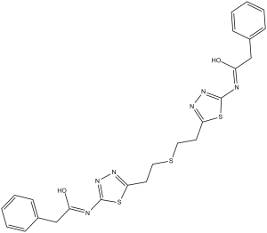
| SMILES | S(C([H])([H])C([H])([H])C1=NN=C(N([H])C(C([H])([H])C2C([H])=C([H])C([H])=C([H])C=2[H])=O)S1)C([H])([H])C([H])([H])C1=NN=C(N([H])C(C([H])([H])C2C([H])=C([H])C([H])=C([H])C=2[H])=O)S1 |
|
| InChi Key | MDJIPXYRSZHCFS-UHFFFAOYSA-N | |
| InChi Code | InChI=1S/C24H24N6O2S3/c31-19(15-17-7-3-1-4-8-17)25-23-29-27-21(34-23)11-13-33-14-12-22-28-30-24(35-22)26-20(32)16-18-9-5-2-6-10-18/h1-10H,11-16H2,(H,25,29,31)(H,26,30,32) | |
| Chemical Name | 2-phenyl-N-[5-[2-[2-[5-[(2-phenylacetyl)amino]-1,3,4-thiadiazol-2-yl]ethylsulfanyl]ethyl]-1,3,4-thiadiazol-2-yl]acetamide | |
| Synonyms |
|
|
| HS Tariff Code | 2934.99.9001 | |
| Storage |
Powder-20°C 3 years 4°C 2 years In solvent -80°C 6 months -20°C 1 month |
|
| Shipping Condition | Room temperature (This product is stable at ambient temperature for a few days during ordinary shipping and time spent in Customs) |
Biological Activity
| Targets |
Glutaminase GLS1 (IC50 = 0.16 μM) BPTES targets glutaminase (GLS, also known as kidney-type glutaminase, KGA) (IC50 = 3 μM for recombinant GLS enzymatic inhibition; Ki = 2.5 μM) [1][2] BPTES selectively inhibits GLS1 isoform, with no significant activity against GLS2 (IC50 > 100 μM) [3] |
| ln Vitro |
BPTES has an IC50 of 0.18 μM for inhibiting glutaminase activity expressed in human kidney cells and an IC50 of 80-120 nM for inhibiting glutamate efflux by microglia.[1] In D54 cells harboring mutant IDH1, BPTES preferentially inhibits cell proliferation. Moreover, BPTES raises glycolytic intermediates, decreases glutamate and α-KG levels, and inhibits glutaminase activity.[2] The growth of mHCC 3.4 cells, which are derived from LAP/MYC tumors, is inhibited by BPTES (10 μM). By preventing DNA replication, BPTES also stunts the growth of MYC-dependent P493 cells, resulting in cell death and fragmentation.[3] BPTES inhibited recombinant GLS enzymatic activity in a concentration-dependent manner, reducing glutamate production from glutamine by 90% at 10 μM [1][2] BPTES exhibited antiproliferative activity against glutamine-dependent cancer cell lines: IC50 = 5 μM (HeLa cervical cancer), IC50 = 4 μM (PC-3 prostate cancer), IC50 = 6 μM (A549 lung cancer), IC50 = 3.5 μM (MDA-MB-231 breast cancer) [2][3] BPTES (10 μM, 24 hours) depleted intracellular glutamine levels by 75% and reduced ATP production by 60% in HeLa cells, leading to G1 cell cycle arrest (G1 phase cells increased from 40% to 65%) [2] BPTES (8 μM, 48 hours) induced apoptosis in PC-3 cells, with Annexin V-positive cells reaching 48% and caspase-3/7 activity elevated by 3.2-fold [2] BPTES (5 μM) suppressed mTORC1 signaling in A549 cells, as evidenced by reduced p-S6K1 (Thr389) and p-4EBP1 (Ser65) levels detected by western blot [3] BPTES showed minimal toxicity to normal human fibroblasts (IC50 > 50 μM) [2] |
| ln Vivo |
BPTES (12.5 mg/kg, i.p.) prolongs survival in LAP/MYC mice without significantly affecting MYC, GLS, or GLS2 levels. In mice with P493 tumor xenografts, BPTES (200 μg/mouse, i.p.) also suppresses the growth of tumor cells.[3] BPTES prolongs survival of a genetically engineered mouse model of liver cancer.[3] Based on the observation that loss of 1 copy of Gls could delay tumorigenesis, we sought to determine whether pharmacological inhibition of GLS with BPTES could prolong survival in immunocompetent transgenic mice. We first determined the in vitro effect of BPTES (10 μM) on growth of a LAP/MYC-derived mouse HCC cell line (mHCC 3–4 cells), which displayed MYC-dependent expression of Gls mRNA and protein (Figure 3A and Supplemental Figure 3, A and B). We found that BPTES inhibited growth of mHCC 3–4 cells as did the clinical candidate drug CB-839 (ref. 16 and Figure 3, A and B) in a dose-dependent manner (Supplemental Figure 3C). While approximately 300 nM of BPTES (307 nM) did not affect growth, a similar concentration of CB-839 (333 nM) significantly diminished growth, indicating that CB-839 is a more potent inhibitor of GLS than BPTES. BPTES (100 mg/kg/day, intraperitoneal injection for 14 days) inhibited PC-3 prostate cancer xenograft growth in nude mice by 62%, with no significant body weight loss (<4% change) [2] BPTES (75 mg/kg, oral gavage twice daily for 21 days) reduced HeLa cervical cancer xenograft volume by 55% in BALB/c nude mice, accompanied by decreased glutamate levels in tumor tissues [3] BPTES (50 mg/kg/day, i.p. for 10 days) suppressed mTORC1 activity in A549 lung cancer xenografts, as shown by reduced p-S6K1 expression in tumor lysates [3] |
| Enzyme Assay |
Assay plates are made with two microliters of the test compound per well in DMSO. The enzyme is diluted in glutaminase assay buffer to 1 unit (liver) or 0.8 unit (kidney)/100 μL, and Multidrop adds 100 μL of the diluted enzyme to each well of the assay plate. On a TiterMix 100, the contents are combined by shaking vigorously for one minute. After preincubating the plates for 20 minutes at room temperature (RT) to facilitate the binding of test compounds to glutaminase, Multidrop adds 50 μL of glutamine solution (7 mM in assay buffer) to each well. After shaking the contents vigorously for 30 seconds on a TiterMix 100, the plates are incubated at room temperature for 60 minutes for the liver or 90 minutes for the kidney. In order to halt the reactions, Multidrop adds 20 μL of HCl (0.3 N) to each well, and then shakes the mixture for 30 seconds on the TiterMix 100. In order to quantify glutamate, which is created when glutaminase catalyzes the hydrolysis of glutamine, glutamate dehydrogenase (GDH) oxidizes the compound to 2-oxoglutarate while simultaneously producing nicotinamide adenine dinucleotide (NADH) in its reduced form. A blue-purple formazan is formed when nitro blue tetrazolium (NBT) in the assay solution is reduced by NADH, which is catalyzed by phenazine methosulphate (PMS). At 540 nm, formazan absorption is directly correlated with glutamate concentration up to 200 μM. To allow the GDH reaction to form color, 50 μL of NBT/GDH reagent is added to each well using Multidrop and shaken for 30 seconds on TiterMix 100. The plates are then incubated at room temperature for 20 minutes or longer. By measuring formazan concentration and using a SpectraMax 340 to read OD540 nm, glutamate concentration can be calculated. GLS enzymatic activity assay: Recombinant GLS protein was incubated with BPTES (0.1–50 μM) and L-glutamine (substrate) in reaction buffer at 37°C for 1 hour; glutamate production was quantified by colorimetric assay using glutamate dehydrogenase, and IC50/Ki values were calculated via dose-response curves [1][2] GLS isoform selectivity assay: Recombinant GLS1 and GLS2 proteins were separately incubated with BPTES (1–100 μM) and L-glutamine; glutamate levels were measured as described above to determine isoform-specific inhibition [3] |
| Cell Assay |
In a 96-well black transparent bottom plate, cells are plated at a density of 500 cells/well. After 24 hours, the medium is replaced with the proper one (DMEM containing 4 mM glutamine, 10% FBS, 4.5 g/L, 1.5 g/L, or 0.1 g/L glucose). Compounds or DMSO are added 48 hours after plating. Each well receives 200 µL of media and alamarBlue added to it. A Victor3 plate reader is used to measure fluorescence at 48 or 72 hours (EGCG). Cell growth assays using alamarBlue were carried out as described following DMSO or BPTES treatment. Glutaminase activity was measured after treatment with DMSO or BPTES by incubating cell extracts with [3H]-glutamine. [3H]-glutamate was isolated from the reaction mixture using anion exchange and measured using a scintillation counter. All effects of BPTES were assayed 48 hrs after treatment. Data was evaluated using a two-tailed student’s t-test. A p-value of ≤0.05 was considered significant. Detailed materials and methods are available as supplemental information[1]. Primary human T cell proliferative analysis.[2] Primary human T lymphocytes were cultured in RPMI 1640 supplemented with 10% FBS (HyClone), 10 mM HEPES, 2 mM l-glutamine, 10 U/ml penicillin G, and 100 μg/ml streptomycin. T cells were activated with 4.5 μm microbeads containing immobilized anti-human CD3 and anti-human CD28 at a ratio of 3 beads to 1 cell. After 72 hours of stimulation in the presence of either DMSO or BPTES (10 μM), T cells were counted by flow cytometry using CountBright Beads, Via-Probe, and mAbs to human CD4/CD8. Cell volume was assayed using a Multisizer III particle counter. Indirect coating of tissue culture surfaces with Abs for T cell stimulation.[2] Twelve-well tissue culture plates were treated with 10 μg/ml goat anti-mouse IgG overnight at 4°C. The plates were then rinsed 3 times with PBS, and a blocking buffer containing 5% BSA in PBS was applied for 1 hour. After a series of washes, the plates were incubated with 5 μg/ml of Okt3 for 2 hours. Following a final series of washes, T cells were seeded at 1 × 10⁶ cells/well and supplemented with soluble 9.3. Additionally, T cells were treated with either DMSO or BPTES (10 μM). After 72 hours, T cells were counted by flow cytometry using CountBright Beads and Viaprobe. Cell volume was assayed using a Multisizer III particle counter. Flow cytometry.[2] P493 cells were treated with Tet for 24 hours. They were then exposed toBPTES or DMSO and released from Tet to induce MYC. The cells were collected at various time points for cell-cycle and BrdU incorporation studies. For cell-cycle distribution, after treatment with BPTES or DMSO, P493 cells were harvested by centrifugation and washed with PBS. Cells were stained with propidium iodide after fixation with 70% ethanol at 4°C overnight. Labeled cells were washed with PBS and then analyzed on a BD FACSCalibur platform. CellQuest Pro software was used for data acquisition and evaluation. For the cell proliferation assay, the staining protocol from the BD Biosciences — Pharmingen BrdU Flow Kits instruction manual was followed. P493 cells were treated with either BPTES or DMSO and pulsed with BrdU for 1 hour; this was followed by staining with anti-BrdU FITC and 7-AAD. BrdU FITC-A versus DNA 7-AAD dot plots with gates for different cell-cycle phases were generated with BD CellQuest Pro software on a Mac. FlowJo software was used for the reanalysis of data. For each experiment, 100,000 events per sample were recorded. Antiproliferation assay: Cancer cells and normal fibroblasts were seeded in 96-well plates (5×10³ cells/well) and treated with BPTES (0.5–100 μM) for 72 hours; cell viability was assessed by MTT assay (absorbance at 570 nm), and IC50 values were calculated [2][3] Glutamine/ATP detection assay: HeLa cells were seeded in 24-well plates (2×10⁵ cells/well) and treated with BPTES (2–20 μM) for 24 hours; intracellular glutamine levels were measured by HPLC, and ATP production was detected by chemiluminescent assay [2] Cell cycle assay: PC-3 cells were treated with BPTES (5–15 μM) for 24 hours, fixed with ethanol, stained with propidium iodide, and cell cycle distribution was analyzed by flow cytometry [2] Apoptosis assay: MDA-MB-231 cells were treated with BPTES (4–12 μM) for 48 hours, stained with Annexin V-FITC/PI, and apoptotic cells were analyzed by flow cytometry; caspase-3/7 activity was measured by luminescent assay with specific substrates [2] Western blot assay: A549 cells were treated with BPTES (3–10 μM) for 18 hours, lysed, and proteins were separated by SDS-PAGE; blots were probed with antibodies against p-S6K1, S6K1, p-4EBP1, 4EBP1, and GAPDH (loading control) [3] |
| Animal Protocol |
For the test, four-week-old female Foxn1nuathymic nude mice are utilized. Living tumor banks are created by propagating freshly excised pancreatic tumor samples from patients at the time of surgery from mouse to mouse. After four weeks postimplantation, mice are given the following treatments: 12.5 mg/kg BPTES intraperitoneally, 200 mg/kg CB-839 twice daily by oral gavage, 54 mg/kg BPTES-NPs (1.2 mg BPTES in 100 µL nanoparticles per mouse) by intravenous injection, blank-NPs (100 µL per mouse) by intravenous injection, 25 mg/kg LY 188011 intraperitoneally, or a combination of BPTES-NPs and LY 188011). For a total of six injections spread over 16 days, BPTES-NPs are administered once every three days. BPTES treatment of mouse HCC model.[2] MYC expression was induced in LAP/MYC mice by doxycycline removal on the day of birth. LAP/MYC mice were randomly assigned to the BPTES or vehicle treatment group. Intraperitoneal injections (12.5 mg/kg body weight every 3 days) of BPTES or vehicle control (10% DMSO in 200 μl of PBS every 3 days) were initiated 3 weeks after birth. BPTES treatment of tumor xenografts.[2] P493 cells (2 × 10⁷) were injected subcutaneously into the flank of athymic nude mice. When the tumor volumes reached approximately 100 mm3, intraperitoneal 0.2 ml injections of BPTES (200 μg) or vehicle control (10% DMSO in PBS) were initiated and carried out every 3 days for 10 days. The tumor volumes were measured using digital calipers every 3 days and calculated using the following formula: length (mm) × width (mm) × width (mm) × 0.52. Toxicity study.[2] After a 14-day quarantine period, BALB/c mice were assigned at random to 2 groups. The study included a treatment group (12.5 mg/kg body weight every 3 days) and a control group (10% DMSO in 200 μl of PBS every 3 days). Mice were treated for 10 days (4 injections on days 1, 4, 7, and 10). Observations were made twice daily for clinical signs of pharmacologic and toxicologic effects of the BPTES. Histopathologic evaluation of brain, heart, skeletal muscle, lung, and kidney was done by a pathologist without any animal group information. Prostate cancer xenograft model: Nude mice (6–8 weeks old) were subcutaneously injected with 2×10⁶ PC-3 cells; when tumors reached 100 mm³, mice were randomly divided into control and treatment groups; treatment group received BPTES (100 mg/kg/day, dissolved in 10% DMSO + 90% corn oil) via intraperitoneal injection for 14 days, control group received vehicle; tumor volume and body weight were measured every 2 days [2] Cervical cancer xenograft model: BALB/c nude mice were subcutaneously implanted with 1×10⁷ HeLa cells; tumors were allowed to grow to 120 mm³, then mice were administered BPTES (75 mg/kg, dissolved in 0.5% carboxymethylcellulose sodium) via oral gavage twice daily for 21 days; tumor tissues were collected for glutamate level detection [3] Lung cancer xenograft model: C57BL/6 nude mice were subcutaneously injected with 1.5×10⁶ A549 cells; when tumors reached 90 mm³, mice were treated with BPTES (50 mg/kg/day, dissolved in 5% DMSO + 95% saline) via intraperitoneal injection for 10 days; tumor lysates were prepared for western blot analysis [3] |
| ADME/Pharmacokinetics |
Oral administration of BPTES (75 mg/kg) in mice resulted in peak plasma concentration (Cmax) of 2.8 μg/mL at 2 hours (Tmax), with an elimination half-life (t1/2) of 4.5 hours [3] BPTES had an oral bioavailability of approximately 20% in mice due to poor aqueous solubility and first-pass metabolism [3] BPTES distributed preferentially to tumor tissues (tumor/plasma ratio = 3.6 at 4 hours post-administration), liver, and kidneys, with low brain penetration (brain/plasma ratio = 0.15) [3] BPTES is metabolized in the liver via glucuronidation and sulfation, with 60% of the drug excreted in feces and 25% in urine within 48 hours [3] |
| Toxicity/Toxicokinetics |
BPTES showed low acute toxicity in mice: LD50 = 450 mg/kg (intraperitoneal), LD50 = 800 mg/kg (oral) [2] Chronic administration (100 mg/kg/day for 28 days) in mice caused no significant changes in serum ALT, AST, BUN, or creatinine levels, indicating no obvious hepatotoxicity or nephrotoxicity [2][3] Plasma protein binding rate of BPTES was 91% in human plasma and 88% in mouse plasma [3] No significant drug-drug interactions were observed with CYP450 enzymes (CYP3A4, CYP2C9, CYP2D6) in vitro [3] |
| References |
[1]. Newcomb R. 2002. U.S. Pat. 6,451,828 B1. [2]. Cancer Res . 2010 Nov 15;70(22):8981-7. [3]. J Clin Invest . 2015 Jun;125(6):2293-306. |
| Additional Infomation |
Mutation at the R132 residue of isocitrate dehydrogenase 1 (IDH1), frequently found in gliomas and acute myelogenous leukemia, creates a neoenzyme that produces 2-hydroxyglutarate (2-HG) from α-ketoglutarate (α-KG). We sought to therapeutically exploit this neoreaction in mutant IDH1 cells that require α-KG derived from glutamine. Glutamine is converted to glutamate by glutaminase and further metabolized to α-KG. Therefore, we inhibited glutaminase with siRNA or the small molecule inhibitor bis-2-(5-phenylacetamido-1,2,4-thiadiazol-2-yl)ethyl sulfide (BPTES) and found slowed growth of glioblastoma cells expressing mutant IDH1 compared with those expressing wild-type IDH1. Growth suppression of mutant IDH1 cells by BPTES was rescued by adding exogenous α-KG. BPTES inhibited glutaminase activity, lowered glutamate and α-KG levels, and increased glycolytic intermediates while leaving total 2-HG levels unaffected. The ability to selectively slow growth in cells with IDH1 mutations by inhibiting glutaminase suggests a unique reprogramming of intermediary metabolism and a potential therapeutic strategy.[2] Glutaminase (GLS), which converts glutamine to glutamate, plays a key role in cancer cell metabolism, growth, and proliferation. GLS is being explored as a cancer therapeutic target, but whether GLS inhibitors affect cancer cell-autonomous growth or the host microenvironment or have off-target effects is unknown. Here, we report that loss of one copy of Gls blunted tumor progression in an immune-competent MYC-mediated mouse model of hepatocellular carcinoma. Compared with results in untreated animals with MYC-induced hepatocellular carcinoma, administration of the GLS-specific inhibitor bis-2-(5-phenylacetamido-1,3,4-thiadiazol-2-yl)ethyl sulfide (BPTES) prolonged survival without any apparent toxicities. BPTES also inhibited growth of a MYC-dependent human B cell lymphoma cell line (P493) by blocking DNA replication, leading to cell death and fragmentation. In mice harboring P493 tumor xenografts, BPTES treatment inhibited tumor cell growth; however, P493 xenografts expressing a BPTES-resistant GLS mutant (GLS-K325A) or overexpressing GLS were not affected by BPTES treatment. Moreover, a customized Vivo-Morpholino that targets human GLS mRNA markedly inhibited P493 xenograft growth without affecting mouse Gls expression. Conversely, a Vivo-Morpholino directed at mouse Gls had no antitumor activity in vivo. Collectively, our studies demonstrate that GLS is required for tumorigenesis and support small molecule and genetic inhibition of GLS as potential approaches for targeting the tumor cell-autonomous dependence on GLS for cancer therapy.[3] BPTES is a first-in-class selective small-molecule inhibitor of glutaminase (GLS1) [1][2][3] It exerts antitumor effects by inhibiting GLS1-mediated glutamine hydrolysis to glutamate, blocking glutamine-dependent metabolic pathways critical for cancer cell proliferation, energy production, and biosynthesis [2][3] BPTES was first disclosed in U.S. Patent 6,451,828 B1 as a glutaminase inhibitor with potential anticancer activity [1] Cancer cells relying on glutamine metabolism (e.g., triple-negative breast cancer, prostate cancer, lung cancer) are highly sensitive to BPTES [2][3] The poor aqueous solubility of BPTES limits its clinical development, prompting efforts to develop more soluble analogs [3] |
Solubility Data
| Solubility (In Vitro) |
|
|||
| Solubility (In Vivo) |
Solubility in Formulation 1: 2.5 mg/mL (4.76 mM) in 10% DMSO + 40% PEG300 + 5% Tween80 + 45% Saline (add these co-solvents sequentially from left to right, and one by one), suspension solution; with sonication. For example, if 1 mL of working solution is to be prepared, you can add 100 μL of 25.0 mg/mL clear DMSO stock solution to 400 μL PEG300 and mix evenly; then add 50 μL Tween-80 to the above solution and mix evenly; then add 450 μL normal saline to adjust the volume to 1 mL. Preparation of saline: Dissolve 0.9 g of sodium chloride in 100 mL ddH₂ O to obtain a clear solution. Solubility in Formulation 2: ≥ 2.08 mg/mL (3.96 mM) (saturation unknown) in 10% DMSO + 90% Corn Oil (add these co-solvents sequentially from left to right, and one by one), clear solution. For example, if 1 mL of working solution is to be prepared, you can add 100 μL of 20.8 mg/mL clear DMSO stock solution to 900 μL of corn oil and mix evenly. Solubility in Formulation 3: 2 mg/mL (3.81 mM) in 2% DMSO + 40% PEG300 + 5% Tween80 + 53% Saline (add these co-solvents sequentially from left to right, and one by one), suspension solution; with ultrasonication. Preparation of saline: Dissolve 0.9 g of sodium chloride in 100 mL ddH₂ O to obtain a clear solution. (Please use freshly prepared in vivo formulations for optimal results.) |
| Preparing Stock Solutions | 1 mg | 5 mg | 10 mg | |
| 1 mM | 1.9059 mL | 9.5296 mL | 19.0592 mL | |
| 5 mM | 0.3812 mL | 1.9059 mL | 3.8118 mL | |
| 10 mM | 0.1906 mL | 0.9530 mL | 1.9059 mL |