Physicochemical Properties
| Molecular Formula | C28H32BF2N7O3 |
| Molecular Weight | 563.41 |
| Exact Mass | 563.26277 |
| CAS # | 175799-93-6 |
| PubChem CID | 171041190 |
| Appearance | Yellow to orange solid powder |
| Hydrogen Bond Donor Count | 1 |
| Hydrogen Bond Acceptor Count | 10 |
| Rotatable Bond Count | 7 |
| Heavy Atom Count | 41 |
| Complexity | 1040 |
| Defined Atom Stereocenter Count | 0 |
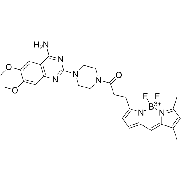
| SMILES | B(N1C(=CC=C1/C=C\2/C(=CC(=N2)C)C)CCC(=O)N3CCN(CC3)C4=NC5=CC(=C(C=C5C(=N4)N)OC)OC)(F)F |
| InChi Key | MIPKRDYUTOIJKI-HMAPJEAMSA-N |
| InChi Code | InChI=1S/C28H32BF2N7O3/c1-17-13-18(2)33-22(17)14-20-6-5-19(38(20)29(30)31)7-8-26(39)36-9-11-37(12-10-36)28-34-23-16-25(41-4)24(40-3)15-21(23)27(32)35-28/h5-6,13-16H,7-12H2,1-4H3,(H2,32,34,35)/b22-14- |
| Chemical Name | 1-[4-(4-amino-6,7-dimethoxyquinazolin-2-yl)piperazin-1-yl]-3-[1-difluoroboranyl-5-[(Z)-(3,5-dimethylpyrrol-2-ylidene)methyl]pyrrol-2-yl]propan-1-one |
| Synonyms | BODIPY FL prazosin; DTXSID901316148; 175799-93-6 |
| HS Tariff Code | 2934.99.9001 |
| Storage |
Powder-20°C 3 years 4°C 2 years In solvent -80°C 6 months -20°C 1 month |
| Shipping Condition | Room temperature (This product is stable at ambient temperature for a few days during ordinary shipping and time spent in Customs) |
Biological Activity
| Targets | α1A-adrenergic receptor (Ki = 14.5 nM); α1B-adrenergic receptor (Ki = 43.3 nM) |
| ln Vitro | BODIPY FL With affinity for different α1-AR ligands, prazosin (10 nM; 100 μl for 30 minutes at room temperature; COS-7 cells) exhibits values of 14.5 and 43.3 nM for α1a-AR and α1b-AR, respectively [1]. BODIPY FL In erythroleukemia cells, prazosin (100 nM, 30 minutes) can be employed as a molecular probe to see where α1-adrenergic drug binding sites are located on non-adrenergic receptors [3]. |
| Enzyme Assay | Even though the erythroleukemia cell lines K562 and HEL do not express α1-adrenoceptors, some α1-adrenergic drugs influence both survival and differentiation of these cell lines. Since Ca2+ is closely related to cellular homeostasis, we examined the capacity of α1-adrenergic drugs to modulate the intracellular Ca2+ content in K562 cells. Because of morphological alterations of mitochondria following α1-adrenergic agonist treatment, we also scrutinized mitochondrial functions. In order to visualize the non-adrenoceptor binding site(s) of α1-adrenergic drugs in erythroleukemia cells, we evaluated the application of the fluorescent α1-adrenergic antagonist BODIPY FL prazosin. We discovered that the α1-adrenergic agonists naphazoline, oxymetazoline and also the α1-adrenergic antagonist benoxathian are able to raise the intracellular Ca2+-content in K562 cells. Furthermore, we demonstrate that naphazoline treatment induces ROS-formation as well as an increase in Δψm in K562 cells. Using BODIPY FL prazosin we were able to visualize the non-adrenoceptor binding site(s) of α1-adrenergic drugs in erythroleukemia cells. Interestingly, the SERCA-inhibitor thapsigargin appears to interfere with the binding of BODIPY FL prazosin. Our data suggest that the effects of α1-adrenergic drugs on erythroleukemia cells are mediated by a thapsigargin sensitive binding site, which controls the fate of erythroleukemia cells towards differentiation, senescence and cell death through modulation of intracellular Ca2+ [3]. |
| Cell Assay |
G protein-coupled receptor (GPCR) subtypes are differentially distributed in the cell; however, it remains unclear how this affects the subtype selectivity of particular drugs. In the present study, we used flow cytometry analysis with the fluorescent ligand, BODIPY FL prazosin, to study the relationship between the subcellular distribution of subtype receptors and the subtype-selective character of ligands using alpha1a and alpha1b-adrenoceptors (ARs). Alpha1a-ARs predominantly localize inside the cell, while alpha1b-ARs on the cell surface. Flow cytometry analysis and confocal laser-scanning micrographs of living cells showed that BODIPY FL prazosin can label not only alpha1-ARs on the cell surface, but also those localized inside the cell. Furthermore, flow cytometry analysis of alpha1A-AR-selective drug, KMD-3213, and alpha1B-AR-selective drug, CEC, revealed that the major determinant of the subtype selectivity of each drug is different. The alpha1A-AR selectivity of KMD-3213 can be explained by its much higher affinity for alpha1a-AR than alpha1b-AR (affinity-dependent selectivity), while the alpha1B-AR selectivity of the hydrophilic alkylating agent CEC is due to preferential inactivation of alpha1-ARs on the cell surface (receptor localization-dependent selectivity). This study illustrates that factors in addition to the affinity of the drug for the receptor, such as subcellular localization of the receptor, should be taken into account in assessing the subtype selectivity of a drug [1]. To study substrate affinity of tested AEDs to BCRP, transport experiments were performed in epithelial BCRP-expressing MDCKII-BCRP and MDCKII-parent cell lines cultured on microporous membrane. For detection of inhibitory potency of AEDs to BCRP, accumulation assays were carried out in MEF3.8-BCRP cells with known BCRP substrates, BODIPY FL prazosin and mitoxantrone[2]. |
| References |
[1]. Differences in the subcellular localization of alpha1-adrenoceptor subtypes can affect the subtype selectivity of drugs in a study with the fluorescent ligand BODIPY FL-prazosin. Life Sci. 2002 Mar 22;70(18):2113-24. [2]. Lack of interactions between breast cancer resistance protein (bcrp/abcg2) and selected antiepileptic agents. Epilepsia. 2006 Mar;47(3):461-8. [3]. α1-adrenergic drugs exhibit affinity to a thapsigargin-sensitive binding site and interfere with the intracellular Ca2+ homeostasis in human erythroleukemia cells. Exp Cell Res. 2011 Dec 10;317(20):2969-80. |
| Additional Infomation | Purpose: Recent studies have indicated constitutive expression of efflux transporter, breast cancer resistance protein (BCRP, ABCG2), in endothelial cells of the blood-brain barrier (BBB). In epileptogenic brain tumors such as ganglioma, astrocytoma, anaplastic astrocytomas, or glioma multiforme, strong expression of BCRP in the microvasculature of the BBB was observed. Therefore it was hypothesized that this phenomenon could critically influence the bioavailability of drugs in these tumors and potentially contribute to the failure of antiepileptic treatment. The aim of this study was to test whether some commonly used antiepileptic drugs (AEDs) are substrates transported by human BCRP. In particular, we focused on phenobarbital, phenytoin, ethosuximide, primidone, valproate, carbamazepine, clonazepam, and lamotrigine. Furthermore, the inhibitory potency of these AEDs to BCRP was examined. Methods: To study substrate affinity of tested AEDs to BCRP, transport experiments were performed in epithelial BCRP-expressing MDCKII-BCRP and MDCKII-parent cell lines cultured on microporous membrane. For detection of inhibitory potency of AEDs to BCRP, accumulation assays were carried out in MEF3.8-BCRP cells with known BCRP substrates, BODIPY FL prazosin and mitoxantrone. Results: No obvious interactions of tested AEDs with BCRP transporter were observed. Therefore these drugs in relevant therapeutic concentrations are neither substrates nor inhibitors of BCRP. Conclusions: Based on our in vitro data we can conclude that resistance to treatment with the tested AEDs probably is not caused by the overexpression of BCRP in the BBB of epileptogenic brain tumors. [2] |
Solubility Data
| Solubility (In Vitro) | May dissolve in DMSO (in most cases), if not, try other solvents such as H2O, Ethanol, or DMF with a minute amount of products to avoid loss of samples |
| Solubility (In Vivo) |
Note: Listed below are some common formulations that may be used to formulate products with low water solubility (e.g. < 1 mg/mL), you may test these formulations using a minute amount of products to avoid loss of samples. Injection Formulations (e.g. IP/IV/IM/SC) Injection Formulation 1: DMSO : Tween 80: Saline = 10 : 5 : 85 (i.e. 100 μL DMSO stock solution → 50 μL Tween 80 → 850 μL Saline) *Preparation of saline: Dissolve 0.9 g of sodium chloride in 100 mL ddH ₂ O to obtain a clear solution. Injection Formulation 2: DMSO : PEG300 :Tween 80 : Saline = 10 : 40 : 5 : 45 (i.e. 100 μL DMSO → 400 μLPEG300 → 50 μL Tween 80 → 450 μL Saline) Injection Formulation 3: DMSO : Corn oil = 10 : 90 (i.e. 100 μL DMSO → 900 μL Corn oil) Example: Take the Injection Formulation 3 (DMSO : Corn oil = 10 : 90) as an example, if 1 mL of 2.5 mg/mL working solution is to be prepared, you can take 100 μL 25 mg/mL DMSO stock solution and add to 900 μL corn oil, mix well to obtain a clear or suspension solution (2.5 mg/mL, ready for use in animals). Injection Formulation 4: DMSO : 20% SBE-β-CD in saline = 10 : 90 [i.e. 100 μL DMSO → 900 μL (20% SBE-β-CD in saline)] *Preparation of 20% SBE-β-CD in Saline (4°C,1 week): Dissolve 2 g SBE-β-CD in 10 mL saline to obtain a clear solution. Injection Formulation 5: 2-Hydroxypropyl-β-cyclodextrin : Saline = 50 : 50 (i.e. 500 μL 2-Hydroxypropyl-β-cyclodextrin → 500 μL Saline) Injection Formulation 6: DMSO : PEG300 : castor oil : Saline = 5 : 10 : 20 : 65 (i.e. 50 μL DMSO → 100 μLPEG300 → 200 μL castor oil → 650 μL Saline) Injection Formulation 7: Ethanol : Cremophor : Saline = 10: 10 : 80 (i.e. 100 μL Ethanol → 100 μL Cremophor → 800 μL Saline) Injection Formulation 8: Dissolve in Cremophor/Ethanol (50 : 50), then diluted by Saline Injection Formulation 9: EtOH : Corn oil = 10 : 90 (i.e. 100 μL EtOH → 900 μL Corn oil) Injection Formulation 10: EtOH : PEG300:Tween 80 : Saline = 10 : 40 : 5 : 45 (i.e. 100 μL EtOH → 400 μLPEG300 → 50 μL Tween 80 → 450 μL Saline) Oral Formulations Oral Formulation 1: Suspend in 0.5% CMC Na (carboxymethylcellulose sodium) Oral Formulation 2: Suspend in 0.5% Carboxymethyl cellulose Example: Take the Oral Formulation 1 (Suspend in 0.5% CMC Na) as an example, if 100 mL of 2.5 mg/mL working solution is to be prepared, you can first prepare 0.5% CMC Na solution by measuring 0.5 g CMC Na and dissolve it in 100 mL ddH2O to obtain a clear solution; then add 250 mg of the product to 100 mL 0.5% CMC Na solution, to make the suspension solution (2.5 mg/mL, ready for use in animals). Oral Formulation 3: Dissolved in PEG400 Oral Formulation 4: Suspend in 0.2% Carboxymethyl cellulose Oral Formulation 5: Dissolve in 0.25% Tween 80 and 0.5% Carboxymethyl cellulose Oral Formulation 6: Mixing with food powders Note: Please be aware that the above formulations are for reference only. InvivoChem strongly recommends customers to read literature methods/protocols carefully before determining which formulation you should use for in vivo studies, as different compounds have different solubility properties and have to be formulated differently. (Please use freshly prepared in vivo formulations for optimal results.) |
| Preparing Stock Solutions | 1 mg | 5 mg | 10 mg | |
| 1 mM | 1.7749 mL | 8.8745 mL | 17.7491 mL | |
| 5 mM | 0.3550 mL | 1.7749 mL | 3.5498 mL | |
| 10 mM | 0.1775 mL | 0.8875 mL | 1.7749 mL |