BLU9931 (BLU-9931; BLU 9931) is an irreversible and selective FGFR4 inhibitor with potential anticancer activity. In a cell-free assay, it inhibits FGFR4 with an IC50 of 3 nM. Compared to other FGFR family kinases like FGFR1/2/3, BLU9931 exhibits high selectivity for inhibiting FGFR4 (297-, 184-, and 50-fold selectivity, respectively). In mice with an overexpressed FGF19 xenograft in the form of an HCC tumor and a liver tumor xenograft model, BLU9931 demonstrates impressive antitumor activity.
Physicochemical Properties
| Molecular Formula | C26H22CL2N4O3 | |
| Molecular Weight | 509.38 | |
| Exact Mass | 508.106 | |
| Elemental Analysis | C, 61.31; H, 4.35; Cl, 13.92; N, 11.00; O, 9.42 | |
| CAS # | 1538604-68-0 | |
| Related CAS # |
|
|
| PubChem CID | 72710839 | |
| Appearance | Off-white to light yellow solid powder | |
| Density | 1.4±0.1 g/cm3 | |
| Index of Refraction | 1.677 | |
| LogP | 5.13 | |
| Hydrogen Bond Donor Count | 2 | |
| Hydrogen Bond Acceptor Count | 6 | |
| Rotatable Bond Count | 7 | |
| Heavy Atom Count | 35 | |
| Complexity | 715 | |
| Defined Atom Stereocenter Count | 0 | |
| SMILES | ClC1C(=C([H])C(=C(C=1C1C([H])=C([H])C2C(=C([H])N=C(N=2)N([H])C2C(=C([H])C([H])=C([H])C=2C([H])([H])[H])N([H])C(C([H])=C([H])[H])=O)C=1[H])Cl)OC([H])([H])[H])OC([H])([H])[H] |
|
| InChi Key | TXEBNKKOLVBTFK-UHFFFAOYSA-N | |
| InChi Code | InChI=1S/C26H22Cl2N4O3/c1-5-21(33)30-18-8-6-7-14(2)25(18)32-26-29-13-16-11-15(9-10-17(16)31-26)22-23(27)19(34-3)12-20(35-4)24(22)28/h5-13H,1H2,2-4H3,(H,30,33)(H,29,31,32) | |
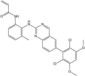
| Chemical Name | N-[2-[[6-(2,6-dichloro-3,5-dimethoxyphenyl)quinazolin-2-yl]amino]-3-methylphenyl]prop-2-enamide | |
| Synonyms | BLU9931; BLU 9931; N-(2-((6-(2,6-dichloro-3,5-dimethoxyphenyl)quinazolin-2-yl)amino)-3-methylphenyl)acrylamide; N-[2-[[6-(2,6-dichloro-3,5-dimethoxyphenyl)quinazolin-2-yl]amino]-3-methylphenyl]prop-2-enamide; UNII-FQK825B5DX; FQK825B5DX; BLU-9931 | |
| HS Tariff Code | 2934.99.9001 | |
| Storage |
Powder-20°C 3 years 4°C 2 years In solvent -80°C 6 months -20°C 1 month |
|
| Shipping Condition | Room temperature (This product is stable at ambient temperature for a few days during ordinary shipping and time spent in Customs) |
Biological Activity
| Targets |
FGFR1 (IC50 = 591 nM); FGFR2 (IC50 = 493 nM); FGFR3 (IC50 = 150 nM); FGFR4 (IC50 = 3 nM) Fibroblast Growth Factor Receptor 4 (FGFR4) (IC50 = 1.6 nM, Ki = 0.9 nM); no significant activity against FGFR1 (IC50 > 1000 nM), FGFR2 (IC50 > 1000 nM), FGFR3 (IC50 > 1000 nM); weak activity against VEGFR2 (IC50 = 850 nM), PDGFRα (IC50 = 920 nM) [1] - Selective binding to FGFR4 ATP-binding pocket; no cross-reactivity with other FGFR subtypes [1] |
| ln Vitro |
BLU9931 significantly suppresses the phosphorylation of the FGFR4 signaling pathway in MDA-MB-453 cells. With an EC50 of less than 1 μM, BLU9931 suppresses the growth of HCC cell lines that express an intact FGFR4 signaling complex, including Hep 3B, HUH-7, and JHH-7 cell lines. Additionally, BLU9931 prevents the growth of PDX-derived cell lines that still have functional FGFR4 signaling.[1] Inhibited recombinant FGFR4 kinase activity: IC50 = 1.6 nM, Ki = 0.9 nM; suppressed FGFR4 autophosphorylation (Tyr642) in Ba/F3-FGFR4 cells (IC50 = 4.8 nM) [1] - Reduced viability of FGFR4-activated hepatocellular carcinoma (HCC) cells: Hep3B (FGF19-overexpressing, IC50 = 12.3 nM), Huh-7 (FGFR4-mutated, IC50 = 15.7 nM), PLC/PRF/5 (IC50 = 18.2 nM); no activity in FGFR4-negative HepG2 cells (IC50 > 500 nM) [1] - Suppressed FGFR4 downstream signaling: 100 nM BLU9931 reduced p-FGFR4 (Tyr642) by 93% in Hep3B cells (2 hours), and downregulated p-ERK1/2 (Thr202/Tyr204) and p-AKT (Ser473) by >88% [1] - Induced apoptosis in Huh-7 cells: 200 nM BLU9931 increased Annexin V-positive cells from 7% (vehicle) to 45% (48 hours); caspase-3/7 activity elevated by 3.9-fold [1] - Reduced colony formation of PLC/PRF/5 cells: 50 nM BLU9931 decreased colony number by 82% compared to vehicle (14-day incubation) [1] |
| ln Vivo |
BLU9931 (300 mg/kg, p.o.) inhibits weight loss caused by tumors and causes tumor regression in mice with FGF19-amplified Hep 3B liver tumors. Tumor regression is also observed in mice treated with BLU9931 (300 mg/kg, p.o.) that bear the FGF19-overexpressing PDX-derived LIXC012 xenografts.[1] In nude mice bearing Hep3B (FGFR4-activated HCC) xenografts: Oral BLU9931 (25 mg/kg/day) for 28 days resulted in 92% tumor growth inhibition (TGI); tumor p-FGFR4 levels reduced by 85% (immunohistochemistry) [1] - In nude mice bearing Huh-7 (FGFR4-mutated HCC) xenografts: Oral BLU9931 (30 mg/kg/day) for 21 days achieved 86% TGI; median tumor doubling time extended from 6 days (vehicle) to 24 days [1] - In mice with orthotopic HCC (Hep3B cell implantation): Oral BLU9931 (20 mg/kg/day) for 35 days improved survival (median survival: 58 days vs. 32 days for vehicle); reduced liver tumor burden by 78% (MRI assessment) [1] |
| Enzyme Assay |
FGFR1–4 Biochemical Assays[1] In assays for FGFR kinase inhibition, ATP is used at KM. Picomolar to low nanomolar concentrations of FGFR proteins are incubated for 90 minutes at 25°C in 1× Kinase Reaction Buffer (KRB) with 1 μM of CSKtide and 50 to 250 μM ATP, either in the presence or absence of a dosed concentration series of inhibitor. The addition of Stop buffer ends each reaction, and plates are read using a Caliper EZReader2. A four-parameter log[Inhibitor] against response model with floating Hill Slope is used to fit IC50 values. Kinase Selectivity Profiling[1] BLU9931 and BGJ398 were screened at a single concentration of 3 μmol/L and LY-2874455 was screened at a single concentration of 1 μmol/L using the KINOMEscan Assay Platform. Kinetics of Inhibition Assays for KI, kinact, and kinact/KI Estimation[1] On-mechanism inhibition constants were collected using the Omnia assay technology, where 22 dose inhibitor titrations were added to a 384-well Nonbinding surface (NBS™) assay plate containing a substrate mixture of 10 μmol/L Y10-sox peptide and 1 mmol/L ATP prepared in 1× KRB. Reactions were initiated with the addition of recombinant FGFR4 protein and fluorescence intensity readings were collected (λex 360 nm/λem 485 nm) every 74 seconds at room temperature. At the conclusion of each assay, raw fluorescence intensity data representing steady-state kinase activity progress curves were fit for kobs using the model for a first-order exponential. kobs versus inhibitor dose curves were then fit for KI and kinact using a model for irreversible inhibition with autoinactivation. GraphPad Prism was used for all curve fitting. FGFR4 kinase activity assay: Recombinant human FGFR4 kinase domain (50 ng/well) was incubated with BLU9931 (0.01-100 nM) in reaction buffer (25 mM HEPES pH 7.5, 10 mM MgCl2, 1 mM DTT) at 30°C for 15 minutes. 10 μM ATP and a fluorescent peptide substrate were added, followed by 60-minute incubation at 30°C. Kinase activity was measured via homogeneous time-resolved fluorescence (HTRF; excitation 340 nm, emission 665 nm); IC50 and Ki values were calculated via nonlinear regression and Lineweaver-Burk plot, respectively [1] - FGFR4 binding assay (SPR): FGFR4 kinase domain (2 μg/mL) was immobilized on a sensor chip. BLU9931 (0.1-100 nM) was injected at 30 μL/min; binding affinity (KD = 0.7 nM) was determined by fitting sensorgrams to a 1:1 binding model [1] |
| Cell Assay |
Established and PDX-derived HCC cell lines are seeded in 96-well plates with the appropriate growth media, given an overnight attachment period, and then subjected to two cell-doubling cycles of treatment with a dilution series of test compounds. CellTiter-Glo is used to assess cell viability; the results are expressed as background-subtracted relative light units normalized to a control that received DMSO treatment. At 50% inhibition, the relative EC50 values are calculated between the top and bottom plateaus of the dose-response curve.
Proliferation Studies[1] Established and PDX-derived HCC cell lines were seeded in 96-well plates in respective growth media, allowed to attach overnight, and treated with a dilution series of test compounds for two cell-doubling times. Cell viability was determined by CellTiter-Glo, and results represented as background-subtracted relative light units normalized to a DMSO-treated control. Relative EC50 values were determined at 50% inhibition between the top and bottom plateau of the dose–response curve. Detection of Activated Caspase-3/7[1] Hep 3B cells were seeded overnight at 2.5 × 10⁴ cells in white 96-well plates in media containing 5% FBS, then treated for 24 hours at indicated compound concentrations, and processed according to the manufacturer's instructions using the Caspase-3/7 Glo assay. Data are expressed relative to DMSO control-treated cells. FGFR4 Turnover Studies[1] Cells were plated at 4 × 10⁵ cells per well of a 6-well plate in media containing 10% serum and allowed to adhere overnight. Cells were treated with 10 μg/mL cycloheximide for indicated times. Samples were subjected to gel electrophoresis followed by immunoblotting as described above. Cell proliferation assay (Hep3B/Huh-7/PLC/PRF/5): Cells were seeded in 96-well plates (5×10³ cells/well) and treated with BLU9931 (0.1 nM-1 μM) for 72 hours. Cell viability was measured via tetrazolium-based colorimetric assay; absorbance at 570 nm was recorded, and IC50 values were determined via four-parameter logistic fitting [1] - Western blot assay (FGFR4/ERK/AKT): Hep3B cells were treated with BLU9931 (10-200 nM) for 2 hours, lysed in RIPA buffer (with protease/phosphatase inhibitors). Lysates (30 μg protein) were separated by 8% SDS-PAGE, transferred to PVDF membranes, and probed with antibodies against p-FGFR4 (Tyr642), total FGFR4, p-ERK1/2, total ERK1/2, p-AKT, total AKT, and GAPDH. Signals were detected via chemiluminescence [1] - Apoptosis assay (Huh-7): Cells were treated with BLU9931 (50-200 nM) for 48 hours, stained with Annexin V-FITC and propidium iodide, and analyzed by flow cytometry. Caspase-3/7 activity was measured via fluorometric assay with a specific substrate [1] - Colony formation assay (PLC/PRF/5): Cells were seeded in 6-well plates (1×10³ cells/well) and treated with BLU9931 (10-50 nM) or vehicle. After 14 days, colonies were fixed with methanol, stained with crystal violet, and counted manually [1] |
| Animal Protocol |
Mice bearing LIXC012 tumors ~300 mg/kg p.o. In Vivo Studies[1] BLU9931 was formulated in 0.5% carboxymethylcellulose/1% Tween 80 and dosed orally as a suspension twice daily. Sorafenib was dissolved in Cremaphor:EtOH (1:1) and diluted with saline or water to yield the stock solution. Sorafenib was dosed orally once daily. All compound doses are expressed as mg/kg free base. For PK–PD studies, 3 mice were included in each treatment group. Mice received four doses of compound or vehicle. Blood and tumors were collected 8, 12, 20, and 24 hours following the last dose. The concentration of BLU9931 in plasma was determined by LC/MS-MS. A section of each tumor was immediately frozen in liquid nitrogen and stored at −80°C. Hep3B subcutaneous xenograft model (nude mice): 6-week-old female nude mice were subcutaneously injected with 5×10⁶ Hep3B cells. When tumors reached 100-120 mm³, mice were randomized to vehicle (0.5% methylcellulose + 0.2% Tween 80) or BLU9931 groups (25 mg/kg/day, oral gavage). Treatments were administered once daily for 28 days; tumor volume (length × width² / 2) and body weight were measured every 3 days [1] - Huh-7 subcutaneous xenograft model (nude mice): Female nude mice were implanted with 1×10⁷ Huh-7 cells subcutaneously. When tumors reached 150 mm³, mice received BLU9931 (30 mg/kg/day, oral gavage) for 21 days. Drug was dissolved in 10% DMSO + 40% PEG400 + 50% normal saline; tumor samples were collected for immunohistochemistry [1] - Orthotopic HCC model (nude mice): 1×10⁶ Hep3B cells were injected into the left liver lobe of 7-week-old nude mice. Seven days later, mice received BLU9931 (20 mg/kg/day, oral gavage) for 35 days. Liver tumor burden was assessed via MRI; survival time was recorded [1] |
| ADME/Pharmacokinetics |
In mice: Oral bioavailability of BLU9931 was 48% (25 mg/kg dose); plasma half-life (t1/2) = 5.1 hours; maximum plasma concentration (Cmax) = 4.3 μM at 1.3 hours post-oral administration [1] - In rats: Intravenous administration (10 mg/kg) showed a clearance rate of 15 mL/min/kg; volume of distribution at steady state (Vss) = 1.0 L/kg [1] - Plasma protein binding: 99.4% binding to human plasma proteins (measured via ultrafiltration method) [1] - Liver penetration: In orthotopic HCC mice, BLU9931 liver/tumor concentration ratio = 2.1 (2 hours post-oral administration) [1] |
| Toxicity/Toxicokinetics |
In 28-day Hep3B xenograft study (25 mg/kg/day, oral): No significant weight loss (>8%); serum ALT (31 ± 5 U/L) and BUN (19 ± 3 mg/dL) were within normal ranges (ALT: 20-40 U/L, BUN: 15-25 mg/dL) [1] - In 21-day Huh-7 xenograft study (30 mg/kg/day, oral): 1/8 mice showed mild diarrhea (resolved within 5 days); no histopathological changes in liver, kidney, or spleen [1] - In 35-day orthotopic HCC study (20 mg/kg/day, oral): No treatment-related mortality; mild hair loss observed in 2/10 mice (reversed post-treatment) [1] |
| References |
[1]. First Selective Small Molecule Inhibitor of FGFR4 for the Treatment of Hepatocellular Carcinomas with an Activated FGFR4 Signaling Pathway. Cancer Discov. 2015 Apr;5(4):424-37. |
| Additional Infomation |
FGFR4 Inhibitor BLU 9931 is an orally bioavailable, selective inhibitor of human fibroblast growth factor receptor 4 (FGFR4), with potential antineoplastic activity. Upon oral administration, FGFR4 antagonist BLU 9931 specifically and irreversibly binds to the cysteine residue at position 552 (Cys 552) that is within the active site of FGFR4. This blocks FGFR4 autophosphorylation and activation of receptor tyrosine kinase activity that would normally occur after binding to its ligand, fibroblast growth factor 19 (FGF19), which both inhibits FGFR4-mediated signaling and leads to an inhibition of tumor cell proliferation in FGF19- and FGFR4-overexpressing cells. FGFR4, a receptor tyrosine kinase, is involved in angiogenesis and in the proliferation, differentiation, and survival of tumor cells. FGFR4 expression is associated with poor prognosis. FGF19 is overexpressed by certain tumor cell types. BLU9931 is the first selective ATP-competitive FGFR4 inhibitor, designed to target hepatocellular carcinoma (HCC) with activated FGFR4 signaling (e.g., FGF19 overexpression, FGFR4 mutation) [1] - Its high selectivity for FGFR4 over FGFR1-3 minimizes off-target effects (e.g., electrolyte imbalance) associated with pan-FGFR inhibitors [1] - Preclinical data support its potential for oral administration in HCC patients with FGFR4 pathway activation, with favorable liver penetration and tumor targeting [1] |
Solubility Data
| Solubility (In Vitro) |
|
|||
| Solubility (In Vivo) |
Solubility in Formulation 1: ≥ 2.08 mg/mL (4.08 mM) (saturation unknown) in 10% DMSO + 40% PEG300 + 5% Tween80 + 45% Saline (add these co-solvents sequentially from left to right, and one by one), clear solution. For example, if 1 mL of working solution is to be prepared, you can add 100 μL of 20.8 mg/mL clear DMSO stock solution to 400 μL PEG300 and mix evenly; then add 50 μL Tween-80 to the above solution and mix evenly; then add 450 μL normal saline to adjust the volume to 1 mL. Preparation of saline: Dissolve 0.9 g of sodium chloride in 100 mL ddH₂ O to obtain a clear solution. Solubility in Formulation 2: ≥ 2.08 mg/mL (4.08 mM) (saturation unknown) in 10% DMSO + 90% (20% SBE-β-CD in Saline) (add these co-solvents sequentially from left to right, and one by one), clear solution. For example, if 1 mL of working solution is to be prepared, you can add 100 μL of 20.8 mg/mL clear DMSO stock solution to 900 μL of 20% SBE-β-CD physiological saline solution and mix evenly. Preparation of 20% SBE-β-CD in Saline (4°C,1 week): Dissolve 2 g SBE-β-CD in 10 mL saline to obtain a clear solution. Solubility in Formulation 3: ≥ 2.08 mg/mL (4.08 mM) (saturation unknown) in 10% DMSO + 90% Corn Oil (add these co-solvents sequentially from left to right, and one by one), clear solution. For example, if 1 mL of working solution is to be prepared, you can add 100 μL of 20.8 mg/mL clear DMSO stock solution to 900 μL of corn oil and mix evenly. Solubility in Formulation 4: 0.5% carboxymethylcellulose+1% Tween 80 as a suspension: 30mg/mL (Please use freshly prepared in vivo formulations for optimal results.) |
| Preparing Stock Solutions | 1 mg | 5 mg | 10 mg | |
| 1 mM | 1.9632 mL | 9.8159 mL | 19.6317 mL | |
| 5 mM | 0.3926 mL | 1.9632 mL | 3.9263 mL | |
| 10 mM | 0.1963 mL | 0.9816 mL | 1.9632 mL |