Physicochemical Properties
| Molecular Formula | C17H13N5 |
| Molecular Weight | 287.33 |
| Exact Mass | 287.117 |
| Elemental Analysis | C, 71.06; H, 4.56; N, 24.37 |
| CAS # | 746667-48-1 |
| Related CAS # | 746667-48-1 |
| PubChem CID | 10469294 |
| Appearance | Light yellow to brown solid powder |
| LogP | 3.39 |
| Hydrogen Bond Donor Count | 1 |
| Hydrogen Bond Acceptor Count | 4 |
| Rotatable Bond Count | 2 |
| Heavy Atom Count | 22 |
| Complexity | 376 |
| Defined Atom Stereocenter Count | 0 |
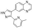
| SMILES | CC1=NC(=CC=C1)C2=C(C=NN2)C3=CC4=NC=CN=C4C=C3 |
| InChi Key | VXJLYXCHOKEODY-UHFFFAOYSA-N |
| InChi Code | InChI=1S/C17H13N5/c1-11-3-2-4-15(21-11)17-13(10-20-22-17)12-5-6-14-16(9-12)19-8-7-18-14/h2-10H,1H3,(H,20,22) |
| Chemical Name | 6-[3-(6-Methylpyridin-2-yl)-1H-pyrazol-4-yl]quinoxaline |
| Synonyms | BIO013077-01; BIO-013077; BIO 013077; BIO-013077-01; BIO 013077-01; 6-(3-(6-Methylpyridin-2-yl)-1H-pyrazol-4-yl)quinoxaline; BIO-013077-01; 6-[3-(6-methylpyridin-2-yl)-1H-pyrazol-4-yl]quinoxaline; 6-[5-(6-methylpyridin-2-yl)-1H-pyrazol-4-yl]quinoxaline; Quinoxaline, 6-[3-(6-methyl-2-pyridinyl)-1H-pyrazol-4-yl]-; SCHEMBL4008166; DTXSID00440482; BIO013077 |
| HS Tariff Code | 2934.99.9001 |
| Storage |
Powder-20°C 3 years 4°C 2 years In solvent -80°C 6 months -20°C 1 month |
| Shipping Condition | Room temperature (This product is stable at ambient temperature for a few days during ordinary shipping and time spent in Customs) |
Biological Activity
| Targets | TGF-β; Alk4/5 |
| ln Vitro | Members of the TGF-β superfamily govern a number of biological processes, including adhesion, migration, apoptosis, differentiation, and proliferation of cells. There are three main TGF-β isoforms that are expressed in mammals: TGF-β1, TGF-β2, and TGF-β3. Type I and type II serine/threonine kinase receptors, which are similar but have different structures and functions, form a complex that allows TGF-β to transduce signals. Numerous human disorders, including as cancer, pancreatitis, and hematological malignancies, have also been linked to dysregulation of TGF-β signaling [1]. |
| Enzyme Assay | The transforming growth factor-β (TGF-β) superfamily members, which include TGF-βs, activins, bone morphogenetic proteins (BMPs), growth and differentiation factors, and Mullerian inhibiting substance are structurally related secreted cytokines found in species ranging from worms and insects to mammals. A wide range of cellular functions such as cell proliferation, differentiation, adhesion, migration, and apoptosis are regulated by TGF-β superfamily members. The TGF-βs include the three major TGF-β isoforms, TGF-β1, TGF-β2, and TGF-β3 which are expressed in mammals. Among them, TGF-β1 is the prototypic member of this family of cytokines and is the major isoform. TGF-β1 transduces signals through a complex of two related but structurally and functionally distinct serine/threonine kinase receptors, termed type 1 and type II. The signaling cascade is promoted by the binding of ligand to the constitutively active type II receptors on the cell surface, which further recruits the type I receptors, also called as activin receptor-like kinase 5 (ALK5), into the complex. Subsequently, ALK5 is phosphorylated in the juxtamembrane GS domain by the type II receptors thereby creating a binding site for Smad proteins and stimulating its kinase activity. The activated ALK5 phosphorylates Smad2 and Smad3 proteins thereby causing their dissociation from the receptor and heteromeric complex formation with Smad4. These Smad complexes translocate to the nucleus, assemble with specific DNA-binding co-factors and co-modulators to finally activate several hundred genes involved in cell differentiation, proliferation, apoptosis, migration, and extracellular matrix production. TGF-β1 plays a critical role in the initiation and progression of fibrosis in various organ systems such as kidney, heart, lung, and liver. Deregulation of TGF-β signaling has been also implicated in various human diseases including cancer, pancreatic diseases, and hematological malignancies [1]. |
| Cell Assay |
Luciferase reporter assay [1] Biological activity of the test compounds was determined by measuring their ability to inhibit TGF-β-induced p3TP-luciferase reporter activity in HaCaT stable cells transfected with p3TP-Luc. HaCaT cells were seeded at concentrations of 3 × 10⁴ in 96-well plates. The next day, when they reach approximately 90% confluency, various concentrations of ALK5 inhibitors and 2 ng/mL of TGF-β were added to the cells. After 24 h, cell lysates were harvested using Luciferase assay kit according to the manufacturer’s instruction, and luminescence was measured by a luminometer Micro Lumat Plus. |
| References |
[1]. Synthesis and biological evaluation of 1-substituted-3(5)-(6-methylpyridin-2-yl)-4-(quinoxalin-6-yl)pyrazoles as transforming growth factor-β type 1 receptor kinase inhibitors. Eur J Med Chem. 2011 Sep;46(9):3917-25. |
| Additional Infomation | A series of 1-substituted-3(5)-(6-methylpyridin-2-yl)-4-(quinoxalin-6-yl)pyrazoles 14a–d, 15a–d, 17a, 17b, 18a–d, 19a, and 19b has been synthesized and evaluated for their ALK5 inhibitory activity in an enzyme assay and in a cell-based luciferase reporter assay. The 2-[3-(6-methylpyridin-2-yl)-4-(quinoxalin-6-yl)-1H-pyrazol-1-yl]-N-phenylethanethioamide (18a) inhibited ALK5 phosphorylation with an IC50 value of 0.013 μM and showed 80% inhibition at 0.1 μM in a luciferase reporter assay using HaCaT cells permanently transfected with p3TP-luc reporter construct.[1] |
Solubility Data
| Solubility (In Vitro) | DMSO : ~100 mg/mL (~348.04 mM) |
| Solubility (In Vivo) |
Solubility in Formulation 1: ≥ 2.5 mg/mL (8.70 mM) (saturation unknown) in 10% DMSO + 40% PEG300 + 5% Tween80 + 45% Saline (add these co-solvents sequentially from left to right, and one by one), clear solution. For example, if 1 mL of working solution is to be prepared, you can add 100 μL of 25.0 mg/mL clear DMSO stock solution to 400 μL PEG300 and mix evenly; then add 50 μL Tween-80 to the above solution and mix evenly; then add 450 μL normal saline to adjust the volume to 1 mL. Preparation of saline: Dissolve 0.9 g of sodium chloride in 100 mL ddH₂ O to obtain a clear solution. Solubility in Formulation 2: ≥ 2.5 mg/mL (8.70 mM) (saturation unknown) in 10% DMSO + 90% (20% SBE-β-CD in Saline) (add these co-solvents sequentially from left to right, and one by one), clear solution. For example, if 1 mL of working solution is to be prepared, you can add 100 μL of 25.0 mg/mL clear DMSO stock solution to 900 μL of 20% SBE-β-CD physiological saline solution and mix evenly. Preparation of 20% SBE-β-CD in Saline (4°C,1 week): Dissolve 2 g SBE-β-CD in 10 mL saline to obtain a clear solution. (Please use freshly prepared in vivo formulations for optimal results.) |
| Preparing Stock Solutions | 1 mg | 5 mg | 10 mg | |
| 1 mM | 3.4803 mL | 17.4016 mL | 34.8032 mL | |
| 5 mM | 0.6961 mL | 3.4803 mL | 6.9606 mL | |
| 10 mM | 0.3480 mL | 1.7402 mL | 3.4803 mL |