Avadomide (formerly known as CC-122) is a novel, orally available pleiotropic pathway modulator with potential with anticancer and immunomodulatory activity. It targets the protein cereblon (CRBN), a substrate receptor of the cullin ring E3 ubiquitin ligase complex CRL4CRBN. Avadomide mimics an interferon response and has antitumor activity in DLBCL (Diffuse large B-cell lymphoma). Avadomide binds CRBN and promotes the degradation of Aiolos and Ikaros resulting in a mimicry of IFN signaling and apoptosis in DLBCL. As a new chemical entity and a pleiotropic pathway modifier, Avadomide has potential application in the treatment of cancer and immune disease.
Physicochemical Properties
| Molecular Formula | C14H14N4O3 |
| Molecular Weight | 286.29 |
| Exact Mass | 286.107 |
| Elemental Analysis | C, 58.74; H, 4.93; N, 19.57; O, 16.77 |
| CAS # | 1015474-32-4 |
| Related CAS # | (S)-Avadomide-d1;1620055-10-8 |
| PubChem CID | 24967599 |
| Appearance | White to gray solid powder |
| LogP | 1.174 |
| Hydrogen Bond Donor Count | 2 |
| Hydrogen Bond Acceptor Count | 5 |
| Rotatable Bond Count | 1 |
| Heavy Atom Count | 21 |
| Complexity | 530 |
| Defined Atom Stereocenter Count | 0 |
| SMILES | O=C1C([H])(C([H])([H])C([H])([H])C(N1[H])=O)N1C(C2=C(C([H])=C([H])C([H])=C2N=C1C([H])([H])[H])N([H])[H])=O |
| InChi Key | RSNPAKAFCAAMBH-UHFFFAOYSA-N |
| InChi Code | InChI=1S/C14H14N4O3/c1-7-16-9-4-2-3-8(15)12(9)14(21)18(7)10-5-6-11(19)17-13(10)20/h2-4,10H,5-6,15H2,1H3,(H,17,19,20) |
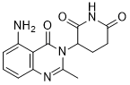
| Chemical Name | 3-(5-amino-2-methyl-4-oxoquinazolin-3(4H)-yl)piperidine-2,6-dione |
| Synonyms | Avadomide; CC 122; CC-122; 1015474-32-4; 3-(5-Amino-2-methyl-4-oxoquinazolin-3(4H)-yl)piperidine-2,6-dione; Avadomide [USAN]; Avadomide(CC-122); CC 122; CC122 |
| HS Tariff Code | 2934.99.9001 |
| Storage |
Powder-20°C 3 years 4°C 2 years In solvent -80°C 6 months -20°C 1 month |
| Shipping Condition | Room temperature (This product is stable at ambient temperature for a few days during ordinary shipping and time spent in Customs) |
Biological Activity
| Targets |
Cereblon E3 ligase
Avadomide targets cereblon (CRBN) with a Ki value of 0.5 nM [2] Avadomide modulates CRBN-mediated ubiquitination pathway [1] |
| ln Vitro |
In ABC and GCB DLBCL, avadomide suppresses proliferation and triggers apoptosis. Aiolos and Ikaros can be degraded by avadomide or knocked down using short hairpin RNA. This leads to apoptosis in both activated B-cell (ABC) and germinal center B-cell DLBCL cell lines and increases transcription of IFN-stimulated genes regardless of IFN-α, -β, and -γ production and/or secretion.[1] In diffuse large B-cell lymphoma (DLBCL) cell lines (OCI-Ly3, SU-DHL-4, SU-DHL-6, WSU-DLCL2), Avadomide exhibited antiproliferative activity with IC50 values ranging from 0.3 to 3.1 nM; it induced G0/G1 cell cycle arrest and apoptosis, as evidenced by increased cleaved caspase-3 and PARP levels [2] - Avadomide upregulated interferon (IFN)-stimulated genes (ISGs) including IRF7, MX1, and OAS1 in DLBCL cells, mimicking an IFN response through activation of the JAK-STAT pathway; it also enhanced the expression of MHC class I molecules on DLBCL cells [2] - In various advanced malignancy cell lines (including solid tumors and hematologic malignancies), Avadomide showed modest antiproliferative activity, with IC50 values mostly above 10 nM in solid tumor cell lines [1] - Avadomide induced degradation of Ikaros (IKZF1) and Aiolos (IKZF3) in DLBCL cells, which are CRBN substrates, contributing to its antitumor effect [2] |
| ln Vivo |
When Avadomide (CC122) was administered once daily at 3 or 30 mg/kg to female CB-17 SCID mice, the tumor growth in OCI-LY10 ABC-DLBCL and WSU-DLCL2 GCB-DLBCL derived xenograft models was significantly reduced (P <.01) in comparison to the vehicle control. We evaluated avadomide (CC122)'scapacityto encourage Ikaros and Aiolos degradation in vivo in a different investigation. The WSU-DLCL2 xenograft transplanted mice used in the 21-day efficacy study had tumors removed 1, 6, or 24 hours after the last dose.Using immunohistochemistry (IHC), the expression of Aiolos and Ikaros was examined. Within the first hour of treatment, there was a reduction of 64% and 30%, respectively, compared with the vehicle; at 6 hours, there was a maximal reduction of 94% and 69%, respectively. Twenty-four hours after dosing, Aiolos and Ikaros levels showed partial recovery, with protein levels at 20% and 34% of vehicle, respectively. The trough compound level after several doses of avadomide is represented by the 24-hour postdose Aiolos and Ikaros expression (CC122).Aiolos expression is significantly lower at the 1-hour time point compared to the 24-hour postdose time point, but not Ikaros expression. Both transcription factors, however, are significantly different at the 6-hour time point compared to the 24-hour time point. Together, these findings show that avadomide (CC122) inhibited the growth of DLBCL tumors in vivo and that this activity was connected to the GCB- and ABC-DLBCL xenograft models' degradation of Ikaros and Aiolos.[1] In SCID mice bearing OCI-Ly3 or SU-DHL-4 DLBCL xenografts, Avadomide administered orally at doses of 0.3, 1, or 3 mg/kg once daily for 21 days dose-dependently inhibited tumor growth; the 3 mg/kg dose achieved tumor growth inhibition rates of 78% (OCI-Ly3) and 65% (SU-DHL-4) compared to vehicle control [2] - In the first-in-human study, Avadomide demonstrated preliminary antitumor activity in patients with advanced malignancies, including stable disease in 25% of evaluable patients (n=40) and partial response in 1 patient with non-Hodgkin lymphoma [1] - Avadomide treatment in DLBCL xenograft mice was associated with increased expression of ISGs and MHC class I molecules in tumor tissues, consistent with in vitro findings [2] |
| Enzyme Assay |
A homogeneous time-resolved fluorescence (HTRF) assay was used to measure the binding affinity of Avadomide to CRBN; recombinant CRBN protein was incubated with Avadomide of various concentrations, and the binding interaction was detected by fluorescence resonance energy transfer (FRET) signal; the Ki value was calculated based on dose-response curves [2] - Kinase activity assays were performed to evaluate the effect of Avadomide on JAK-STAT pathway kinases; purified JAK1, JAK2, STAT1, and STAT3 proteins were incubated with Avadomide and their respective substrates, and kinase activity was measured by detecting phosphorylated substrates via ELISA; Avadomide was found to enhance JAK-STAT phosphorylation in a CRBN-dependent manner [2] |
| Cell Assay |
CC-122 inhibits proliferation and induces apoptosis in ABC and GCB DLBCL[1] To explore the antiproliferative activity of CC-122 in DLBCL cell lines, thymidine incorporation assays were performed in a panel of DLBCL cell lines after 5 days of treatment with CC-122. Exposure of 4 ABC-DLBCL lines (TMD8, U2932, Riva, and OCI-LY10) and 5 GCB-DLBCL lines (Karpas 422, WSU-DLCL2, SUDHL-4, OCI-LY19, and Pfeiffer) cell lines with 0.01 to 10 000 nM CC-122 for 5 days led to a marked decrease in proliferation (Figure 1A). The ABC cell lines were more sensitive than the GCB cell lines (ABC 50% inhibition concentration range, 8 nM to 6 μM; GCB 50% inhibition concentration range, 1 μM to >10 μM). CC-122 promotes CRBN-Ikaros interaction and subsequent proteasomal degradation of Aiolos and Ikaros in vitro[1] The thalidomide binding domain of CRBN was recently shown to contain a hydrophobic pocket in which 3 tryptophan residues govern the binding with the glutarimide moiety within thalidomide, lenalidomide, and pomalidomide.9 As the chemical structure of CC-122 contains a glutarimide ring, we explored if CC-122 binds CRBN. As shown in Figure 2A, CRBN from U266 multiple myeloma cell extracts interacted with FG affinity beads coupled to an immobilized thalidomide analog (DMSO control lane). Furthermore, incubation of the complex with increasing concentration of free CC-122 resulted in the displacement of CRBN from the thalidomide analog–immobilized beads, consistent with CC-122 competing with thalidomide for binding CRBN. Additionally, fluorescence quenching studies on a purified C terminus fragment of CRBN (amino acids 321-440) confirmed direct binding of CC-122 with CRBN (supplemental Figure 1). DLBCL cell lines were cultured in RPMI 1640 medium supplemented with fetal bovine serum and antibiotics; cells were seeded in 96-well plates at 5×10³ cells/well and treated with Avadomide at concentrations ranging from 0.01 to 100 nM for 72 hours; cell viability was assessed using a colorimetric assay based on mitochondrial dehydrogenase activity, and IC50 values were calculated from dose-response curves [2] - For cell cycle analysis, DLBCL cells were treated with Avadomide (1 nM) for 48 hours, fixed with ethanol, stained with propidium iodide, and analyzed by flow cytometry to determine the proportion of cells in G0/G1, S, and G2/M phases [2] - Apoptosis was evaluated by Annexin V-FITC/PI double staining; DLBCL cells were treated with Avadomide (1 nM) for 72 hours, stained with Annexin V-FITC and PI, and apoptotic cells (Annexin V-positive) were quantified by flow cytometry [2] - Western blot analysis was performed to detect protein expression; cells were lysed in RIPA buffer, proteins were separated by SDS-PAGE, transferred to PVDF membranes, and probed with antibodies against IKZF1, IKZF3, cleaved caspase-3, PARP, STAT1, phospho-STAT1, and GAPDH (loading control); immunoreactive bands were visualized using chemiluminescence [2] - Quantitative real-time PCR (qPCR) was used to measure ISG expression; total RNA was extracted from Avadomide-treated DLBCL cells, reverse-transcribed to cDNA, and amplified with specific primers for IRF7, MX1, OAS1, and GAPDH (reference gene); relative gene expression was calculated using the ΔΔCt method [2] |
| Animal Protocol |
On the first day of these studies, the female SCID mice (CB17/Icr-Prkdcscid, Charles River) were 8 weeks old and weighed between 15.0 and 23.2 g. Each SCID mouse received a subcutaneous injection of 5x10⁶ OCI-LY10 cells (0.2 ml cell suspension) in the right flank. In order to track growth, tumors were classified in two dimensions as their mean volume got closer to 100–150 mm3. Mice were divided into treatment groups (n=10) after tumor cell implantation, either twenty-one days (OCI-LY10) or fourteen days (WSU-DLCL2). Throughout the study, tumors were called in twice a week.In 0.5% carboxymethyl cellulose:0.25% Tween-80 in de-ionized water, apadomide (CC122) was suspended. For twenty-eight days (qd x28), avadomide (CC122) and the vehicle were each given by oral gavage (p.o.) once daily. [1] Female SCID mice (6-8 weeks old) were subcutaneously inoculated with 5×10⁶ OCI-Ly3 or SU-DHL-4 DLBCL cells into the right flank; when tumors reached a volume of ~100 mm³, mice were randomly divided into 4 groups (n=6 per group) [2] - Avadomide was formulated in 0.5% methylcellulose and 0.1% Tween 80 in water; mice were administered Avadomide orally at doses of 0.3, 1, or 3 mg/kg once daily, or vehicle control, for 21 consecutive days [2] - Tumor volume was measured twice weekly using calipers (volume = length × width² / 2), and body weight was recorded to monitor toxicity; at the end of the study, mice were euthanized, tumors were excised and weighed, and tumor tissues were collected for protein and RNA analysis [2] - For the first-in-human study, patients with advanced malignancies received Avadomide orally once daily in escalating doses (0.15 to 2.5 mg/day) in 28-day cycles; dose escalation followed a 3+3 design to evaluate safety and tolerability [1] |
| ADME/Pharmacokinetics |
Pharmacokinetics, pharmacodynamics, and biomarkers[1] At all dose levels, the avadomide plasma concentration versus time profiles were characterized by a rapid absorption phase and similar median time to maximum concentration (Fig. 1). After attainment of maximum observed concentration in plasma, avadomide appeared to decrease in a monophasic manner at all dose levels. By visual inspection of mean plasma concentrations versus time profiles, avadomide plasma exposures increased in a dose-dependent manner across the 0.5- to 3.5-mg dose range. All 7 dose levels showed mild-to-moderate accumulation of avadomide plasma exposure after multiple doses. Supplementary Table S1 summarizes avadomide plasma pharmacokinetic parameters by day and dose level. In general, as assessed from the geometric coefficient of variation percentage, interpatient variability was noted for both avadomide area under the concentration–time curve and maximum observed concentration in plasma. The mean total recovery of avadomide in urine within 24 hours ranged from 18% to 35% across the 0.5- to 3.5-mg dose range. The mean avadomide renal clearance ranged from 0.53 to 1.31 L/h across the 0.5- to 3.5-mg dose range. The t1/2 ranged from 7.68 to 27.91 hours. In humans, after oral administration of Avadomide, peak plasma concentrations (Cmax) were achieved at a median time (Tmax) of 2-4 hours; Cmax and area under the plasma concentration-time curve (AUC) increased proportionally with doses from 0.15 to 2.5 mg/day [1] - The terminal elimination half-life (t1/2) of Avadomide in humans was approximately 12-16 hours [1] - Plasma protein binding of Avadomide was 97-99% in human plasma, as determined by equilibrium dialysis [1] - In preclinical studies (mice), Avadomide showed good oral bioavailability (~70%) and distributed widely into tissues, with tumor tissue concentrations approximately 2-3 times higher than plasma concentrations [2] - Avadomide was primarily metabolized by cytochrome P450 (CYP) 3A4 in human liver microsomes; no major active metabolites were identified [1] |
| Toxicity/Toxicokinetics |
Most patients (85%) had ≥1 TEAE that was suspected by the investigators of being related to avadomide. Across all cohorts, the most common TEAEs (≥15%) were fatigue (44%), neutropenia (29%), and diarrhea (15%). Avadomide-related grade ≥3 TEAEs occurred in 14 patients (41%). The most common grade ≥3 TEAEs were neutropenia (2 patients in the 1.0-mg cohort; 1 patient each in the 1.5-, 2.0-, 2.5-, and 3.5-mg cohorts; and 3 patients in the 3.0-mg cohort) and pneumonia (2 patients in the 3.0-mg cohort). Table 2 summarizes the TEAEs in the treated population. One death occurred within 28 days of the last dose of avadomide; 1 patient with pancreatic carcinoma in the 3.5-mg cohort died due to disease progression. In the first-in-human study, the most common treatment-related adverse events (TRAEs) were fatigue (45%), nausea (35%), diarrhea (30%), and decreased appetite (25%); the majority of TRAEs were grade 1-2 [1] - Grade 3 TRAEs included neutropenia (10%), anemia (5%), and thrombocytopenia (5%) [1] - Avadomide did not cause significant changes in liver function tests (ALT, AST, bilirubin) or renal function tests (creatinine, BUN) in patients [1] - In preclinical mouse studies, Avadomide at doses up to 3 mg/kg/day for 21 days did not cause significant weight loss (>10%) or gross pathological changes in major organs (liver, kidney, heart, lung, spleen) [2] - No drug-drug interaction potential was identified for Avadomide based on in vitro CYP inhibition and induction studies [1] |
| References |
[1].A First-in-Human Study of Novel Cereblon Modulator Avadomide (CC-122) in Advanced Malignancies. Clin Cancer Res. 2019 Jan 1;25(1):90-98. [2].CC-122, a pleiotropic pathway modifier, mimics an IFN response and has antitumor activity in DLBCL.Blood.Aug 6;126(6):779-89. |
| Additional Infomation |
Avadomide is under investigation in clinical trial NCT02031419 (Novel Combinations of CC-122, CC-223, CC-292, and Rituximab in Diffuse Large B-cell Lymphoma and Follicular Lymphoma). Avadomide is a novel, small molecule cereblon-modulating agent with potential antineoplastic, antiangiogenic and immunomodulatory activities. Upon oral administration, avadomide binds to and modulates cereblon to promote recruitment of the hematopoietic transcription factors Aiolos and Ikaros to the Cullin-4 RING E3 ubiquitin ligase complex. This binding results in the ubiquitination and rapid proteasomal degradation of Aiolos and Ikaros and the derepression of interferon (IFN)-stimulated genes, including DDX58 and IRF7, leading to apoptosis of certain tumor cells. Additionally, Aiolos degredation leads to derepression of the IL2 gene, thereby enhancing interleukin-2 production, costimulation of T-lymphocytes and IL-2-induced T-cell proliferation. Avadomide may also promote the activation of natural killer (NK) cells, potentially enhancing tumor cell killing. Aiolos and Ikaros are transcriptional repressors known to play an important role in normal B- and T-cell function. Drug Indication Treatment of mature B-cell neoplasms Avadomide is a novel oral cereblon modulator with pleiotropic antitumor activities, including direct antiproliferation, induction of apoptosis, modulation of the immune response, and enhancement of antitumor immunity [1][2] - The antitumor mechanism of Avadomide involves CRBN-mediated degradation of IKZF1 and IKZF3, activation of the JAK-STAT pathway, and induction of an IFN-like response, which synergistically inhibit tumor cell growth [2] - The first-in-human study enrolled patients with advanced solid tumors, non-Hodgkin lymphoma, and multiple myeloma who had failed prior standard therapies [1] - Avadomide showed promising activity in patients with relapsed/refractory non-Hodgkin lymphoma, supporting further development in this indication [1] |
Solubility Data
| Solubility (In Vitro) |
DMSO : 33.33~57 mg/mL ( 116.42~199.09 mM ) Ethanol : ~1 mg/mL |
| Solubility (In Vivo) |
Solubility in Formulation 1: ≥ 2.5 mg/mL (8.73 mM) (saturation unknown) in 10% DMSO + 40% PEG300 +5% Tween-80 + 45% Saline (add these co-solvents sequentially from left to right, and one by one), clear solution. For example, if 1 mL of working solution is to be prepared, you can add 100 μL of 25.0 mg/mL clear DMSO stock solution to 400 μL PEG300 and mix evenly; then add 50 μL Tween-80 + to the above solution and mix evenly; then add 450 μL normal saline to adjust the volume to 1 mL. Preparation of saline: Dissolve 0.9 g of sodium chloride in 100 mL ddH₂ O to obtain a clear solution. (Please use freshly prepared in vivo formulations for optimal results.) |
| Preparing Stock Solutions | 1 mg | 5 mg | 10 mg | |
| 1 mM | 3.4930 mL | 17.4648 mL | 34.9296 mL | |
| 5 mM | 0.6986 mL | 3.4930 mL | 6.9859 mL | |
| 10 mM | 0.3493 mL | 1.7465 mL | 3.4930 mL |