Atglistatin is a highly potent and selective inhibitor of adipose triglyceride lipase (ATGL) with IC50 of 0.7 μM, it showed high selectivity over other key metabolic lipases. In lysates from E. coli overexpressing ATGL, atglistatin inhibited ATGL activity with IC50 value of 0.7 μM. Atglistatin at concentrations up to 50 μM had no cytotoxicity. Also, atglistatin inhibited ATGL with Ki value of 355 nM in a competitive way. In white adipose tissue (WAT) lysates of wild-type mice, atglistatin inhibited triacylglycerol (TG) hydrolase activity by 78%. The combination of Atglistatin and the HSL inhibitor Hi 76-007917 inhibited TG hydrolase activity by 95%.
Physicochemical Properties
| Molecular Formula | C17H21N3O | |
| Molecular Weight | 283.37 | |
| Exact Mass | 283.168 | |
| Elemental Analysis | C, 72.06; H, 7.47; N, 14.83; O, 5.65 | |
| CAS # | 1469924-27-3 | |
| Related CAS # |
|
|
| PubChem CID | 71699712 | |
| Appearance | White to yellow solid powder | |
| Density | 1.1±0.1 g/cm3 | |
| Boiling Point | 484.4±45.0 °C at 760 mmHg | |
| Flash Point | 246.7±28.7 °C | |
| Vapour Pressure | 0.0±1.2 mmHg at 25°C | |
| Index of Refraction | 1.621 | |
| LogP | 2.83 | |
| Hydrogen Bond Donor Count | 1 | |
| Hydrogen Bond Acceptor Count | 2 | |
| Rotatable Bond Count | 3 | |
| Heavy Atom Count | 21 | |
| Complexity | 335 | |
| Defined Atom Stereocenter Count | 0 | |
| InChi Key | AWOPBSAJHCUSAS-UHFFFAOYSA-N | |
| InChi Code | InChI=1S/C17H21N3O/c1-19(2)16-10-8-13(9-11-16)14-6-5-7-15(12-14)18-17(21)20(3)4/h5-12H,1-4H3,(H,18,21) | |
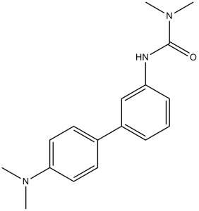
| Chemical Name | 3-(4-(dimethylamino)-[1,1-biphenyl]-3-yl)-1,1-dimethylurea | |
| Synonyms |
|
|
| HS Tariff Code | 2934.99.9001 | |
| Storage |
Powder-20°C 3 years 4°C 2 years In solvent -80°C 6 months -20°C 1 month |
|
| Shipping Condition | Room temperature (This product is stable at ambient temperature for a few days during ordinary shipping and time spent in Customs) |
Biological Activity
| Targets | Adipose triglyceride lipase/ATGL (IC50 = 0.7 μM) | ||
| ln Vitro | Triacylglycerol (TG) hydrolase activity in wild-type white adipose tissue (WAT) is inhibited by atglistatin in a dose-dependent manner; at the maximum concentration, this inhibition reaches 78%. When compared to wild-type preparations, the TG hydrolase activity in WAT lysates from ATGL-ko mice was roughly 70% lower, while atglistatin only moderately affected the residual activity. The TG hydrolase activity of WAT was nearly completely inhibited (-95%) by the combination of atglistatin and the hormone-sensitive lipase (HSL) inhibitor Hi 76-0079, indicating that HSL is primarily responsible for the majority of the non-ATGL activity[1]. | ||
| ln Vivo | Animals were given oral gavages with dissolved agostatin in olive oil. Following application, tissue and blood were taken in order to measure the inhibitor concentrations, tissue triacylglycerol (TG) levels, and plasma parameters. Fatty acids (FA) and glycerol, the lipolytic parameters, dropped at 4 and 8 hours after application and recovered to normal after 12 hours, according to time course experiments. Eight hours after treatment, there was a dose-dependent drop in FA and glycerol levels of up to 50% and 62%, respectively. Additionally, atglistatin significantly reduced plasma TG levels (-43%) while having no discernible effect on blood sugar, total cholesterol, ketone bodies, or insulin levels. Intraperitoneal injection of Atglistatin was likewise found to inhibit lipolysis in a dose- and time-dependent manner [1]. | ||
| Cell Assay |
Lipolysis of 3T3-L1 cells[1] 3T3-L1 fibroblasts (CL-173) were obtained from ATCC and cultivated in DMEM containing 4.5 g/l glucose and L-glutamine supplemented with 10% FCS and antibiotics under standard conditions. Cells were seeded in 12 well plates and two days after confluency medium was changed to DMEM supplemented with 10% FCS containing 10 μg/ml insulin, 0.25 μM (0.4 μg/ml) dexamethasone, and 500 μM isobutylmethylxanthine. After 3 and 5 days, medium was changed to DMEM supplemented with 10% FCS containing 10 μg/ml and 0.5 μg/ml insulin, respectively. On day 7 of differentiation the cells were incubated overnight in the absence of insulin. Cells were used at day 8 of differentiation. For experiments, cells were preincubated with 0, 0.1, 1, 10, or 50 μM of Atglistatin in the presence or absence of 10 μM Hi 76-0079 for 2 h. Then, the medium was replaced by DMEM containing 2% BSA, 20 μM forskolin, and 0, 0.1, 1, 10, or 50 μM of Atglistatin in the presence or absence of 10 μM Hi 76-0079 for 1h. The release of FA and glycerol in the media was determined using commercial kits. Protein concentration was determined using BCA reagent after extracting total lipids using hexane:isopropanol (3:2), and lysing the cells using 0.3 N NaOH/0.1 % SDS.[1] Lipolysis of isolated WAT organ cultures[1] Gonadal fat pads of wild-type and ATGL-ko mice were surgically removed and washed several times with PBS. Tissue pieces (~15 mg) were preincubated in DMEM containing 0, 0.1, 1, 10, and 50 μM Atglistatin for 8 h at C37°C, 5% CO2, 95% humidified atmosphere. Thereafter, the medium was replaced by DMEM containing 2 % BSA (fatty acid-free) either in the presence or in the absence of 20 μM forskolin and 0, 0.1, 1, 10, and 50 μM, and incubated for another 60 min at 37°C. Then, aliquots of the medium were collected and analyzed for FA and glycerol content using commercial kits (HR Series NEFA-HR(2)). For protein determinations, fat pads were washed extensively with PBS and lysed in 0.3 N NaOH/0.1 % SDS. Protein measurements were performed using the BCA reagent.[1] Toxicity test for Atglistatin in AML-12 mouse hepatocytes[1] For MTT-based in vitro viability assays, cells were seeded at an initial density of 1×10⁴ cells per well in 96-well plates and cultured under standard conditions for 24 hours. The next day, cells were pretreated with different concentrations of Atglistatin dissolved in DMSO or Cisplatin dissolved in dimethylformamide (DMF) as positive control for two hours. Medium was replaced by an identical fresh medium and incubated again for the indicated time-points. Thereafter cells were incubated for 3 hours with 100 μl Thiazolyl Blue Tetrazolium Bromide (MTT). The resulting violet formazan crystals were dissolved by adding 100 μl of MTT solubilization solution (0.1% NP-40, 4 mM HCl and anhydrous isopropanol). After complete dissolution of the formazan product, absorbance was measured at 595 nm using 690 nm as reference wavelength. |
||
| Animal Protocol |
|
||
| Toxicity/Toxicokinetics | Cytotoxicity assays for compound 4 revealed virtually no toxicity up to a concentration of 50 μM (Supplementary Fig. 2). This compound appeared suitable as a chemical tool for detailed biological characterization and was named Atglistatin.[1] | ||
| References |
[1]. Development of small-molecule inhibitors targeting adipose triglyceride lipase. (2013) Nat Chem Biol. 9(12):785-7. |
||
| Additional Infomation | Atglistatin is a biphenyl that is 1,1'-biphenyl substituted by (dimethylcarbamoyl)amino and dimethylamino groups at positions 3 and 4', respectively. It is a potent inhibitor of adipose triglyceride lipase activity (IC50 = 700nM). It has a role as an EC 3.1.1.3 (triacylglycerol lipase) inhibitor and a cardioprotective agent. It is a member of biphenyls, a member of ureas, a tertiary amino compound and an aromatic amine. |
Solubility Data
| Solubility (In Vitro) |
|
|||
| Solubility (In Vivo) |
Solubility in Formulation 1: ≥ 2.5 mg/mL (8.82 mM) (saturation unknown) in 10% DMSO + 40% PEG300 + 5% Tween80 + 45% Saline (add these co-solvents sequentially from left to right, and one by one), clear solution. For example, if 1 mL of working solution is to be prepared, you can add 100 μL of 25.0 mg/mL clear DMSO stock solution to 400 μL PEG300 and mix evenly; then add 50 μL Tween-80 to the above solution and mix evenly; then add 450 μL normal saline to adjust the volume to 1 mL. Preparation of saline: Dissolve 0.9 g of sodium chloride in 100 mL ddH₂ O to obtain a clear solution. Solubility in Formulation 2: ≥ 2.5 mg/mL (8.82 mM) (saturation unknown) in 10% DMSO + 90% Corn Oil (add these co-solvents sequentially from left to right, and one by one), clear solution. For example, if 1 mL of working solution is to be prepared, you can add 100 μL of 25.0 mg/mL clear DMSO stock solution to 900 μL of corn oil and mix evenly. Solubility in Formulation 3: 6.25 mg/mL (22.06 mM) in Corn Oil (add these co-solvents sequentially from left to right, and one by one), suspension solution; with ultrasonication. (Please use freshly prepared in vivo formulations for optimal results.) |
| Preparing Stock Solutions | 1 mg | 5 mg | 10 mg | |
| 1 mM | 3.5290 mL | 17.6448 mL | 35.2896 mL | |
| 5 mM | 0.7058 mL | 3.5290 mL | 7.0579 mL | |
| 10 mM | 0.3529 mL | 1.7645 mL | 3.5290 mL |