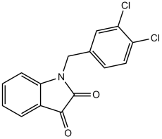
Apoptosis Activator 2 is a novel and potent small molecule apoptosis activator with an IC50 value of about 4 μM. Apoptosis Activator 2 is a cell permeable substance that encourages apoptosis by triggering caspases in a way that depends on cytochrome c and Apaf-1. When there is 0.15 mM cytochrome c, 20 μM apoptosis activator 2 increases the fraction of Apaf-1 in the apoptosome. It was discovered that Apaf-1 oligomerization was induced by Apoptosis Activator 2. Apoptosis Activator 2 prominently causes DNA fragmentation, PARP cleavage, and caspase-3 activation through the induction of apoptosis.
Physicochemical Properties
| Molecular Formula | C15H9CL2NO2 | |
| Molecular Weight | 306.14 | |
| Exact Mass | 305.001 | |
| Elemental Analysis | C, 58.85; H, 2.96; Cl, 23.16; N, 4.58; O, 10.45 | |
| CAS # | 79183-19-0 | |
| Related CAS # |
|
|
| PubChem CID | 1901244 | |
| Appearance | Solid powder | |
| Density | 1.5±0.1 g/cm3 | |
| Boiling Point | 470.6±55.0 °C at 760 mmHg | |
| Flash Point | 238.4±31.5 °C | |
| Vapour Pressure | 0.0±1.2 mmHg at 25°C | |
| Index of Refraction | 1.671 | |
| LogP | 3.42 | |
| Hydrogen Bond Donor Count | 0 | |
| Hydrogen Bond Acceptor Count | 2 | |
| Rotatable Bond Count | 2 | |
| Heavy Atom Count | 20 | |
| Complexity | 415 | |
| Defined Atom Stereocenter Count | 0 | |
| SMILES | O=C1N(CC2=CC=C(Cl)C(Cl)=C2)C3=C(C=CC=C3)C1=O |
|
| InChi Key | KGRJPLRFGLMQMV-UHFFFAOYSA-N | |
| InChi Code | InChI=1S/C15H9Cl2NO2/c16-11-6-5-9(7-12(11)17)8-18-13-4-2-1-3-10(13)14(19)15(18)20/h1-7H,8H2 | |
| Chemical Name | 1-[(3,4-dichlorophenyl)methyl]indole-2,3-dione | |
| Synonyms | Apoptosis Activator 2; AAII; N-(3,4-dichlorobenzyl) Isatin. MDK-83190; MDK 83190; MDK83190 | |
| HS Tariff Code | 2934.99.9001 | |
| Storage |
Powder-20°C 3 years 4°C 2 years In solvent -80°C 6 months -20°C 1 month |
|
| Shipping Condition | Room temperature (This product is stable at ambient temperature for a few days during ordinary shipping and time spent in Customs) |
Biological Activity
| Targets | Caspase-3 | ||
| ln Vitro | Apoptosis Activator 2 (20 μM) at the reduced cyto c concentration increases the fraction of Apaf-1 in the apoptosome by 1.5-fold to 33%. At the decreased level of cytochrome c and caspase-3 activation, Apoptosis Activator 2 causes a 4-fold increase in the extent of caspase-3 activation. With an IC50 of 4 μM, Apoptosis Activator 2 kills cells by strongly inducing caspase-3 activation, PARP cleavage, and DNA fragmentation. With an IC50 of 50 μM, 43 μM, 4 μM, 6 μM, 9 μM, 20 μM, 44 μM and 35 μM. , Apoptosis Activator 2 induces apoptosis in PBL, HUVEC, Jurkat, Molt-4, CCRF-CEM, BT-549, MDA-MB-468, and NCI-H23. The majority of tumor cell lines tested exhibit a cytostatic response to apoptosis activator 2, which inhibits cell growth by 50–100% at 10 M. In 40 of the 48 tumor cell lines examined, Apoptosis Activator 2 inhibits cell growth by 50–100% at 10 μM, having a cytostatic effect on the majority of the tumor cell lines examined. [1] Through the induction of apoptosome formation, Apoptosis Activator 2 causes cell death. The survival rates of Ventral midbrain cultures for Apoptosis Activator 2 (-8.1 ± 6.0%) are not significantly influenced by En1 expression levels. Use of the other three reagents has no appreciable impact on the survival rate for Apoptosis Activator 2 (-10.7 ± 4.7%). [2] The Tunel assay and apoptotic DNA ladder are used to determine whether or not apoptosis activator 2 (10 μM) induces apoptosis in AGS cells. Anti TROP2 conjugated liposomes induce apoptosis more effectively when apoptosis activator 2 (10 μM) is added. [3] In neuronal cultures, zVAD (50 μM) or cyclohexamide (10 μg/mL) significantly reduce the toxicity of Apoptosis Activator 2. Numerous neurones with pyknotic nuclei suggestive of cell death involving apoptosis are produced in response to apoptosis activator 2 (3 μM). In neuronal cultures, DHT (10 nM) or E2 (10 nM) significantly reduce the toxicity of Apoptosis Activator 2. [4] | ||
| ln Vivo |
|
||
| Enzyme Assay | According to previously published reports, HeLa cell cytoplasmic extracts are created. Apoptosis Activator 2 is diluted in DMSO to a final concentration of 1 mM, with a final DMSO concentration of 1% vol/vol, and then distributed into 96-well microtiter plates. 250 μg of total protein from cytoplasmic extracts in HEB buffer (50 mM Hepes, pH 7.4/50 mM KCl/5 mM EGTA/2 mM MgCl) are added to each well along with 150 μL of the DEVD-AFC (Asp-Glu-Val-Asp-7-amino-4-trifluoromethylcoumarin) substrate. Fluorescence is measured in a LJL Biosystems plate reader at 10-minute intervals while the plates are incubated at 37 °C. | ||
| Cell Assay | An experimenter blinded to condition counts all viable cells using a manual mechanical counter within the defined field of a microscope reticle grid. The vital dye calcein acetoxymethyl ester and the morphological criterion of a smooth, spherical soma are both used to determine whether a cell is viable. Per culture well, counts of viable cells are performed in four non-overlapping fields, with three separate wells for each condition. For vehicle-treated control conditions, there were 100–200 viable cell counts per well. At least three different culture preparations are used for each experiment. One-way ANOVA is used to statistically analyze the raw cell count data, and the Fisher LSD test is used to compare between groups (significance is denoted by P < 0.05). Cell viability is represented graphically as a proportion of alive cells in the vehicle-treated control condition. | ||
| Animal Protocol |
|
||
| References |
[1]. Proc Natl Acad Sci U S A . 2003 Jun 24;100(13):7533-8. [2]. Neural Dev . 2009 Mar 16:4:11. [3]. N Am J Med Sci . 2012 Nov;4(11):582-5. [4]. J Neuroendocrinol . 2010 Sep;22(9):1013-22. |
||
| Additional Infomation | 1-[(3,4-dichlorophenyl)methyl]indole-2,3-dione is a member of indoles. |
Solubility Data
| Solubility (In Vitro) |
|
|||
| Solubility (In Vivo) |
Solubility in Formulation 1: ≥ 2.5 mg/mL (8.17 mM) (saturation unknown) in 10% DMSO + 40% PEG300 +5% Tween-80 + 45% Saline (add these co-solvents sequentially from left to right, and one by one), clear solution. For example, if 1 mL of working solution is to be prepared, you can add 100 μL of 25.0 mg/mL clear DMSO stock solution to 400 μL PEG300 and mix evenly; then add 50 μL Tween-80 + to the above solution and mix evenly; then add 450 μL normal saline to adjust the volume to 1 mL. Preparation of saline: Dissolve 0.9 g of sodium chloride in 100 mL ddH₂ O to obtain a clear solution. (Please use freshly prepared in vivo formulations for optimal results.) |
| Preparing Stock Solutions | 1 mg | 5 mg | 10 mg | |
| 1 mM | 3.2665 mL | 16.3324 mL | 32.6648 mL | |
| 5 mM | 0.6533 mL | 3.2665 mL | 6.5330 mL | |
| 10 mM | 0.3266 mL | 1.6332 mL | 3.2665 mL |
