Apcin A HCL is the hydrochloride salt of Apcin-A, which is a novel and potent anaphase-promoting complex (APC) inhibitor. References: A novel strategy to block mitotic progression for targeted therapy.
Physicochemical Properties
| Molecular Formula | C10H15CL4N5O2 |
| Molecular Weight | 379.070397615433 |
| Exact Mass | 378.995 |
| CAS # | 1683535-53-6 |
| Related CAS # | 1683617-62-0 (freebase) |
| PubChem CID | 155906331 |
| Appearance | Typically exists as solid at room temperature |
| Hydrogen Bond Donor Count | 4 |
| Hydrogen Bond Acceptor Count | 6 |
| Rotatable Bond Count | 7 |
| Heavy Atom Count | 21 |
| Complexity | 297 |
| Defined Atom Stereocenter Count | 0 |
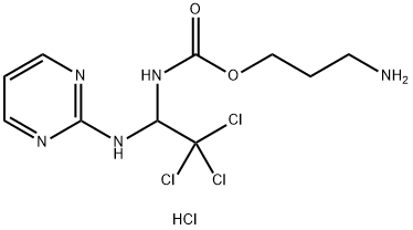
| SMILES | C1=CN=C(N=C1)NC(C(Cl)(Cl)Cl)NC(=O)OCCCN.Cl |
| InChi Key | NEYHFUCNZUYAJK-UHFFFAOYSA-N |
| InChi Code | InChI=1S/C10H14Cl3N5O2.ClH/c11-10(12,13)7(17-8-15-4-2-5-16-8)18-9(19)20-6-1-3-14;/h2,4-5,7H,1,3,6,14H2,(H,18,19)(H,15,16,17);1H |
| Chemical Name | 3-aminopropyl N-[2,2,2-trichloro-1-(pyrimidin-2-ylamino)ethyl]carbamate;hydrochloride |
| Synonyms | 1683535-53-6; 3-Aminopropyl (2,2,2-trichloro-1-(pyrimidin-2-ylamino)ethyl)carbamate hydrochloride; Apcin A hydrochloride; Apcin-A (monohydrochloride); 3-aminopropyl N-[2,2,2-trichloro-1-(pyrimidin-2-ylamino)ethyl]carbamate;hydrochloride; Apcin A HCL; starbld0007153; C10H15Cl4N5O2; |
| HS Tariff Code | 2934.99.9001 |
| Storage |
Powder-20°C 3 years 4°C 2 years In solvent -80°C 6 months -20°C 1 month |
| Shipping Condition | Room temperature (This product is stable at ambient temperature for a few days during ordinary shipping and time spent in Customs) |
Biological Activity
| Targets |
- Anaphase-Promoting Complex/Cyclosome (APC/C) bound to Cdc20 (APC/Cⁿᵈᶜ²⁰)
- Apcin-A is a selective inhibitor of APC/Cⁿᵈᶜ²⁰, with an IC₅₀ of 1.2 μM for recombinant human APC/Cⁿᵈᶜ²⁰ in HTRF-based activity assays [2] - It shows no significant inhibition of APC/C bound to Cdh1 (APC/Cᶜᵈʰ¹) even at concentrations up to 50 μM [2,3] |
| ln Vitro |
- Mitotic progression modulation: 1. Mitotic arrest in cancer cells: Treatment with Apcin-A (5–20 μM) induced G₂/M phase arrest in HeLa, MCF-7, and A549 cancer cells. At 10 μM, the percentage of mitotic cells (phospho-histone H3-positive) increased from ~5% (control) to ~35% after 24 hours [1] 2. Paradoxical mitotic exit in non-transformed cells: In RPE-1 (normal retinal pigment epithelial) cells, Apcin-A (10–15 μM) caused premature mitotic exit without proper chromosome segregation, leading to tetraploidy (4N DNA content) in ~40% of cells after 48 hours [2] - Antiproliferative activity: 1. Cancer cell viability reduction: Apcin-A inhibited proliferation of HeLa cells with an EC₅₀ of 8.5 μM (CellTiter-Glo assay, 72-hour treatment). For MCF-7 cells, the EC₅₀ was 12.3 μM [1] 2. Normal cell selectivity: RPE-1 cells showed higher tolerance to Apcin-A, with a CC₅₀ (concentration causing 50% cytotoxicity) of >30 μM, indicating ~3.5-fold selectivity for cancer cells [2] - Synergistic mitotic blockade: 1. Combination with proTAME: Co-treatment of HeLa cells with Apcin-A (5 μM) and proTAME (another APC/C inhibitor, 5 μM) resulted in synergistic G₂/M arrest, with mitotic cell percentage reaching ~60% (vs. ~20% and ~18% for single agents). The combination index (CI) was 0.45, confirming synergism [3] 2. Substrate accumulation: Apcin-A (10 μM) alone increased Cyclin B1 and securin (APC/Cⁿᵈᶜ²⁰ substrates) levels by 2.5-fold and 3-fold, respectively (western blot). Co-treatment with proTAME further elevated these levels by 4-fold and 5-fold [3] |
| ln Vivo |
- Tumor growth inhibition in xenograft models: 1. HeLa xenografts: Nude mice (6–8 weeks old, female BALB/c nu/nu) were subcutaneously inoculated with 1×10⁶ HeLa cells. When tumors reached ~100 mm³, mice were treated with Apcin-A (100 mg/kg, ip, twice daily) or vehicle. After 21 days, Apcin-A reduced tumor volume by ~55% and tumor weight by ~50% compared to vehicle. Tumor Ki-67 (proliferation marker) positive cells decreased by ~40% [1] 2. Combination efficacy: Mice bearing HeLa xenografts treated with Apcin-A (50 mg/kg, ip, twice daily) + proTAME (50 mg/kg, ip, twice daily) showed ~80% tumor volume reduction (vs. ~55% for Apcin-A alone). No significant increase in toxicity was observed compared to single-agent treatment [3] |
| Enzyme Assay |
- APC/Cⁿᵈᶜ²⁰ activity assay (HTRF) [2]: 1. Reagent preparation: Recombinant human APC/C (purified from insect cells) was complexed with Cdc20 (APC/Cⁿᵈᶜ²⁰) in assay buffer (50 mM Tris-HCl pH 7.5, 10 mM MgCl₂, 1 mM ATP, 1 mM DTT). A biotinylated Cyclin B1 peptide (APC/C substrate) and Eu³⁺-labeled anti-ubiquitin antibody were prepared as detection reagents. 2. Reaction setup: 20 μL reaction mixtures contained APC/Cⁿᵈᶜ²⁰ (20 nM), biotinylated Cyclin B1 peptide (50 nM), Eu³⁺-antibody (10 nM), and Apcin-A (0.1–50 μM, vehicle: DMSO). Mixtures were incubated at 37°C for 90 minutes to allow ubiquitination of the substrate. 3. Detection: Streptavidin-conjugated allophycocyanin (SA-APC) was added to bind biotinylated substrates. HTRF signal (665 nm/620 nm emission ratio) was measured using a microplate reader. IC₅₀ was calculated by nonlinear regression of signal inhibition vs. Apcin-A concentration. - APC/Cᶜᵈʰ¹ selectivity assay [2]: 1. Reaction setup: The assay was performed as above, but APC/C was complexed with Cdh1 (APC/Cᶜᵈʰ¹) instead of Cdc20. Apcin-A concentrations up to 50 μM were tested. 2. Result: No significant inhibition of APC/Cᶜᵈʰ¹ activity was observed (signal reduction <10% at 50 μM), confirming selectivity for APC/Cⁿᵈᶜ²⁰ [2] |
| Cell Assay |
- Mitotic cell cycle analysis [1,2]: 1. Cell preparation: HeLa or RPE-1 cells were seeded in 6-well plates (2×10⁵ cells/well) and cultured overnight. 2. Drug treatment: Cells were treated with Apcin-A (0.1–30 μM) or vehicle for 24–48 hours. For combination experiments, proTAME (5 μM) was added simultaneously with Apcin-A. 3. Staining and detection: Cells were harvested, fixed with 70% ethanol at -20°C for 2 hours, permeabilized with 0.2% Triton X-100, and stained with anti-phospho-histone H3 antibody (1:1000) and propidium iodide (PI, 50 μg/mL). Flow cytometry was used to quantify phospho-histone H3-positive (mitotic) cells and DNA content (cell cycle phase) [1,2] - Western blot for APC/C substrates [3]: 1. Protein extraction: HeLa cells treated with Apcin-A (5–20 μM) or combination for 24 hours were lysed in RIPA buffer containing protease/phosphatase inhibitors. 2. Analysis: Equal amounts of protein (30 μg) were separated by 10% SDS-PAGE, transferred to PVDF membranes, and probed with anti-Cyclin B1, anti-securin, and anti-GAPDH (loading control) antibodies. Band intensity was quantified using ImageJ, with relative levels normalized to GAPDH [3] - Apoptosis detection [1]: 1. Annexin V/PI staining: HeLa cells treated with Apcin-A (10–20 μM) for 48 hours were stained with Annexin V-FITC and PI. Flow cytometry was used to count apoptotic cells (Annexin V-positive/PI-negative or double-positive). At 20 μM, Apcin-A induced apoptosis in ~35% of HeLa cells (vs. ~5% in control) [1] |
| Animal Protocol |
- HeLa xenograft experiment [1]: 1. Tumor inoculation: 1×10⁶ HeLa cells were suspended in 100 μL PBS + 50% Matrigel and subcutaneously injected into the right flank of nude mice (6–8 weeks old, female BALB/c nu/nu). 2. Drug formulation: Apcin-A was dissolved in DMSO (100 mg/mL stock) and diluted with sterile saline containing 5% Tween 80 to a final concentration of 10 mg/mL (for 100 mg/kg dose, 10 μL/g body weight). 3. Treatment groups and schedule: Mice were randomized into 3 groups (n=6/group) when tumors reached ~100 mm³: - Vehicle group: Ip injection of 10 μL/g DMSO + saline + Tween 80 (same ratio as drug group), twice daily for 21 days. - Apcin-A group: Ip injection of 100 mg/kg Apcin-A, twice daily for 21 days. - Combination group (for [3]): Ip injection of Apcin-A (50 mg/kg) + proTAME (50 mg/kg), twice daily for 21 days. 4. Monitoring and endpoint: Tumor volume (length × width² / 2) and mouse body weight were measured every 3 days. On day 21, mice were euthanized; tumors were excised, weighed, and fixed in 4% paraformaldehyde for Ki-67 immunohistochemistry [1,3] |
| ADME/Pharmacokinetics |
- Mouse pharmacokinetics: 1. Plasma concentration profile: Nude mice received a single ip injection of Apcin-A (100 mg/kg). Plasma samples were collected at 0.5, 1, 2, 4, 8, and 24 hours post-dose. Apcin-A reached a Cmax of 25.3 μM at 1 hour, with a terminal half-life (t₁/₂) of 3.8 hours [1] 2. Tissue distribution: At 2 hours post-dose, Apcin-A concentrations in tumor tissue were 18.7 μM (tumor/plasma ratio = 0.74), while liver and kidney concentrations were 32.5 μM and 28.9 μM, respectively. Brain concentration was <2 μM, indicating limited blood-brain barrier penetration [1] 3. Excretion: Within 24 hours, ~60% of the administered Apcin-A dose was excreted unchanged in urine, and ~15% in feces. Metabolism was minimal, with <10% converted to inactive metabolites (LC-MS/MS analysis) [1] |
| Toxicity/Toxicokinetics |
- In vitro toxicity: 1. Cytotoxicity in normal cells: RPE-1 cells treated with Apcin-A (30 μM) for 72 hours showed ~30% viability reduction (MTT assay), while HeLa cells showed ~70% reduction at the same concentration [2] 2. Genotoxicity: Apcin-A (10–20 μM) did not increase micronucleus formation in RPE-1 cells (cytokinesis-block micronucleus assay), with micronucleus frequency <1% (vs. ~0.8% in control) [2] - In vivo toxicity: 1. General toxicity: Mice treated with Apcin-A (100 mg/kg, ip, twice daily) for 21 days showed no significant body weight loss (<5% vs. control) or clinical signs of toxicity (e.g., lethargy, diarrhea). Serum ALT, AST, creatinine, and BUN levels were within normal ranges [1] 2. Organ histology: Histological analysis of liver, kidney, spleen, heart, and lung from Apcin-A-treated mice revealed no pathological changes (e.g., hepatocyte necrosis, renal tubular injury) [1] 3. Combination toxicity: Mice treated with Apcin-A + proTAME showed similar toxicity profiles to single-agent Apcin-A, with no exacerbation of organ damage [3] |
| References |
[1]. A novel strategy to block mitotic progression for targeted therapy. EBioMedicine. 2019 Oct 25. pii: S2352-3964(19)30677-2. [2]. Paradoxical mitotic exit induced by a small molecule inhibitor of APC/CCdc20. Nat Chem Biol. 2020 May;16(5):546-555. [3]. Synergistic blockade of mitotic exit by two chemical inhibitors of the APC/C. Nature. 2014 Oct 30;514(7524):646-9. |
| Additional Infomation |
- Mechanism of action: Apcin-A binds to the substrate-recognition domain of APC/Cⁿᵈᶜ²⁰, blocking the interaction between APC/Cⁿᵈᶜ²⁰ and its substrates (e.g., Cyclin B1, securin). This prevents substrate ubiquitination and degradation, leading to mitotic arrest in cancer cells or paradoxical mitotic exit in normal cells [2,3] - Therapeutic potential: Apcin-A is a promising candidate for targeted cancer therapy, especially in combination with other APC/C inhibitors (e.g., proTAME), due to its selectivity for cancer cells and synergistic efficacy. It is being investigated for treatment of solid tumors with high mitotic activity (e.g., cervical cancer, lung cancer) [1,3] - Development background: Apcin-A was identified through high-throughput screening of small-molecule libraries targeting mitotic regulators. Its selectivity for APC/Cⁿᵈᶜ²⁰ over APC/Cᶜᵈʰ¹ avoids off-target effects on post-mitotic cells, reducing potential toxicity [2] |
Solubility Data
| Solubility (In Vitro) | May dissolve in DMSO (in most cases), if not, try other solvents such as H2O, Ethanol, or DMF with a minute amount of products to avoid loss of samples |
| Solubility (In Vivo) |
Note: Listed below are some common formulations that may be used to formulate products with low water solubility (e.g. < 1 mg/mL), you may test these formulations using a minute amount of products to avoid loss of samples. Injection Formulations (e.g. IP/IV/IM/SC) Injection Formulation 1: DMSO : Tween 80: Saline = 10 : 5 : 85 (i.e. 100 μL DMSO stock solution → 50 μL Tween 80 → 850 μL Saline) *Preparation of saline: Dissolve 0.9 g of sodium chloride in 100 mL ddH ₂ O to obtain a clear solution. Injection Formulation 2: DMSO : PEG300 :Tween 80 : Saline = 10 : 40 : 5 : 45 (i.e. 100 μL DMSO → 400 μLPEG300 → 50 μL Tween 80 → 450 μL Saline) Injection Formulation 3: DMSO : Corn oil = 10 : 90 (i.e. 100 μL DMSO → 900 μL Corn oil) Example: Take the Injection Formulation 3 (DMSO : Corn oil = 10 : 90) as an example, if 1 mL of 2.5 mg/mL working solution is to be prepared, you can take 100 μL 25 mg/mL DMSO stock solution and add to 900 μL corn oil, mix well to obtain a clear or suspension solution (2.5 mg/mL, ready for use in animals). Injection Formulation 4: DMSO : 20% SBE-β-CD in saline = 10 : 90 [i.e. 100 μL DMSO → 900 μL (20% SBE-β-CD in saline)] *Preparation of 20% SBE-β-CD in Saline (4°C,1 week): Dissolve 2 g SBE-β-CD in 10 mL saline to obtain a clear solution. Injection Formulation 5: 2-Hydroxypropyl-β-cyclodextrin : Saline = 50 : 50 (i.e. 500 μL 2-Hydroxypropyl-β-cyclodextrin → 500 μL Saline) Injection Formulation 6: DMSO : PEG300 : castor oil : Saline = 5 : 10 : 20 : 65 (i.e. 50 μL DMSO → 100 μLPEG300 → 200 μL castor oil → 650 μL Saline) Injection Formulation 7: Ethanol : Cremophor : Saline = 10: 10 : 80 (i.e. 100 μL Ethanol → 100 μL Cremophor → 800 μL Saline) Injection Formulation 8: Dissolve in Cremophor/Ethanol (50 : 50), then diluted by Saline Injection Formulation 9: EtOH : Corn oil = 10 : 90 (i.e. 100 μL EtOH → 900 μL Corn oil) Injection Formulation 10: EtOH : PEG300:Tween 80 : Saline = 10 : 40 : 5 : 45 (i.e. 100 μL EtOH → 400 μLPEG300 → 50 μL Tween 80 → 450 μL Saline) Oral Formulations Oral Formulation 1: Suspend in 0.5% CMC Na (carboxymethylcellulose sodium) Oral Formulation 2: Suspend in 0.5% Carboxymethyl cellulose Example: Take the Oral Formulation 1 (Suspend in 0.5% CMC Na) as an example, if 100 mL of 2.5 mg/mL working solution is to be prepared, you can first prepare 0.5% CMC Na solution by measuring 0.5 g CMC Na and dissolve it in 100 mL ddH2O to obtain a clear solution; then add 250 mg of the product to 100 mL 0.5% CMC Na solution, to make the suspension solution (2.5 mg/mL, ready for use in animals). Oral Formulation 3: Dissolved in PEG400 Oral Formulation 4: Suspend in 0.2% Carboxymethyl cellulose Oral Formulation 5: Dissolve in 0.25% Tween 80 and 0.5% Carboxymethyl cellulose Oral Formulation 6: Mixing with food powders Note: Please be aware that the above formulations are for reference only. InvivoChem strongly recommends customers to read literature methods/protocols carefully before determining which formulation you should use for in vivo studies, as different compounds have different solubility properties and have to be formulated differently. (Please use freshly prepared in vivo formulations for optimal results.) |
| Preparing Stock Solutions | 1 mg | 5 mg | 10 mg | |
| 1 mM | 2.6380 mL | 13.1902 mL | 26.3804 mL | |
| 5 mM | 0.5276 mL | 2.6380 mL | 5.2761 mL | |
| 10 mM | 0.2638 mL | 1.3190 mL | 2.6380 mL |