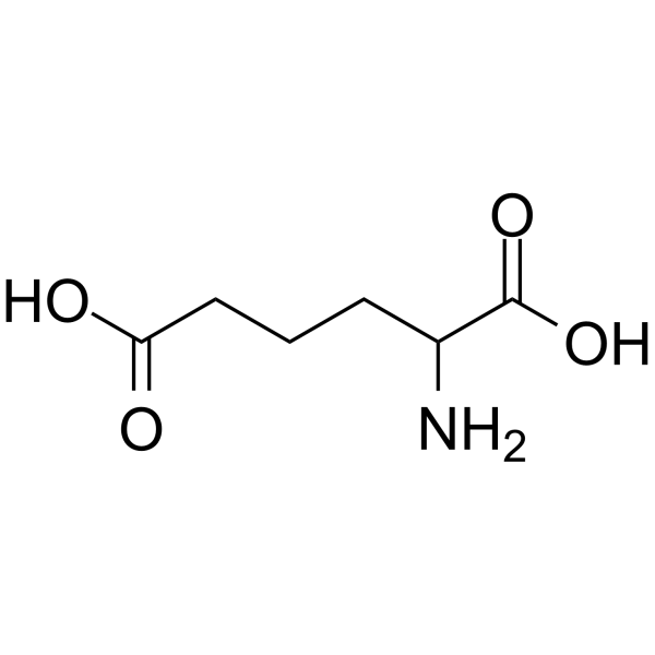
Physicochemical Properties
| Molecular Formula | C6H11NO4 |
| Molecular Weight | 161.15584 |
| Exact Mass | 161.068 |
| CAS # | 542-32-5 |
| PubChem CID | 469 |
| Appearance | White to off-white solid powder |
| Density | 1.3±0.1 g/cm3 |
| Boiling Point | 364.0±32.0 °C at 760 mmHg |
| Melting Point | 196-198ºC |
| Flash Point | 173.9±25.1 °C |
| Vapour Pressure | 0.0±1.7 mmHg at 25°C |
| Index of Refraction | 1.515 |
| LogP | -0.54 |
| Hydrogen Bond Donor Count | 3 |
| Hydrogen Bond Acceptor Count | 5 |
| Rotatable Bond Count | 5 |
| Heavy Atom Count | 11 |
| Complexity | 157 |
| Defined Atom Stereocenter Count | 0 |
| InChi Key | OYIFNHCXNCRBQI-UHFFFAOYSA-N |
| InChi Code | InChI=1S/C6H11NO4/c7-4(6(10)11)2-1-3-5(8)9/h4H,1-3,7H2,(H,8,9)(H,10,11) |
| Chemical Name | 2-aminohexanedioic acid |
| HS Tariff Code | 2934.99.9001 |
| Storage |
Powder-20°C 3 years 4°C 2 years In solvent -80°C 6 months -20°C 1 month Note: This product requires protection from light (avoid light exposure) during transportation and storage. |
| Shipping Condition | Room temperature (This product is stable at ambient temperature for a few days during ordinary shipping and time spent in Customs) |
Biological Activity
| Targets |
- Aminoadipic acid targets Müller cell gliosis (modulates glial fibrillary acidic protein, GFAP, expression) in retinal tissue [2] - Aminoadipic acid is a plasma biomarker for type 2 diabetes risk [1] |
| ln Vitro |
- In primary rat Müller cells, treatment with Aminoadipic acid (50–200 μM) for 24 h dose-dependently reduced lipopolysaccharide (LPS)-induced GFAP protein expression (by 35–68% vs. LPS group) (Western blot) and decreased GFAP-positive cell count (by 30–62%) (immunofluorescence). It also reduced LPS-induced IL-6 secretion (by 28–55%) in cell supernatants (ELISA) [2] - Higher plasma levels of Aminoadipic acid (≥0.8 μM) were associated with a 2.3-fold increased risk of developing type 2 diabetes over 10 years in non-diabetic participants [1] |
| ln Vivo |
- In a rat model of acute ocular hypertension (AOH), Aminoadipic acid (10 nmol/eye, intravitreal injection immediately post-AOH) reduced GFAP-positive Müller cell count in the inner retinal layer (by ~52% vs. AOH model), increased retinal ganglion cell (RGC) survival rate (by ~45% vs. AOH model), and preserved inner plexiform layer thickness (by ~38% vs. AOH model) at 7 days post-AOH [2] - In 2,484 non-diabetic participants, baseline plasma Aminoadipic acid levels >0.8 μM predicted incident diabetes (hazard ratio = 2.3, 95% CI: 1.5–3.5) independent of BMI and fasting glucose [1] |
| Cell Assay |
- Primary rat Müller cells were isolated from retinas, cultured to passage 3, and seeded in 6-well/24-well plates. Cells were pre-treated with Aminoadipic acid (50–200 μM) for 2 h, then stimulated with LPS (1 μg/mL) for 24 h. GFAP protein was detected by Western blot; GFAP-positive cells were counted via immunofluorescence (Alexa Fluor 488-conjugated secondary antibody, DAPI nuclear staining); IL-6 in supernatants was measured by ELISA [2] |
| Animal Protocol |
- 8-week-old male Sprague-Dawley rats (n=6/group) were anesthetized. AOH was induced by infusing saline into the anterior chamber to maintain IOP at 110 mmHg for 60 min. Aminoadipic acid (10 nmol/eye, dissolved in 5 μL PBS) was administered via intravitreal injection post-AOH; controls received 5 μL PBS. IOP was measured daily for 7 days. Retinas were dissected for GFAP detection (Western blot) and RGC labeling (immunofluorescence); ocular tissues were fixed for retinal thickness measurement (H&E staining) [2] |
| Toxicity/Toxicokinetics |
- Aminoadipic acid (up to 200 μM, 24 h) did not affect viability of primary rat Müller cells (MTT assay: viability >92% vs. control) [2] - Intravitreal injection of Aminoadipic acid (10 nmol/eye) in rats caused no ocular adverse effects (e.g., inflammation, retinal detachment) or systemic toxicity (body weight, ALT, creatinine comparable to controls) over 7 days [2] |
| References |
[1]. 2-Aminoadipic acid is a biomarker for diabetes risk. J Clin Invest. 2013 Oct;123(10):4309-17. [2]. α-Aminoadipic acid protects against retinal disruption through attenuating Müller cell gliosis in a rat model of acute ocular hypertension. Drug Des Devel Ther. 2016 Oct 20;10:3449-3457. |
| Additional Infomation |
2-aminoadipic acid is an alpha-amino acid that is adipic acid bearing a single amino substituent at position 2. An intermediate in the formation of lysine. It has a role as a mammalian metabolite and a Caenorhabditis elegans metabolite. It is an amino dicarboxylic acid, a non-proteinogenic alpha-amino acid and a dicarboxylic fatty acid. It is a conjugate acid of a 2-aminoadipate(2-). Aminoadipic acid is a metabolite found in or produced by Escherichia coli (strain K12, MG1655). 2-Aminohexanedioic acid has been reported in Drosophila melanogaster, Psophocarpus tetragonolobus, and other organisms with data available. Alpha-Aminoadipic Acid is an alpha-amino acid that is an intermediate in the lysine degradation pathway. It is synthesized from homoisocitrate and then converted to a semialdehyde that reacts with glutamic acid to form saccharopine. Alpha-aminoadipic acid levels are elevated in tissues of certain cancers and may potentially be used as a cancer biomarker. Aminoadipic acid (2-aminoadipate) is a metabolite in the principal biochemical pathway of lysine. It is an intermediate in the metabolism (i.e. breakdown or degradation) of lysine and saccharopine. It antagonizes neuroexcitatory activity modulated by the glutamate receptor, N-methyl-D-aspartate; (NMDA). Aminoadipic has also been shown to inhibit the production of kynurenic acid in brain tissue slices. Kynurenic acid is a broad spectrum excitatory amino acid receptor antagonist. Recent studies have shown that aminoadipic acid is elevated in prostate biopsy tissues from prostate cancer patients. Mutations in DHTKD1 (dehydrogenase E1 and transketolase domain-containing protein 1) have been shown to cause human 2-aminoadipic and 2-oxoadipic aciduria via impaired turnover of decarboxylation 2-oxoadipate to glutaryl-CoA, which is the last step in the lysine degradation pathway. Aging, diabetes, sepsis and renal failure are known to catalyze the oxidation of lysyl residues to 2-aminoadipic acid in human skin collagen and potentially other tissues. Proteolytic breakdown of these tissues can lead to the release of free 2-aminoadipic acid. Studies in rats indicate that aminoadipic acid (along with the 3 branched chain amino acid - Leu, Val and Ile) levels are elevated in the pre-diabetic phase and so aminoadipic acid may serve as a predictive biomarker for the development of diabetes. Long-term hyperglycemia of endothelial cells leads to elevated levels of aminoadipate which is though to be a sign of lysine breakdown through oxidative stress and reactive oxygen species (ROS). 2-aminoadipate is a potential small-molecule marker of oxidative stress. (A3343, A3344, A3345, A3346, A3347, A3348, A3349) A metabolite in the principal biochemical pathway of lysine. It antagonizes neuroexcitatory activity modulated by the glutamate receptor, N-METHYL-D-ASPARTATE; (NMDA). - Aminoadipic acid is an intermediate in lysine metabolism; elevated plasma levels reflect impaired lysine catabolism, correlating with insulin resistance and β-cell dysfunction (key drivers of type 2 diabetes) [1] - Aminoadipic acid inhibits Müller cell gliosis via downregulating GFAP and pro-inflammatory cytokines, preserving retinal structure and RGC survival—suggesting potential in retinal degenerative diseases (e.g., glaucoma) [2] |
Solubility Data
| Solubility (In Vitro) |
H2O : ~5 mg/mL (~31.03 mM) DMSO : ~2 mg/mL (~12.41 mM) |
| Solubility (In Vivo) |
Solubility in Formulation 1: 4 mg/mL (24.82 mM) in PBS (add these co-solvents sequentially from left to right, and one by one), clear solution; with heating and sonication. (Please use freshly prepared in vivo formulations for optimal results.) |
| Preparing Stock Solutions | 1 mg | 5 mg | 10 mg | |
| 1 mM | 6.2050 mL | 31.0251 mL | 62.0501 mL | |
| 5 mM | 1.2410 mL | 6.2050 mL | 12.4100 mL | |
| 10 mM | 0.6205 mL | 3.1025 mL | 6.2050 mL |