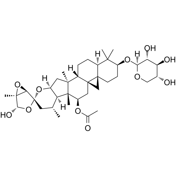
Actein is a naturally occurring triterpene glycoside isolated from the rhizomes of Cimicifuga foetida, acting by suppressing cell proliferation, inducing autophagy and apoptosis through promoting ROS/JNK activation, and blunting AKT pathway in human bladder cancer.
Physicochemical Properties
| Molecular Formula | C37H56O11 |
| Molecular Weight | 676.8340 |
| Exact Mass | 676.382 |
| CAS # | 18642-44-9 |
| PubChem CID | 10032468 |
| Appearance | White to off-white solid powder |
| Density | 1.4±0.1 g/cm3 |
| Melting Point | 246-250ºC |
| Index of Refraction | 1.610 |
| LogP | 5.67 |
| Hydrogen Bond Donor Count | 4 |
| Hydrogen Bond Acceptor Count | 11 |
| Rotatable Bond Count | 4 |
| Heavy Atom Count | 48 |
| Complexity | 1390 |
| Defined Atom Stereocenter Count | 19 |
| SMILES | C[C@@H]1C[C@@]2([C@H]3[C@](O3)([C@H](O2)O)C)O[C@@H]4[C@H]1[C@]5([C@@H](C[C@@]67C[C@@]68CC[C@@H](C([C@@H]8CC[C@H]7[C@@]5(C4)C)(C)C)O[C@H]9[C@@H]([C@H]([C@@H](CO9)O)O)O)OC(=O)C)C |
| InChi Key | NEWMWGLPJQHSSQ-PSDKAYTQSA-N |
| InChi Code | InChI=1S/C37H56O11/c1-17-12-37(29-34(7,47-29)30(42)48-37)46-20-13-32(5)22-9-8-21-31(3,4)23(45-28-27(41)26(40)19(39)15-43-28)10-11-35(21)16-36(22,35)14-24(44-18(2)38)33(32,6)25(17)20/h17,19-30,39-42H,8-16H2,1-7H3/t17-,19-,20+,21+,22+,23+,24-,25+,26+,27-,28+,29-,30+,32+,33-,34+,35-,36+,37-/m1/s1 |
| Chemical Name | [(1S,1'R,2S,3'R,4R,4'R,5R,5'R,6'R,10'S,12'S,13'S,16'R,18'S,21'R)-2-hydroxy-1,4',6',12',17',17'-hexamethyl-18'-[(2S,3R,4S,5R)-3,4,5-trihydroxyoxan-2-yl]oxyspiro[3,6-dioxabicyclo[3.1.0]hexane-4,8'-9-oxahexacyclo[11.9.0.01,21.04,12.05,10.016,21]docosane]-3'-yl] acetate |
| HS Tariff Code | 2934.99.9001 |
| Storage |
Powder-20°C 3 years 4°C 2 years In solvent -80°C 6 months -20°C 1 month Note: This product requires protection from light (avoid light exposure) during transportation and storage. |
| Shipping Condition | Room temperature (This product is stable at ambient temperature for a few days during ordinary shipping and time spent in Customs) |
Biological Activity
| ln Vitro |
Actein alone dose-dependently inhibited the viability of human gastric cancer cell lines SNU-216 and AGS, as shown by MTT assay. The half-maximal inhibitory concentration was not specified. [1] Actein (15 µM) alone induced minimal cleavage of Caspase-8, -9, and -3 in SNU-216 and AGS cells, as determined by Western blot. [1] Actein alone (15 µM) inhibited clonogenic growth of SNU-216 and AGS cells in colony formation assays. [1] Actein alone up-regulated p53 protein expression in SNU-216 and AGS cells in a dose- and time-dependent manner. [1] Actein alone significantly increased the protein levels of decoy receptors DcR1 and DcR2 in SNU-216 and AGS cells. [1] The combination of Actein (15 µM) and TRAIL (30 ng/mL) exhibited a synergistic effect, showing stronger anti-proliferative activity against SNU-216 and AGS cells than either agent alone in MTT assays. This combination showed no cytotoxicity on the normal human gastric epithelial cell line GES1. [1] The combination of Actein and TRAIL significantly increased apoptosis in SNU-216 and AGS cells compared to single-agent treatments, as measured by Annexin V-FITC/PI flow cytometry. [1] The combination of Actein and TRAIL significantly impeded the clonogenic growth of SNU-216 and AGS cells in colony formation assays. [1] Western blot analysis showed that the combination of Actein and TRAIL markedly increased the cleavage (activation) of Caspase-8, Caspase-9, Caspase-3, and PARP in SNU-216 and AGS cells. [1] The combination treatment decreased the protein levels of anti-apoptotic Bcl-2 family members (Bcl-2, Mcl-1) and increased the levels of pro-apoptotic members (Bad, Bak) in SNU-216 and AGS cells. [1] The combination of Actein and TRAIL enhanced Caspase-3 and Caspase-9 enzymatic activities in SNU-216 and AGS cells, which were abolished by specific Caspase inhibitors. [1] The combination of Actein and TRAIL up-regulated the protein levels of DcR1, DcR2, FADD, p73, and p53 more potently than Actein alone in SNU-216 and AGS cells. [1] Pre-treatment with the p53 inhibitor PFT-α reversed the up-regulation of p53, DcR1, DcR2, FADD, Cleaved Caspase-3, and Cleaved PARP induced by the Actein and TRAIL combination. It also decreased the apoptosis induced by the combination treatment. [1] |
| ln Vivo |
In a xenograft mouse model established by subcutaneous injection of SNU-216 cells, intraperitoneal administration of Actein (15 mg/kg) alone every two days for 21 days showed some inhibitory effect on tumor growth. [1] The combination of Actein (15 mg/kg, i.p.) and TRAIL (100 µg/mouse, i.p.) administered every two days for 21 days markedly inhibited tumor growth (volume and weight) in the SNU-216 xenograft model compared to monotherapy groups. [1] The combination therapy significantly increased the mRNA levels of DcR1, DcR2, FADD, and p53 in tumor tissues compared to the control group. [1] Immunohistochemical analysis of tumor tissues showed that the combination therapy increased the number of p53-positive cells and TUNEL-positive (apoptotic) cells, while decreasing the expression of the proliferation marker KI-67, compared to monotherapies. [1] |
| Enzyme Assay |
Caspase-3 and Caspase-9 activities were determined using colorimetric activity assay kits. Cells were lysed and centrifuged. The supernatant was incubated with specific chromogenic peptide substrates (DEVD-pNA for Caspase-3 and LEHD-pNA for Caspase-9) at 37°C for 2 hours. The cleavage of substrates was measured by absorbance at 405 nm using an ELISA reader. [1] |
| Cell Assay |
For MTT assay, 5 x 10^3 cells were seeded per well in a 96-well plate. After 24 hours, cells were treated with Actein (0–30 µM), TRAIL (0–60 ng/mL), or their combination for 24 hours. MTT reagent was then added, and the absorbance was measured at 570 nm to determine cell viability. [1] For colony formation assay, 300 cells were seeded on a 60 mm culture plate. After 48 hours, fresh medium containing Actein, TRAIL, or their combination was added for 48 hours. Then cells were washed and incubated in drug-free medium (changed every 5 days) for an additional 2 weeks. Colonies were fixed with methanol, stained with Giemsa, and counted (groupings of ≥30 cells) under a microscope. [1] For apoptosis analysis by flow cytometry, cells were treated, harvested, and stained using an Annexin V-FITC/PI kit according to the manufacturer's instructions. Stained cells were resuspended in binding buffer and analyzed immediately by flow cytometry. [1] For Western blot analysis, treated cells were collected, lysed in hypotonic buffer, and centrifuged. Protein concentration was determined. Equal amounts of protein were separated by SDS-PAGE, transferred to a membrane, and probed with specific primary and secondary antibodies for detection. [1] For RT-qPCR analysis, total RNA was extracted from cells or tissues, quantified, and reverse transcribed. mRNA levels of target genes were measured using specific primers and calculated relative to an endogenous control (cyclophilin or GAPDH) using the 2^(-ΔΔCt) method. [1] |
| Animal Protocol |
A xenograft tumor model was established by subcutaneously injecting 5 x 10^5 SNU-216 gastric cancer cells into the dorsal flanks of 6-week-old male athymic nude mice. [1] Mice were randomly divided into four groups: Control (normal saline, i.p.), Actein alone (15 mg/kg, i.p.), TRAIL alone (100 µg per mouse, i.p.), and Actein + TRAIL combination. [1] Treatments were administered via intraperitoneal injection every two days for a total of 21 days. [1] Tumor volume was measured every three days by calculating two maximum perpendicular diameters. At the end of the study, mice were sacrificed, tumors were harvested and weighed. Kidneys and livers were also collected for histopathological examination. [1] |
| Toxicity/Toxicokinetics |
In vitro, Actein (up to 30 µM) and TRAIL (up to 60 ng/mL), alone or in combination, showed no significant cytotoxicity on the normal human gastric epithelial cell line GES1 in MTT assays. [1] In vivo, no significant change in body weight was observed among mice treated with Actein alone, TRAIL alone, or their combination compared to the control group. [1] Histological examination (H&E staining) of kidney and liver tissues from all treatment groups showed normal architecture, indicating no apparent organ toxicity. [1] |
| References |
[1]. Actein enhances TRAIL effects on suppressing gastric cancer progression by activating p53/Caspase-3 signaling. Biochem Biophys Res Commun. 2018 Mar 18;497(4):1177-1183. [2]. Actein induces autophagy and apoptosis in human bladder cancer by potentiating ROS/JNK and inhibiting AKT pathways. Oncotarget. 2017 Nov 1;8(68):112498-112515. |
| Additional Infomation |
Actein is a triterpenoid. It has a role as a metabolite. Actein has been reported in Actaea elata, Actaea cimicifuga, and other organisms with data available. See also: Black Cohosh (part of). Actein is a tetracyclic triterpenoid glycoside compound isolated from the rhizome of Cimicifuga foetida. [1] It has been reported to have inhibitory effects on breast cancer cells and can synergize with other chemotherapeutic agents. [1] This study is the first to investigate the effects of Actein on gastric cancer both in vitro and in vivo. [1] The study demonstrates that Actein sensitizes TRAIL-resistant human gastric cancer cells to TRAIL-induced apoptosis, at least in part, by activating the p53 signaling pathway, leading to upregulation of death receptors (DcR1/DcR2) and the downstream apoptosis cascade. [1] The combination of Actein and TRAIL presents a potential therapeutic strategy against gastric cancer. [1] |
Solubility Data
| Solubility (In Vitro) | DMSO : ~100 mg/mL (~147.75 mM) |
| Solubility (In Vivo) |
Note: Listed below are some common formulations that may be used to formulate products with low water solubility (e.g. < 1 mg/mL), you may test these formulations using a minute amount of products to avoid loss of samples. Injection Formulations (e.g. IP/IV/IM/SC) Injection Formulation 1: DMSO : Tween 80: Saline = 10 : 5 : 85 (i.e. 100 μL DMSO stock solution → 50 μL Tween 80 → 850 μL Saline) *Preparation of saline: Dissolve 0.9 g of sodium chloride in 100 mL ddH ₂ O to obtain a clear solution. Injection Formulation 2: DMSO : PEG300 :Tween 80 : Saline = 10 : 40 : 5 : 45 (i.e. 100 μL DMSO → 400 μLPEG300 → 50 μL Tween 80 → 450 μL Saline) Injection Formulation 3: DMSO : Corn oil = 10 : 90 (i.e. 100 μL DMSO → 900 μL Corn oil) Example: Take the Injection Formulation 3 (DMSO : Corn oil = 10 : 90) as an example, if 1 mL of 2.5 mg/mL working solution is to be prepared, you can take 100 μL 25 mg/mL DMSO stock solution and add to 900 μL corn oil, mix well to obtain a clear or suspension solution (2.5 mg/mL, ready for use in animals). Injection Formulation 4: DMSO : 20% SBE-β-CD in saline = 10 : 90 [i.e. 100 μL DMSO → 900 μL (20% SBE-β-CD in saline)] *Preparation of 20% SBE-β-CD in Saline (4°C,1 week): Dissolve 2 g SBE-β-CD in 10 mL saline to obtain a clear solution. Injection Formulation 5: 2-Hydroxypropyl-β-cyclodextrin : Saline = 50 : 50 (i.e. 500 μL 2-Hydroxypropyl-β-cyclodextrin → 500 μL Saline) Injection Formulation 6: DMSO : PEG300 : castor oil : Saline = 5 : 10 : 20 : 65 (i.e. 50 μL DMSO → 100 μLPEG300 → 200 μL castor oil → 650 μL Saline) Injection Formulation 7: Ethanol : Cremophor : Saline = 10: 10 : 80 (i.e. 100 μL Ethanol → 100 μL Cremophor → 800 μL Saline) Injection Formulation 8: Dissolve in Cremophor/Ethanol (50 : 50), then diluted by Saline Injection Formulation 9: EtOH : Corn oil = 10 : 90 (i.e. 100 μL EtOH → 900 μL Corn oil) Injection Formulation 10: EtOH : PEG300:Tween 80 : Saline = 10 : 40 : 5 : 45 (i.e. 100 μL EtOH → 400 μLPEG300 → 50 μL Tween 80 → 450 μL Saline) Oral Formulations Oral Formulation 1: Suspend in 0.5% CMC Na (carboxymethylcellulose sodium) Oral Formulation 2: Suspend in 0.5% Carboxymethyl cellulose Example: Take the Oral Formulation 1 (Suspend in 0.5% CMC Na) as an example, if 100 mL of 2.5 mg/mL working solution is to be prepared, you can first prepare 0.5% CMC Na solution by measuring 0.5 g CMC Na and dissolve it in 100 mL ddH2O to obtain a clear solution; then add 250 mg of the product to 100 mL 0.5% CMC Na solution, to make the suspension solution (2.5 mg/mL, ready for use in animals). Oral Formulation 3: Dissolved in PEG400 Oral Formulation 4: Suspend in 0.2% Carboxymethyl cellulose Oral Formulation 5: Dissolve in 0.25% Tween 80 and 0.5% Carboxymethyl cellulose Oral Formulation 6: Mixing with food powders Note: Please be aware that the above formulations are for reference only. InvivoChem strongly recommends customers to read literature methods/protocols carefully before determining which formulation you should use for in vivo studies, as different compounds have different solubility properties and have to be formulated differently. (Please use freshly prepared in vivo formulations for optimal results.) |
| Preparing Stock Solutions | 1 mg | 5 mg | 10 mg | |
| 1 mM | 1.4775 mL | 7.3874 mL | 14.7748 mL | |
| 5 mM | 0.2955 mL | 1.4775 mL | 2.9550 mL | |
| 10 mM | 0.1477 mL | 0.7387 mL | 1.4775 mL |