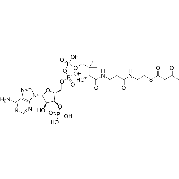
Physicochemical Properties
| Molecular Formula | C25H40N7O18P3S |
| Exact Mass | 851.136 |
| CAS # | 1420-36-6 |
| PubChem CID | 92153 |
| Appearance | Typically exists as solid at room temperature |
| LogP | 0.006 |
| Hydrogen Bond Donor Count | 9 |
| Hydrogen Bond Acceptor Count | 23 |
| Rotatable Bond Count | 22 |
| Heavy Atom Count | 54 |
| Complexity | 1490 |
| Defined Atom Stereocenter Count | 5 |
| SMILES | CC(CC(SCCNC(CCNC([C@H](C(COP(OP(OC[C@@H]1O[C@@H](N2C=NC3=C(N=CN=C23)N)[C@H](O)[C@H]1OP(=O)(O)O)(O)=O)(O)=O)(C)C)O)=O)=O)=O)=O |
| InChi Key | OJFDKHTZOUZBOS-CITAKDKDSA-N |
| InChi Code | InChI=1S/C25H40N7O18P3S/c1-13(33)8-16(35)54-7-6-27-15(34)4-5-28-23(38)20(37)25(2,3)10-47-53(44,45)50-52(42,43)46-9-14-19(49-51(39,40)41)18(36)24(48-14)32-12-31-17-21(26)29-11-30-22(17)32/h11-12,14,18-20,24,36-37H,4-10H2,1-3H3,(H,27,34)(H,28,38)(H,42,43)(H,44,45)(H2,26,29,30)(H2,39,40,41)/t14-,18-,19-,20+,24-/m1/s1 |
| Chemical Name | S-[2-[3-[[(2R)-4-[[[(2R,3S,4R,5R)-5-(6-aminopurin-9-yl)-4-hydroxy-3-phosphonooxyoxolan-2-yl]methoxy-hydroxyphosphoryl]oxy-hydroxyphosphoryl]oxy-2-hydroxy-3,3-dimethylbutanoyl]amino]propanoylamino]ethyl] 3-oxobutanethioate |
| Synonyms | acetoacetyl-CoA; Acetoacetyl coenzyme A; Acetoacetyl coa; 3-acetoacetyl-CoA; 1420-36-6; S-Acetoacetyl-CoA; S-acetoacetyl-coenzyme A; acetoacetyl-S-CoA; |
| HS Tariff Code | 2934.99.9001 |
| Storage |
Powder-20°C 3 years 4°C 2 years In solvent -80°C 6 months -20°C 1 month |
| Shipping Condition | Room temperature (This product is stable at ambient temperature for a few days during ordinary shipping and time spent in Customs) |
Biological Activity
| Targets | Human Endogenous Metabolite |
| ln Vitro | With a substrate specificity of 2.6 μM and 5 μM, respectively, the refined recombinant Msed_0389 and Msed_1423 catalyze the NAD-dependent oxidation of (S)-3-hydroxybutyryl-CoA to Acetoacetyl-CoA[2]. When it comes to autotrophic Desulfurococcales (Ignicoccus hospitalis) and Thermoproteales (Pyrobaculum neutrophilus) that fix CO2 using the dicarboxylate/4-hydroxybutyrate cycle, the homologous bifunctional protein is the only enzyme that can convert crotonyl-CoA into Acetoacetyl-CoA[2]. |
| ln Vivo | Autotrophic Crenarchaeota use two different cycles for carbon dioxide fixation. Members of the Sulfolobales use the 3-hydroxypropionate/4-hydroxybutyrate (HP/HB) cycle, whereas Desulfurococcales and Thermoproteales use the dicarboxylate/4-hydroxybutyrate cycle. While these two cycles differ in the carboxylation reactions resulting in the conversion of acetyl-CoA + 2 CO2 to succinyl-CoA, they have a common regeneration part in which succinyl-CoA is reconverted to two acetyl-CoA molecules. This common part includes crotonyl-CoA conversion to acetoacetyl-CoA, which has unequivocally been shown in Ignicoccus hospitalis (Desulfurococcales) and Pyrobaculum neutrophilus (Thermoproteales) to be catalyzed by a bifunctional crotonase/3-hydroxybutyryl-CoA dehydrogenase. It is a fusion protein consisting of an enoyl-CoA hydratase and a dehydrogenase domain. As the homologous bifunctional protein is present in Sulfolobales as well, its common functioning in the conversion of crotonyl-CoA to acetoacetyl-CoA was proposed. Here we show that a model autotrophic member of Sulfolobales, Metallosphaera sedula, possesses in addition to the bifunctional protein (Msed_0399) several separate genes coding for crotonyl-CoA hydratase and (S)-3-hydroxybutyryl-CoA dehydrogenase. Their genes were previously shown to be transcribed under autotrophic and mixotrophic conditions. The dehydrogenase Msed_1423 (and not the bifunctional protein Msed_0399) appears to be the main enzyme catalyzing the (S)-3-hydroxybutyryl-CoA dehydrogenase reaction. Homologs of this dehydrogenase are the only (S)-3-hydroxybutyryl-CoA dehydrogenases present in all autotrophic Sulfolobales, strengthening this conclusion. Two uncharacterized crotonase homologs present in M. sedula genome (Msed_0336 and Msed_0384) were heterologously produced and characterized. Both proteins were highly efficient crotonyl-CoA hydratases and may contribute (or be responsible) for the corresponding reaction in the HP/HB cycle in vivo [2]. |
| References |
[1]. Miziorko HM. Enzymes of the mevalonate pathway of isoprenoid biosynthesis. Arch Biochem Biophys. 2011 Jan 15;505(2):131-43. [2]. Enzymes Catalyzing Crotonyl-CoA Conversion to Acetoacetyl-CoA During the Autotrophic CO2 Fixation in Metallosphaera sedula. Front Microbiol. 2020 Mar 11;11:354. [3]. Enzymes of fatty acid metabolism. Biochimica et Biophysica Acta. Volume 12, Issues 1-2, October 1953, Pages 299-314. |
| Additional Infomation |
Acetoacetyl-CoA is a 3-oxoacyl-CoA that results from the formal condensation of the thiol group of coenzyme A with the carboxy group of acetoacetic acid. It has a role as an Escherichia coli metabolite and a mouse metabolite. It is functionally related to a butyryl-CoA and an acetoacetic acid. It is a conjugate acid of an acetoacetyl-CoA(4-). Acetoacetyl-CoA is a metabolite found in or produced by Escherichia coli (strain K12, MG1655). acetoacetyl-CoA has been reported in Homo sapiens and Bos taurus with data available. acetoacetyl-CoA is a metabolite found in or produced by Saccharomyces cerevisiae. The mevalonate pathway accounts for conversion of acetyl-CoA to isopentenyl 5-diphosphate, the versatile precursor of polyisoprenoid metabolites and natural products. The pathway functions in most eukaryotes, archaea, and some eubacteria. Only recently has much of the functional and structural basis for this metabolism been reported. The biosynthetic acetoacetyl-CoA thiolase and HMG-CoA synthase reactions rely on key amino acids that are different but are situated in active sites that are similar throughout the family of initial condensation enzymes. Both bacterial and animal HMG-CoA reductases have been extensively studied and the contrasts between these proteins and their interactions with statin inhibitors defined. The conversion of mevalonic acid to isopentenyl 5-diphosphate involves three ATP-dependent phosphorylation reactions. While bacterial enzymes responsible for these three reactions share a common protein fold, animal enzymes differ in this respect as the recently reported structure of human phosphomevalonate kinase demonstrates. There are significant contrasts between observations on metabolite inhibition of mevalonate phosphorylation in bacteria and animals. The structural basis for these contrasts has also recently been reported. Alternatives to the phosphomevalonate kinase and mevalonate diphosphate decarboxylase reactions may exist in archaea. Thus, new details regarding isopentenyl diphosphate synthesis from acetyl-CoA continue to emerge. [1] The intermediates in the biological breakdown and synthesis of fatty acids are S-acyl derivatives of coenzyme A. Fatty acid synthesis is accomplished through repetition of a cycle of four consecutive reactions: a. Condensation of two molecules of acetyl CoA to form acetoacetyl CoA and coenzyme A (CoASH); b. reduction of acetoacetyl CoA to β-hydroxybutyryl CoA; c. dehydration of β-hydroxybutyryl CoA to crotonyl CoA, and d. reduction of crotonyl CoA to butyryl CoA. A new cycle is started by the reaction of butyryl CoA with another molecule of acetyl CoA to form β-keto-caproyl CoA + CoA-SH, and so forth. The cycle is repeated eight times until stearyl CoA is formed. All four reactions of the fatty acid cycle are reversible and fatty acid oxidation, once the fatty acid is activated through conversion to the corresponding S-acyl CoA derivative, proceeds by a reversal of the above sequence. There are two main mechanisms for activation of fatty acids: (a) By a reaction with ATP and CoA to form S-acyl CoA, adenosine monophosphate and pyrophosphate, and (b) by transfer of CoA from certain acyl CoA compounds such as acetyl CoA or succinyl CoA. The isolation and identification of some of the key enzymes of fatty acid metabolism is outlined and their mechanism of action discussed.[3] |
Solubility Data
| Solubility (In Vitro) | May dissolve in DMSO (in most cases), if not, try other solvents such as H2O, Ethanol, or DMF with a minute amount of products to avoid loss of samples |
| Solubility (In Vivo) |
Note: Listed below are some common formulations that may be used to formulate products with low water solubility (e.g. < 1 mg/mL), you may test these formulations using a minute amount of products to avoid loss of samples. Injection Formulations (e.g. IP/IV/IM/SC) Injection Formulation 1: DMSO : Tween 80: Saline = 10 : 5 : 85 (i.e. 100 μL DMSO stock solution → 50 μL Tween 80 → 850 μL Saline) *Preparation of saline: Dissolve 0.9 g of sodium chloride in 100 mL ddH ₂ O to obtain a clear solution. Injection Formulation 2: DMSO : PEG300 :Tween 80 : Saline = 10 : 40 : 5 : 45 (i.e. 100 μL DMSO → 400 μLPEG300 → 50 μL Tween 80 → 450 μL Saline) Injection Formulation 3: DMSO : Corn oil = 10 : 90 (i.e. 100 μL DMSO → 900 μL Corn oil) Example: Take the Injection Formulation 3 (DMSO : Corn oil = 10 : 90) as an example, if 1 mL of 2.5 mg/mL working solution is to be prepared, you can take 100 μL 25 mg/mL DMSO stock solution and add to 900 μL corn oil, mix well to obtain a clear or suspension solution (2.5 mg/mL, ready for use in animals). Injection Formulation 4: DMSO : 20% SBE-β-CD in saline = 10 : 90 [i.e. 100 μL DMSO → 900 μL (20% SBE-β-CD in saline)] *Preparation of 20% SBE-β-CD in Saline (4°C,1 week): Dissolve 2 g SBE-β-CD in 10 mL saline to obtain a clear solution. Injection Formulation 5: 2-Hydroxypropyl-β-cyclodextrin : Saline = 50 : 50 (i.e. 500 μL 2-Hydroxypropyl-β-cyclodextrin → 500 μL Saline) Injection Formulation 6: DMSO : PEG300 : castor oil : Saline = 5 : 10 : 20 : 65 (i.e. 50 μL DMSO → 100 μLPEG300 → 200 μL castor oil → 650 μL Saline) Injection Formulation 7: Ethanol : Cremophor : Saline = 10: 10 : 80 (i.e. 100 μL Ethanol → 100 μL Cremophor → 800 μL Saline) Injection Formulation 8: Dissolve in Cremophor/Ethanol (50 : 50), then diluted by Saline Injection Formulation 9: EtOH : Corn oil = 10 : 90 (i.e. 100 μL EtOH → 900 μL Corn oil) Injection Formulation 10: EtOH : PEG300:Tween 80 : Saline = 10 : 40 : 5 : 45 (i.e. 100 μL EtOH → 400 μLPEG300 → 50 μL Tween 80 → 450 μL Saline) Oral Formulations Oral Formulation 1: Suspend in 0.5% CMC Na (carboxymethylcellulose sodium) Oral Formulation 2: Suspend in 0.5% Carboxymethyl cellulose Example: Take the Oral Formulation 1 (Suspend in 0.5% CMC Na) as an example, if 100 mL of 2.5 mg/mL working solution is to be prepared, you can first prepare 0.5% CMC Na solution by measuring 0.5 g CMC Na and dissolve it in 100 mL ddH2O to obtain a clear solution; then add 250 mg of the product to 100 mL 0.5% CMC Na solution, to make the suspension solution (2.5 mg/mL, ready for use in animals). Oral Formulation 3: Dissolved in PEG400 Oral Formulation 4: Suspend in 0.2% Carboxymethyl cellulose Oral Formulation 5: Dissolve in 0.25% Tween 80 and 0.5% Carboxymethyl cellulose Oral Formulation 6: Mixing with food powders Note: Please be aware that the above formulations are for reference only. InvivoChem strongly recommends customers to read literature methods/protocols carefully before determining which formulation you should use for in vivo studies, as different compounds have different solubility properties and have to be formulated differently. (Please use freshly prepared in vivo formulations for optimal results.) |