AZ-876 is a novel, potent high-affinity agonist of Liver X Receptor (LXR) with Ki values of 0.007 μM and 0.011 μM for human LXRα and LXRβ respectively. AZ876 protects against pathological cardiac hypertrophy and fibrosis without lipogenic side effects. LXR activation with AZ876 attenuated this increase, and significantly reduced TAC-induced increases in heart weight, myocardial fibrosis, and cardiac dysfunction without affecting blood pressure. Liver X receptors (LXRs) transcriptionally regulate inflammation, metabolism, and immunity.
Physicochemical Properties
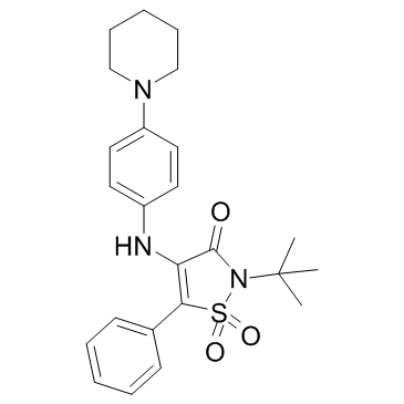
| Molecular Formula | C24H29N3O3S |
| Molecular Weight | 439.5704 |
| Exact Mass | 439.192 |
| CAS # | 898800-26-5 |
| PubChem CID | 11655079 |
| Appearance | Yellow to orange solid powder |
| Density | 1.3±0.1 g/cm3 |
| Boiling Point | 605.3±65.0 °C at 760 mmHg |
| Flash Point | 319.9±34.3 °C |
| Vapour Pressure | 0.0±1.7 mmHg at 25°C |
| Index of Refraction | 1.642 |
| LogP | 3.27 |
| Hydrogen Bond Donor Count | 1 |
| Hydrogen Bond Acceptor Count | 5 |
| Rotatable Bond Count | 5 |
| Heavy Atom Count | 31 |
| Complexity | 789 |
| Defined Atom Stereocenter Count | 0 |
| InChi Key | IVANYIPLGFVBGR-UHFFFAOYSA-N |
| InChi Code | InChI=1S/C24H29N3O3S/c1-24(2,3)27-23(28)21(22(31(27,29)30)18-10-6-4-7-11-18)25-19-12-14-20(15-13-19)26-16-8-5-9-17-26/h4,6-7,10-15,25H,5,8-9,16-17H2,1-3H3 |
| Chemical Name | 2-(1,1-Dimethylethyl)-5-phenyl-4-[[4-(1-piperidinyl)phenyl]amino]-3(2H)-isothiazolone 1,1-dioxide |
| Synonyms | AZ876; AZ 876; AZ-876, AZ12260493; AZ 12260493; AZ-12260493. |
| HS Tariff Code | 2934.99.9001 |
| Storage |
Powder-20°C 3 years 4°C 2 years In solvent -80°C 6 months -20°C 1 month |
| Shipping Condition | Room temperature (This product is stable at ambient temperature for a few days during ordinary shipping and time spent in Customs) |
Biological Activity
| Targets | In isolated cardiac myocytes and fibroblasts, immunocytochemistry confirmed nuclear expression of LXRα in both these cell types. In cardiomyocytes, phenylephrine-stimulated cellular hypertrophy was significantly decreased in AZ876-treated cells. In cardiac fibroblasts, AZ876 prevented TGFβ- and angiotensin II-induced fibroblast collagen synthesis, and inhibited up-regulation of the myofibroblastic marker, α-smooth muscle actin. Plasma triglycerides and liver weight were unaltered following AZ876 treatment.[1] |
| ln Vitro |
In isolated cardiac myocytes and fibroblasts, immunocytochemistry confirmed nuclear expression of LXRα in both these cell types. In cardiomyocytes, phenylephrine-stimulated cellular hypertrophy was significantly decreased in AZ876-treated cells. In cardiac fibroblasts, AZ876 prevented TGFβ- and angiotensin II-induced fibroblast collagen synthesis, and inhibited up-regulation of the myofibroblastic marker, α-smooth muscle actin. Plasma triglycerides and liver weight were unaltered following AZ876 treatment.[1] In scintillation proximity binding assays, AZ876 was 25-fold and 2.5-fold more potent than GW3965 in displacing a radioligand from human LXRα and LXRβ proteins, respectively. [2] In reporter gene transactivation assays using U2/OS cells co-transfected with Gal4-chimeric LXR constructs, AZ876 was 196-fold and 5-fold more potent than GW3965 on human LXRα and LXRβ, respectively. It was also 248-fold and 10.5-fold more potent on mouse LXRα and LXRβ, respectively. [2] AZ876 increased the mRNA expression of the reverse cholesterol transport (RCT) gene ABCA1 in hamster and human peripheral blood polymorphonuclear cells in vitro, being 4- to 7-fold more potent than GW3965. [2] |
| ln Vivo |
Cardiac hypertrophy was induced in C57Bl6/J mice via transverse aortic constriction (TAC) for 6 weeks. During this period, mice received chow supplemented or not with AZ876 (20 µmol/kg/day). In murine hearts, LXRα protein expression was up-regulated ∼7-fold in response to TAC. LXR activation with AZ876 attenuated this increase, and significantly reduced TAC-induced increases in heart weight, myocardial fibrosis, and cardiac dysfunction without affecting blood pressure. At the molecular level, AZ876 suppressed up-regulation of hypertrophy- and fibrosis-related genes, and further inhibited prohypertrophic and profibrotic transforming growth factor β (TGFβ)-Smad2/3 signalling. [1] APOE*3Leiden mice were fed an atherogenic diet alone or supplemented with either AZ876 (5 or 20µmol·kg(-1) ·day(-1) ) for 20 weeks. Total cholesterol and triglyceride levels were measured using commercial kits. Plasma cytokines were determined by using bead-based multiplex suspension array kits with the Luminex technology. Atherosclerosis was assessed histochemically and lesion composition was assessed by immunohistochemical methods. Low-dose AZ876 had no effect on plasma , whereas high-dose AZ876 increased plasma triglycerides (+110%) and reduced cholesterol (-16%) compared with controls. Low-dose AZ876 reduced lesion area (-47%); and high-dose AZ876 strongly decreased lesion area (-91%), lesion number (-59%) and severity. In either dose, AZ876 did not affect lesion composition. High-dose AZ876 , but not low-dose AZ876, reduced inflammation as reflected by lower cytokine levels and vessel wall activation.[2] In hyperlipidemic APOE3Leiden transgenic mice fed an atherogenic diet for 20 weeks, a low dose of AZ876 (5 µmol·kg⁻¹·day⁻¹) reduced atherosclerotic lesion area in the aortic root by 47% compared to controls, without affecting plasma or liver triglyceride levels. [2] A high dose of AZ876 (20 µmol·kg⁻¹·day⁻¹) strongly decreased atherosclerotic lesion area (-91%), lesion number (-59%), and the abundance of severe lesions. This dose also increased plasma HDL levels (appearance of large HDL-1 particles) and reduced plasma VLDL-cholesterol. [2] The high dose of AZ876 (20 µmol·kg⁻¹·day⁻¹), but not the low dose, reduced vascular inflammation as evidenced by decreased plasma levels of cytokines (TNF-α, IL-1β) and reduced monocyte adherence to the aortic endothelium. [2] In a separate in vivo reverse cholesterol transport (RCT) assay in C57BL/6J mice, AZ876 (20 µmol·kg⁻¹·day⁻¹) administered for 8 days increased the recovery of radiolabeled cholesterol from injected macrophages in plasma (total lipids +75%, free cholesterol +100%) and feces (total lipids +94%, free cholesterol +195%) compared to vehicle controls. [2] Treatment with the high dose of AZ876 (20 µmol·kg⁻¹·day⁻¹) increased mRNA expression of the RCT genes Abca1 and Abcg1 in the aorta of APOE3Leiden mice. The low dose showed a tendency to increase their expression. [2] The high dose of AZ876 (20 µmol·kg⁻¹·day⁻¹) increased plasma triglycerides by 110% and liver triglyceride content by 53%, and raised plasma alanine aminotransferase (ALT) levels, indicating hepatic steatosis and potential liver damage. The low dose had no such effects. [2] AZ876 did not affect the collagen or smooth muscle cell content of atherosclerotic lesions, whereas GW3965 reduced collagen content, suggesting AZ876 treatment resulted in a more stable lesion phenotype at an equimolar dose. [2] |
| Enzyme Assay |
Binding assays for LXRα and LXRβ were performed using purified ligand-binding domain proteins expressed in E. coli. The assay mixture contained assay buffer, polylysine-coated scintillation proximity assay (SPA) beads, LXR protein, a fixed concentration of tritium-labeled reference ligand (T0901317), and the test compound in a 10-point dose-response dilution. The mixture was shaken gently for 2 hours, allowed to settle for 1 hour, and then counted to determine displacement of the radioligand. [2] Reporter gene transactivation assays were conducted to measure LXR activation. Vectors containing the ligand-binding domain of human or mouse LXRα or LXRβ fused to the yeast Gal4 DNA-binding domain were co-transfected with a luciferase reporter plasmid containing Gal4 recognition sites into U2/OS osteosarcoma cells. After transfection, ligands were added in a 10-point dose-response curve. Luciferase activity was measured 48 hours later to determine transcriptional activation potency (EC50). [2] |
| Cell Assay |
An in vitro macrophage reverse cholesterol transport assay was performed using J774A1 macrophages. The macrophages were cultured and loaded with radiolabeled [³H]cholesterol by incubation with acetylated LDL and BSA for 48 hours. After washing, the cholesterol-loaded, radiolabeled macrophages were injected into mice for the in vivo RCT experiment. The viability of cells before injection was estimated using Trypan blue staining. [2] Quantitative real-time PCR (qPCR) was used to measure gene expression in mouse tissues. Total RNA was extracted from tissues, treated with DNase, and reverse transcribed into cDNA. Gene expression levels of targets like Abca1 and Abcg1 were quantified using specific FAM/TAMRA-labeled probes on a real-time PCR system. Expression data were normalized to an internal control gene (mouse acidic ribosomal phosphoprotein P0, m36B4), and relative expression was calculated using the 2^(-ΔΔCt) method. [2] |
| Animal Protocol |
AZ876 (20 µmol·kg−1·day−1,∼9·mg·kg−1·day−1)by oral gavage twice daily for 8 days[2] After matching into four groups, based on age, plasma cholesterol and triglyceride levels, the mice received Western-type diet either alone (control group) or supplemented with AZ876 in a low dose (5 µmol·kg−1·day−1; ∼2 mg·kg−1·day−1) or high dose (20 µmol·kg−1·day−1; ∼9 mg·kg−1·day−1) or with GW3965 (17 µmol·kg−1·day−1; ∼10 mg·kd−1·day−1) (Joseph et al., 2002). Initially, the high dose of AZ876 was 10 µmol·kg−1·day−1; however, after 4 weeks of treatment, GW3965 induced hypertriglyceridaemia but AZ876 (10 µmol·kg−1·day−1) did not. Therefore, it was decided to increase the dose of AZ876 to 20 µmol·kg−1·day−1. After 20 weeks, mice were killed and the hearts, aortic root, livers and small intestine (duodenum) were isolated.[2] Study 1 - Macrophage Reverse Cholesterol Transport (RCT): Body weight-matched 7-week-old male C57BL/6J mice were treated with vehicle (amorphous nanoparticles with 3.4% dimethylacetamide), AZ876 (20 µmol·kg⁻¹·day⁻¹, ~9 mg·kg⁻¹·day⁻¹), or GW3965 (34 µmol·kg⁻¹·day⁻¹, ~20 mg·kg⁻¹·day⁻¹) by oral gavage twice daily for 8 days. On day 6, each mouse was injected intraperitoneally with a suspension of [³H]cholesterol-labeled J774A1 macrophages. Feces were collected 24-48 hours post-injection. At 48 hours post-injection, mice were anesthetized, plasma was collected, and livers were perfused and collected. Lipids were extracted from feces, liver, and plasma, and radiolabel content in different lipid fractions was quantified. [2] Study 2 - Atherosclerosis in APOE3Leiden Mice: Female heterozygous APOE3Leiden transgenic mice (~13 weeks old) were fed a semi-synthetic Western-type diet (high in cholesterol and fat) for a 3-week run-in period. Mice were then matched into groups based on age and plasma lipids and received the same diet alone (control) or supplemented with AZ876 at a low dose (5 µmol·kg⁻¹·day⁻¹, ~2 mg·kg⁻¹·day⁻¹), a high dose (20 µmol·kg⁻¹·day⁻¹, ~9 mg·kg⁻¹·day⁻¹; initially 10 µmol·kg⁻¹·day⁻¹ for 4 weeks, then increased), or GW3965 (17 µmol·kg⁻¹·day⁻¹, ~10 mg·kg⁻¹·day⁻¹) for 20 weeks. Compounds were mixed into the diet. After 20 weeks, mice were euthanized, and hearts (with aortic root), livers, and small intestines were collected for analysis. Blood was collected after a 4-hour fast at various time points for plasma analysis. [2] |
| ADME/Pharmacokinetics |
The study measured markers related to cholesterol metabolism rather than classical PK parameters. Serum lanosterol levels, a marker of cholesterol synthesis, were increased by 310% in APOE3Leiden mice treated with the high dose of AZ876 (20 µmol·kg⁻¹·day⁻¹). [2] The absorption of cholesterol, as indicated by serum β-sitosterol levels, was not affected by AZ876 treatment. [2] Faecal output of neutral sterols (cholesterol) and bile acids was not significantly changed by AZ876 treatment. [2] |
| Toxicity/Toxicokinetics |
In APOE3Leiden mice, the high dose of AZ876 (20 µmol·kg⁻¹·day⁻¹) for 20 weeks induced hepatic steatosis, as evidenced by a 53% increase in liver triglyceride content and a 29% increase in liver weight. [2] Concomitant with hepatic steatosis, plasma alanine aminotransferase (ALT) levels were increased by 114% in the high-dose AZ876 group, indicating potential liver damage. [2] The high dose of AZ876 also induced hypertriglyceridemia, elevating plasma triglyceride levels by 110%. [2] The low dose of AZ876 (5 µmol·kg⁻¹·day⁻¹) did not cause significant changes in liver triglycerides, liver weight, plasma ALT, or plasma triglycerides. [2] |
| References |
[1]. The liver X receptor agonist AZ876 protects against pathological cardiac hypertrophy and fibrosis without lipogenic side effects. Eur J Heart Fail. 2015 Mar;17(3):273-82. [2]. Low dose of the liver X receptor agonist, AZ876, reduces atherosclerosis in APOE*3Leiden mice without affecting liver or plasma triglyceride levels. Br J Pharmacol. 2011 Apr;162(7):1553-63. |
| Additional Infomation |
AZ876 is a novel synthetic liver X receptor (LXR) agonist investigated for its potential to treat atherosclerosis. A major challenge in developing LXR agonists has been their tendency to cause hypertriglyceridemia and hepatic steatosis as side effects. [2] The primary proposed mechanism for the atheroprotective effect of low-dose AZ876 is the promotion of reverse cholesterol transport (RCT), facilitating the removal of cholesterol from macrophages in the arterial wall, rather than via anti-inflammatory effects. [2] The study demonstrates that the effects of AZ876 are dose-dependent. A low dose can inhibit atherosclerosis progression without adverse effects on plasma/liver lipids or inflammation, while a high dose provides stronger efficacy but triggers the typical LXR-mediated lipogenic side effects. [2] The APOE3Leiden transgenic mouse model was used because it responds to lipid-modifying drugs in a manner similar to humans. [2] |
Solubility Data
| Solubility (In Vitro) | DMSO : ~100 mg/mL (~227.50 mM) |
| Solubility (In Vivo) |
Solubility in Formulation 1: 2.5 mg/mL (5.69 mM) in 10% DMSO + 90% (20% SBE-β-CD in Saline) (add these co-solvents sequentially from left to right, and one by one), suspension solution; with sonication. For example, if 1 mL of working solution is to be prepared, you can add 100 μL of 25.0 mg/mL clear DMSO stock solution to 900 μL of 20% SBE-β-CD physiological saline solution and mix evenly. Preparation of 20% SBE-β-CD in Saline (4°C,1 week): Dissolve 2 g SBE-β-CD in 10 mL saline to obtain a clear solution. (Please use freshly prepared in vivo formulations for optimal results.) |
| Preparing Stock Solutions | 1 mg | 5 mg | 10 mg | |
| 1 mM | 2.2750 mL | 11.3748 mL | 22.7495 mL | |
| 5 mM | 0.4550 mL | 2.2750 mL | 4.5499 mL | |
| 10 mM | 0.2275 mL | 1.1375 mL | 2.2750 mL |