AZ20 is a novel potent and selective inhibitor of ATR (ATM and Rad3-related) protein kinase with potential antitumor activity. In a cell-free assay, it inhibits ATR with an IC50 of 5 nM and demonstrates 8-fold selectivity for ATR over mTOR. ATR is a promising new target for anticancer drugs, and its inhibitors may be used as monotherapy in tumors that are dependent on specific DNA-repair pathways or as sensitizers to chemotherapy or radiation. HT29 colorectal adenocarcinoma tumor cells' ATR-mediated phosphorylation of Chk1 and ATR immunoprecipitated from HeLa nuclear extracts are both inhibited by AZ20, with an IC50 of 5 nM and 50 nM, respectively.
Physicochemical Properties
| Molecular Formula | C21H24N4O3S | |
| Molecular Weight | 412.51 | |
| Exact Mass | 412.156 | |
| Elemental Analysis | C, 61.14; H, 5.86; N, 13.58; O, 11.64; S, 7.77 | |
| CAS # | 1233339-22-4 | |
| Related CAS # |
|
|
| PubChem CID | 46244454 | |
| Appearance | White solid powder | |
| Density | 1.4±0.1 g/cm3 | |
| Boiling Point | 634.6±55.0 °C at 760 mmHg | |
| Flash Point | 337.6±31.5 °C | |
| Vapour Pressure | 0.0±1.9 mmHg at 25°C | |
| Index of Refraction | 1.687 | |
| LogP | 0.5 | |
| Hydrogen Bond Donor Count | 1 | |
| Hydrogen Bond Acceptor Count | 6 | |
| Rotatable Bond Count | 4 | |
| Heavy Atom Count | 29 | |
| Complexity | 707 | |
| Defined Atom Stereocenter Count | 1 | |
| SMILES | S(C([H])([H])[H])(C1(C2=C([H])C(=NC(C3C([H])=C([H])C([H])=C4C=3C([H])=C([H])N4[H])=N2)N2C([H])([H])C([H])([H])OC([H])([H])[C@@]2([H])C([H])([H])[H])C([H])([H])C1([H])[H])(=O)=O |
|
| InChi Key | SCGCBAAYLFTIJU-CQSZACIVSA-N | |
| InChi Code | InChI=1S/C21H24N4O3S/c1-14-13-28-11-10-25(14)19-12-18(21(7-8-21)29(2,26)27)23-20(24-19)16-4-3-5-17-15(16)6-9-22-17/h3-6,9,12,14,22H,7-8,10-11,13H2,1-2H3/t14-/m1/s1 | |
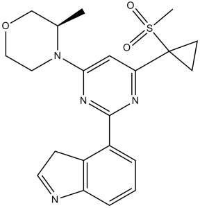
| Chemical Name | (3R)-4-[2-(1H-indol-4-yl)-6-(1-methylsulfonylcyclopropyl)pyrimidin-4-yl]-3-methylmorpholine | |
| Synonyms |
|
|
| HS Tariff Code | 2934.99.9001 | |
| Storage |
Powder-20°C 3 years 4°C 2 years In solvent -80°C 6 months -20°C 1 month |
|
| Shipping Condition | Room temperature (This product is stable at ambient temperature for a few days during ordinary shipping and time spent in Customs) |
Biological Activity
| Targets | ATR ( IC50 = 5 nM ); mTOR ( IC50 = 38 nM ); PI3Kα ( IC50 = 13000 nM ) | |
| ln Vitro |
|
|
| ln Vivo |
|
|
| Enzyme Assay | In order to obtain ATR for the in vitro enzyme assay, rabbit polyclonal antiserum raised to amino acids 400–480 of ATR was immunoprecipitated from HeLa nuclear extract. This buffer contained the ATR. pH 7.4, 250 mM NaCl, 0.5 mM EDTA, 0.1 mM Na3VO4, 10% v/v glycerol, and 0.01% v/v Tween 20 are combined to make 25 mM HEPES (pH 7.4). Protein A-Sepharose beads were incubated with ATR-antibody complexes from nuclear extract for one hour, after which the beads were recovered by centrifugation. ATR assay buffer (50 mM HEPES (pH 7.4), 150 mM NaCl, 6 mM MgCl2, 4 mM MnCl2, 0.1 mM Na3VO4, 0.1 mM DTT, and 10% (v/v) glycerol) was incubated with 1 μg of substrate glutathione S-transferase-p53N66 in a well of a 96-well plate. This was done in the presence or absence of an inhibitor. The reaction proceeded at 37°C for an extra hour after 10 minutes of moderate shaking, during which time ATP was added to a final concentration of 3 μM. A 96-well plate coated with white opaque glutathione was used to transfer the reaction and incubate it overnight at 4°C. The reaction was stopped by adding 100 μL of PBS. Following a PBS/0.05% (v/v) Tween 20 wash, this plate was blotted dry and subjected to an ELISA analysis using a phosphoserine 15 p53 antibody. Utilizing a secondary antibody conjugated with goat anti-mouse horseradish peroxidase, the phosphorylated glutathione S-transferase?p53N66 substrate was detected. A TopCount plate reader was utilized for chemiluminescent detection after a signal was generated using an enhanced chemiluminescence solution. IC50 values for the compounds were then calculated using the resulting calculated percentage of enzyme activity. | |
| Cell Assay | Using a Labcyte Echo acoustic dispensing device, compound dose ranges are created by dilution in 100% DMSO and then further into assay medium (EMEM, 10% FCS, 1% glutamine). Plates of cells are grown for 24 hours in 40 μL of EMEM, 10% FCS, and 1% glutamine. The cells are plated at 9×10⁴ cells per mL in 384-well Costar plates. After the compound is added, the cells are incubated for sixty minutes. Using the Labcyte Echo, a final concentration of 3 μM 4NQO (prepared in 100% DMSO) is added, and the cells are incubated for an additional 60 minutes. For 20 minutes, 40 μL of 3.7% v/v formaldehyde solution is added to the cells to fix them. The fix is removed from the cells, and they are then permeabilized in 40 μL of PBS containing 0.1% Triton X-100 and cleaned with PBS. Following a wash, 15 μL of pChk1 Ser345 primary antibody solution is added to the cells. The trays are incubated for the entire night at 4°C. After eliminating the primary antibody, 20 μL of secondary antibody solution and 1 μM Hoechst 33258 are added and allowed to sit at room temperature for 90 minutes. After being cleaned, the plates are placed in 40 μL of PBS. After that, staining intensities are measured on the plates using an ArrayScan VTI instrument. Dose responses are then collected and utilized to calculate the IC50 values for the compounds. | |
| Animal Protocol |
|
|
| References |
[1]. Cancer Res 2012;72(8 Suppl):Abstract nr 1823. [2]. J Med Chem . 2013 Mar 14;56(5):2125-38. |
|
| Additional Infomation | (3R)-4-[2-(1H-indol-4-yl)-6-(1-methylsulfonylcyclopropyl)-4-pyrimidinyl]-3-methylmorpholine is a member of indoles. |
Solubility Data
| Solubility (In Vitro) |
|
|||
| Solubility (In Vivo) |
Solubility in Formulation 1: ≥ 2.5 mg/mL (6.06 mM) (saturation unknown) in 10% DMSO + 40% PEG300 + 5% Tween80 + 45% Saline (add these co-solvents sequentially from left to right, and one by one), clear solution. For example, if 1 mL of working solution is to be prepared, you can add 100 μL of 25.0 mg/mL clear DMSO stock solution to 400 μL PEG300 and mix evenly; then add 50 μL Tween-80 to the above solution and mix evenly; then add 450 μL normal saline to adjust the volume to 1 mL. Preparation of saline: Dissolve 0.9 g of sodium chloride in 100 mL ddH₂ O to obtain a clear solution. Solubility in Formulation 2: ≥ 2.5 mg/mL (6.06 mM) (saturation unknown) in 10% DMSO + 90% (20% SBE-β-CD in Saline) (add these co-solvents sequentially from left to right, and one by one), clear solution. For example, if 1 mL of working solution is to be prepared, you can add 100 μL of 25.0 mg/mL clear DMSO stock solution to 900 μL of 20% SBE-β-CD physiological saline solution and mix evenly. Preparation of 20% SBE-β-CD in Saline (4°C,1 week): Dissolve 2 g SBE-β-CD in 10 mL saline to obtain a clear solution. (Please use freshly prepared in vivo formulations for optimal results.) |
| Preparing Stock Solutions | 1 mg | 5 mg | 10 mg | |
| 1 mM | 2.4242 mL | 12.1209 mL | 24.2418 mL | |
| 5 mM | 0.4848 mL | 2.4242 mL | 4.8484 mL | |
| 10 mM | 0.2424 mL | 1.2121 mL | 2.4242 mL |