AZ191 is a potent and selective DYRK1B (Dual-specificity tyrosine phosphorylation-regulated kinase) inhibitor with IC50 of 17 nM in a cell-free assay, about 5- and 110-fold selectivity over DYRK1A and DYRK2, respectively. Tyrosine kinase autophosphorylation is unaffected by AZ191, which specifically inhibits DYRK1B serine/threonine kinase activity. Moreover, AZ191 inhibits CCND1 phosphorylation in HEK-293 cells with significantly more potency against DYRK1B than DYRK1A. AZ191 accelerates cell-cycle progression in HD1B cells by potently suppressing the levels of the cell-cycle regulators, p21Cip1 and p27Kip1.
Physicochemical Properties
| Molecular Formula | C24H27N7O | |
| Molecular Weight | 429.52 | |
| Exact Mass | 429.228 | |
| Elemental Analysis | C, 67.11; H, 6.34; N, 22.83; O, 3.72 | |
| CAS # | 1594092-37-1 | |
| Related CAS # |
|
|
| PubChem CID | 72716071 | |
| Appearance | Brown solid powder | |
| LogP | 3.61 | |
| Hydrogen Bond Donor Count | 1 | |
| Hydrogen Bond Acceptor Count | 7 | |
| Rotatable Bond Count | 5 | |
| Heavy Atom Count | 32 | |
| Complexity | 601 | |
| Defined Atom Stereocenter Count | 0 | |
| SMILES | O(C([H])([H])[H])C1=C(C([H])=C([H])C(=C1[H])N1C([H])([H])C([H])([H])N(C([H])([H])[H])C([H])([H])C1([H])[H])N([H])C1=NC([H])=C([H])C(C2=C([H])N(C([H])([H])[H])C3C([H])=NC([H])=C([H])C2=3)=N1 |
|
| InChi Key | ZYVXTMKTGDARKR-UHFFFAOYSA-N | |
| InChi Code | InChI=1S/C24H27N7O/c1-29-10-12-31(13-11-29)17-4-5-21(23(14-17)32-3)28-24-26-9-7-20(27-24)19-16-30(2)22-15-25-8-6-18(19)22/h4-9,14-16H,10-13H2,1-3H3,(H,26,27,28) | |
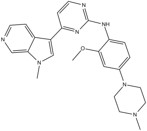
| Chemical Name | N-[2-methoxy-4-(4-methylpiperazin-1-yl)phenyl]-4-(1-methylpyrrolo[2,3-c]pyridin-3-yl)pyrimidin-2-amine | |
| Synonyms |
|
|
| HS Tariff Code | 2934.99.9001 | |
| Storage |
Powder-20°C 3 years 4°C 2 years In solvent -80°C 6 months -20°C 1 month |
|
| Shipping Condition | Room temperature (This product is stable at ambient temperature for a few days during ordinary shipping and time spent in Customs) |
Biological Activity
| Targets |
DYRK1B (IC50 = 17 nM); DYRK1A (IC50 = 88 nM) AZ191 specifically targets dual-specificity tyrosine-phosphorylation-regulated kinase 1B (DYRK1B) (IC50 = 13 nM) [1] AZ191 shows no significant inhibition of glycogen synthase kinase 3β (GSK3β) even at concentrations up to 10 μM [1] |
| ln Vitro |
AZ191 blocks DYRK1B serine/threonine kinase activity specifically; tyrosine kinase autophosphorylation is unaffected. Azzanol also inhibits CCND1 phosphorylation in HEK-293 cells, with much greater potency against DYRK1B than against DYRK1A. The levels of the cell-cycle regulators, p21Cip1 and p27Kip1, are significantly inhibited by AZ191 in HD1B cells, which also promotes cell expansion. AZ191 (0.1–10 μM) dose-dependently inhibited DYRK1B-mediated phosphorylation of cyclin D1 at Thr286 in HEK293 cells overexpressing DYRK1B and cyclin D1, without affecting Thr288 phosphorylation. This inhibition was independent of GSK3β, as AZ191 did not alter GSK3β activity or GSK3β-mediated cyclin D1 phosphorylation [1] - In liposarcoma cell lines (SW872, HT1080, and MLS 402), AZ191 suppressed cell proliferation with IC50 values of 2.5 μM (SW872), 3.1 μM (HT1080), and 3.8 μM (MLS 402) after 72 hours of treatment. It induced G1 phase cell cycle arrest, as evidenced by increased proportion of G1 phase cells (from ~45% to ~70% in SW872 cells at 5 μM) and decreased S phase cells [2] - AZ191 (1–5 μM) inhibited migration of SW872 and HT1080 cells in scratch assay: at 5 μM, migration closure rate was reduced from ~85% to ~30% (SW872) and ~80% to ~28% (HT1080) after 24 hours. It also suppressed colony formation, with colony numbers decreased by ~65% (SW872) and ~58% (HT1080) at 5 μM [2] - Western blot analysis showed that AZ191 (2.5–5 μM) downregulated cyclin D1 protein levels (by ~50% at 5 μM in SW872 cells) and upregulated p27Kip1 expression (by ~2.3-fold at 5 μM), while having no effect on cyclin E1 or CDK4 levels [2] |
| ln Vivo |
In nude mouse xenograft model of liposarcoma (SW872 cells), intraperitoneal administration of AZ191 (25 mg/kg, 5 times per week for 3 weeks) significantly inhibited tumor growth. Tumor volume was reduced by ~60% and tumor weight was decreased by ~55% compared to the vehicle control group. Immunohistochemical staining of tumor tissues showed reduced Ki-67 proliferation index (from ~75% to ~30%) and decreased cyclin D1 phosphorylation at Thr286 [2] - No significant changes in body weight of mice were observed during AZ191 treatment, indicating no obvious acute toxicity at the tested dose [2] |
| Enzyme Assay |
DYRK1B kinase activity assay: Recombinant human DYRK1B protein was incubated with a synthetic peptide substrate (e.g., GSK3βtide or cyclin D1-derived peptide containing Thr286) in reaction buffer. AZ191 was serially diluted (0.001–10 μM) and added to the reaction system, followed by addition of [γ-32P]ATP. After incubation at 30°C for 30 minutes, the reaction was terminated by adding stop buffer, and the mixture was spotted onto phosphocellulose paper. Unbound radioactivity was washed off, and the radioactivity of the bound substrate was measured by liquid scintillation counting to calculate the inhibition rate and IC50 value [1] - Kinase selectivity assay: AZ191 (1 μM) was tested against a panel of 30 different kinases (including GSK3α, CDK2, CDK4, JAK2, etc.). The kinase activity was measured using the same radioactive assay as DYRK1B, and the inhibition rate for each kinase was calculated to evaluate the selectivity of AZ191 [1] |
| Cell Assay |
AZ191 at 1uM was unable to selectively inhibit DYRK1B serine/threonine kinase activity or inhibit DYRK1B autophospho-Tyr273 in HEK-293 cells. At doses as low as 30–100 nM, AZ191 inhibited phosphorylation of CCND1 with greater potency for DYRK1B over DYRK1A in HEK-293 cells co-expressing CCND1 with either DYRK1A or DYRK1B. With approximately 100-fold selectivity over DYRK2 and 5–10-fold selectivity over DYRK1A, AZ191 is a novel inhibitor that selectively targets DYRK1B. To identify DYRK1B substrates and functions, AZ191 will be a helpful probe. Cyclin D1 phosphorylation assay: HEK293 cells were transfected with expression plasmids for DYRK1B and cyclin D1. After 24 hours of transfection, cells were serum-starved for 12 hours, then treated with AZ191 (0.1–10 μM) for 4 hours. Cell lysates were prepared, and Western blot was performed to detect phosphorylated cyclin D1 (Thr286 and Thr288), total cyclin D1, DYRK1B, and GSK3β (as internal controls) [1] - Cell proliferation assay: Liposarcoma cells (SW872, HT1080, MLS 402) were seeded in 96-well plates at 5×10³ cells/well. After 24 hours of adherence, AZ191 (0.1–20 μM) was added, and cells were cultured for 72 hours. MTT reagent was added to each well, incubated for 4 hours, then the supernatant was removed and formazan crystals were dissolved. Absorbance at 570 nm was measured to calculate cell viability and IC50 values [2] - Cell migration assay (scratch assay): SW872 and HT1080 cells were seeded in 6-well plates and cultured to confluence. A straight scratch was made with a 200 μL pipette tip, and floating cells were removed by washing with PBS. AZ191 (1–5 μM) was added, and images of the scratch area were captured at 0 and 24 hours. The migration closure rate was calculated by measuring the remaining gap width [2] - Cell cycle and colony formation assay: SW872 cells were treated with AZ191 (2.5–5 μM) for 24 hours, then harvested, fixed with ethanol, stained with propidium iodide, and analyzed by flow cytometry to determine cell cycle distribution. For colony formation, treated cells were seeded in 6-well plates at 500 cells/well, cultured for 14 days, stained with crystal violet, and visible colonies were counted [2] |
| Animal Protocol |
Liposarcoma xenograft model: Female nude mice (6–8 weeks old) were subcutaneously injected with SW872 cells (5×10⁶ cells/mouse) into the right flank. When tumors reached a volume of ~100 mm³, mice were randomly divided into control and treatment groups. AZ191 was dissolved in DMSO and diluted with normal saline (final DMSO concentration = 10%), then administered intraperitoneally at a dose of 25 mg/kg, 5 times per week (Monday to Friday) for 3 weeks. The control group received the same volume of vehicle (DMSO/saline). Tumor volume (measured by caliper every 3 days) and body weight (measured weekly) were recorded throughout the experiment. At the end of the experiment, mice were sacrificed, tumors were excised, weighed, and fixed in formalin for immunohistochemical analysis [2] |
| Toxicity/Toxicokinetics |
In the nude mouse xenograft study, intraperitoneal administration of AZ191 (25 mg/kg, 5 times/week for 3 weeks) did not cause significant changes in body weight (control vs. treatment: ~20 g vs. ~19.5 g) or obvious toxic symptoms (e.g., lethargy, loss of appetite, organ abnormalities) [2] |
| References |
[1]. A novel DYRK1B inhibitor AZ191 demonstrates that DYRK1B acts independently of GSK3β to phosphorylate cyclin D1 at Thr(286), not Thr(288). Biochem J. 2014 Jan 1;457(1):43-56. [2]. Targeting DYRK1B suppresses the proliferation and migration of liposarcoma cells. Oncotarget. 2017 Nov 28;9(17):13154-13166. |
| Additional Infomation |
AZ191 is a novel and selective small-molecule inhibitor of DYRK1B [1,2] - The mechanism of AZ191 involves specific inhibition of DYRK1B-mediated cyclin D1 Thr286 phosphorylation, leading to stabilization of cyclin D1 and subsequent cell cycle arrest in G1 phase [1] - AZ191 exhibits antitumor activity against liposarcoma cells both in vitro and in vivo, suggesting potential therapeutic value for the treatment of liposarcoma [2] - AZ191 does not cross-react with GSK3β, distinguishing it from non-selective DYRK/GSK3 inhibitors and reducing potential off-target effects [1] |
Solubility Data
| Solubility (In Vitro) |
|
|||
| Solubility (In Vivo) |
Solubility in Formulation 1: ≥ 2.5 mg/mL (5.82 mM) (saturation unknown) in 10% DMSO + 40% PEG300 + 5% Tween80 + 45% Saline (add these co-solvents sequentially from left to right, and one by one), clear solution. For example, if 1 mL of working solution is to be prepared, you can add 100 μL of 25.0 mg/mL clear DMSO stock solution to 400 μL PEG300 and mix evenly; then add 50 μL Tween-80 to the above solution and mix evenly; then add 450 μL normal saline to adjust the volume to 1 mL. Preparation of saline: Dissolve 0.9 g of sodium chloride in 100 mL ddH₂ O to obtain a clear solution. Solubility in Formulation 2: 2.5 mg/mL (5.82 mM) in 10% DMSO + 90% (20% SBE-β-CD in Saline) (add these co-solvents sequentially from left to right, and one by one), suspension solution; with ultrasonication. For example, if 1 mL of working solution is to be prepared, you can add 100 μL of 25.0 mg/mL clear DMSO stock solution to 900 μL of 20% SBE-β-CD physiological saline solution and mix evenly. Preparation of 20% SBE-β-CD in Saline (4°C,1 week): Dissolve 2 g SBE-β-CD in 10 mL saline to obtain a clear solution. (Please use freshly prepared in vivo formulations for optimal results.) |
| Preparing Stock Solutions | 1 mg | 5 mg | 10 mg | |
| 1 mM | 2.3282 mL | 11.6409 mL | 23.2818 mL | |
| 5 mM | 0.4656 mL | 2.3282 mL | 4.6564 mL | |
| 10 mM | 0.2328 mL | 1.1641 mL | 2.3282 mL |