Physicochemical Properties
| Molecular Formula | C19H21N3OS2 |
| Molecular Weight | 371.52 |
| Exact Mass | 371.113 |
| CAS # | 524708-03-0 |
| PubChem CID | 3581333 |
| Appearance | White to yellow solid powder |
| LogP | 4.76 |
| Hydrogen Bond Donor Count | 1 |
| Hydrogen Bond Acceptor Count | 5 |
| Rotatable Bond Count | 3 |
| Heavy Atom Count | 25 |
| Complexity | 503 |
| Defined Atom Stereocenter Count | 0 |
| InChi Key | JMSPCTGDYFVMJZ-UHFFFAOYSA-N |
| InChi Code | InChI=1S/C19H21N3OS2/c1-11(2)22-9-8-13-16(10-22)25-18(20-12(3)23)17(13)19-21-14-6-4-5-7-15(14)24-19/h4-7,11H,8-10H2,1-3H3,(H,20,23) |
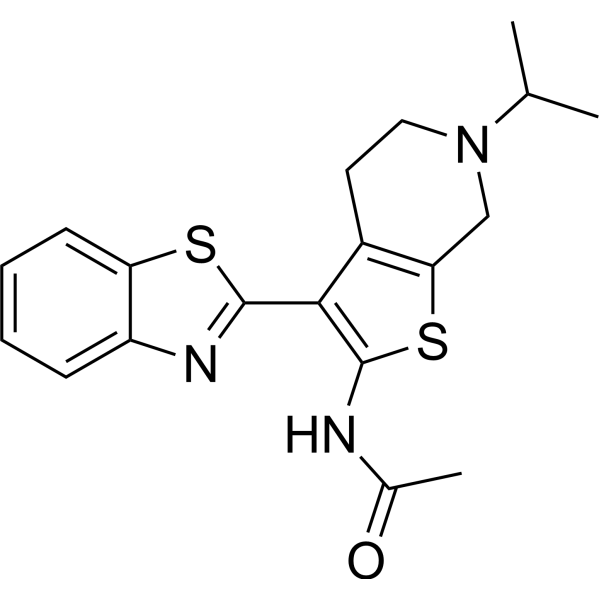
| Chemical Name | N-[3-(1,3-benzothiazol-2-yl)-6-propan-2-yl-5,7-dihydro-4H-thieno[2,3-c]pyridin-2-yl]acetamide |
| Synonyms | APE1 Inhibitor III; 524708-03-0; APE1-IN-1; N-[3-(1,3-benzothiazol-2-yl)-6-propan-2-yl-5,7-dihydro-4H-thieno[2,3-c]pyridin-2-yl]acetamide; CHEMBL1617574; N-[3-(1,3-benzothiazol-2-yl)-6-isopropyl-4,5,6,7-tetrahydrothieno[2,3-c]pyridin-2-yl]acetamide; MLS000419194; N-[3-(1,3-benzothiazol-2-yl)-6-(propan-2-yl)-4,5,6,7-tetrahydrothieno[2,3-c]pyridin-2-yl]acetamide; |
| HS Tariff Code | 2934.99.9001 |
| Storage |
Powder-20°C 3 years 4°C 2 years In solvent -80°C 6 months -20°C 1 month |
| Shipping Condition | Room temperature (This product is stable at ambient temperature for a few days during ordinary shipping and time spent in Customs) |
Biological Activity
| Targets | APE1 [apurinic/apyrimidinic (AP) endonuclease 1] (IC50 = 2 μM) |
| ln Vitro | In the qHTS assay, APE1-IN-1 (Compound 3) has an IC50 of 2 μM, while in the radiotracer incision assay (RIA), it is 12 μM [1]. In HeLa whole cell extracts, APE1-IN-1 (0, 1, 3, 10, 30 or 100 μM; 15 min) suppresses AP site nicking in a dose-dependent manner [1]. APE1-IN-1 (5-30 μM; 24 hours) increases the action of methyl mesylate and temozolomide and shows cytotoxic effect on HeLa cells [1]. |
| ln Vivo | APE1-IN-1 has good pharmacokinetic characteristics (30 mpk; IP; single dose) [1]. APE1-IN-1 (compound 3) (IP; 30 mpk) pharmacokinetic parameters in CD1 mice [1]. Brain/plasma 21 Cmax (μM) 16 217 tmax (h) 0.25 0.25 CLogP 2.83 Plasma Brain t1/2 (h) 2.1 |
| Enzyme Assay |
Enzyme Kinetic Studies [1] Ten pg of APE1 (~28 pM) was incubated without (positive control, 1% DMSO) or with 5, 10 or 20 µM of the indicated inhibitor at room temperature in RIA buffer (see above) for 15 min. Varying concentrations of 32P radiolabeled AP-DNA substrate (i.e. 5, 10, 25, 50, or 100 nM) were then added to a 10 µL final volume, and the reactions were incubated at 37 °C for 5 min, and stopped by adding stop buffer and heating at 95 °C for 10 min. The reaction velocity (nmolar substrate incised per min) at each substrate concentration was calculated as described above. Lineweaver – Burk plots of 1/V versus 1/[S] were used to determine KM and kcat, and the mode of inhibition. EMSA [1] Ten ng of APE1 (~28 nM) was incubated without inhibitor (positive control, 1% DMSO) or with increasing concentrations of inhibitor (1, 3, 10, 30 and 100 µM) in binding buffer (50 mM Tris pH 7.5, 25 mM NaCl, 1 mM EDTA, 1 mM DTT, 10% glycerol, 0.01% Tween 20) for 10 min on ice, and then radiolabeled 32P AP-DNA substrate (100 fmol) was added to a 10 µL final volume. Following incubation on ice for 5 min, samples were subjected to non-denaturing polyacrylamide gel electrophoresis (20 mM Tris pH 7.5, 10 mM sodium acetate, 0.5 mM EDTA, 8% polyacrylamide, 2.5% glycerol) for 2 h at 120 V in electrophoresis buffer (20 mM Tris pH 7.5, 10 mM sodium acetate, 0.5 mM EDTA) to separate the APE1-DNA complex from unbound radiolabeled DNA. After electrophoresis, the gel was subjected to standard phosphoimager analysis as above, and the percentage of substrate DNA in complex with APE1 was determined. |
| Cell Assay |
Cytotoxicity assay [1] Cell Types: HeLa cells Tested Concentrations: 5-30 μM Incubation Duration: 24 hrs (hours) Experimental Results: demonstrated cytotoxic activity against HeLa cells, with a 50% reduction in cell viability at approximately 15 μM. The activity of methyl mesylate (0.4 mM) and temozolomide (1 mM) was greatly enhanced, producing optimal synergy at ~5 μM and ~10 μM, respectively. HeLa Whole Cell Extract Incision Assays [1] To prepare protein extracts, HeLa cells maintained in DMEM with 10% fetal bovine serum and 1% penicillin-streptomycin were harvested, washed with 1X PBS, and re-suspended in ice cold hypotonic lysis buffer (50 mM Tris pH 7.4, 1 mM EDTA, 1 mM DTT, 10% glycerol, 0.5 mM PMSF). The suspension was frozen at −80 °C for at least 30 min and then slowly thawed at 4 °C for ~1 h. KCl was added to the cell suspension to a final concentration of 222 mM, followed by incubation on ice for 30 min and clarification by centrifugation at 12,000 xg for 15 min at 4 °C. The supernatant (whole cell extract) was retained, the protein concentration determined using the Bio-Rad Bradford reagent, and aliquots were stored until needed at −80 °C. For the incision assays, 300 ng of HeLa whole cell extract was incubated with 0, 1, 3, 10, 30 or 100 µM of the indicated inhibitor at room temperature for 15 min prior to the addition of 0.5 pmol of 32P radiolabeled AP-DNA substrate (final volume of 10 µL). The reaction mix was then transferred to 37 µC for 5 min to allow for incision. Following addition of stop buffer and heat denaturation, the reaction products were analyzed as above. Genomic AP site accumulation in cells [1] HeLa cells with 80% confluency in a 25 cm2 flask were treated with DMSO, 275 µM MMS, or 7.5 µM APE1 inhibitor alone, or with a combination of 275 µM MMS and 7.5 µM inhibitor for 24 h at 37 °C. Cells were then harvested, and genomic DNA of each sample was isolated according to Qiagen Genomic DNA isolation kit. The concentration of genomic DNA was measured and adjusted to 100 ng/µL. Ten µL of purified DNA was further labeled with an Aldehyde Reactive Probe (ARP) reagent (N’ aminooxymethylcarbonylhydrazino-D-biotin), and AP sites were measured using the DNA Damage Quantification kit. MMS and TMZ potentiation assay [1] HeLa cells were plated by multichannel pipette or Multidrop Combi dispenser at 6K/ 25 µL/well in DMEM culture medium with 10% FBS into white solid bottom 384-well cell culture plates. Cells were cultured at 37 µC overnight to allow for cell attachment. The following day, the entire cell medium in the well was replaced with fresh medium containing serial dilutions of the compounds of interest (5–30 µM) in the presence or absence of MMS (0.4 mM) or TMZ (1 mM). The plates were incubated for 24 h at 37 µC. Cell viability was then evaluated via luminescence detection by adding 15 µL of CellTiter Glo reagent to each well and incubating at room temperature for 30 min, and subsequently measuring the luminescence using a ViewLux reader. Percent viability was calculated for each concentration of the tested compounds in duplicate relative to the luminescence of the negative DMSO control. |
| Animal Protocol |
Animal/Disease Models: CD1 Male Mice (n = 3)[1] Doses: 30 mpk Route of Administration: IP; single dose Experimental Results: Displays lipophilicity (CLogP = 2.8) and readily crosses the BBB, resulting in a B/P ratio of 21. In vivo PK analysis [1] compound 3 (APE1-IN-1) was dissolved in PEG 400 and cremophor with vortexing and sonification, then saline was gradually added with vortexing and sonification to obtain a final concentration of 3 mg/mL of compound 3 (APE1-IN-1) in 50% PEG 400 and 10% cremophor. Compound 52 was dissolved in PEG 200 cremophor with vortexing and sonification, then saline was gradually added as above to obtain a final concentration of 3 mg/mL of 52 in 50% PEG 200 and 10% cremophor. The dose for both compounds was administered via IP. All blood samples were collected through a cardiac puncture per sampling time point (0.25, 0.5 h, 1 h, 2 h, 4 h, 8 h, 12 h, and 24 h post dose). Approximately 0.12 mL blood was collected at each time point. All blood samples were transferred into plastic micro centrifuge tubes containing Heparin and placed at −80 °C until processed (see below). At each time point (see above), the brain was harvested immediately after euthanasia by carbon dioxide. The brain was rinsed with saline and wiped clean and then weighed in a sterilized plastic tube. The tissue sample was then homogenized in water with a brain weight (g):water (mL) ratio of 1:4 (g:mL). The detected values were then multiplied by 5 to achieve the final concentration of the compound in the brain. Blood samples were processed for plasma by centrifugation at 4 °C at 4000 g for 5 min. Plasma samples were then stored in tubes, quickly frozen in a freezer and kept at −80 °C until LC/MS/MS analysis. Plasma concentration of compound 3 (APE1-IN-1) or 52 at the various time points (data obtained from the LC/MS/MS studies) was analyzed using the WinNonlin software program. |
| ADME/Pharmacokinetics | The two lead compounds (52 and compound 3 (APE1-IN-1)) were analyzed for their in vivo PK properties (Table 7) via intraperitoneal (IP) administration at 30 mg/kg body weight in 6–8 week old CD1 mice. Both compounds were well tolerated by the animals, with no adverse effects noted after a 24 hour observation. Compound 52, the more hydrophilic analog (CLogP ~ 1), had a favorable plasma t(1/2) of 5 hours and a drug concentration (ng/mL) that exceeded the IC50 value for over 12 hours. This compound also had reasonable blood brain barrier (BBB) penetration, with a high initial concentration that quickly tailed off, resulting in a brain/plasma (B/P) ratio of 1.4. In contrast, analog 3, which is more lipophilic (CLogP = 2.8), crosses the BBB quite readily, giving rise to a (B/P) ratio of 21. This result correlates with expectations, as reduction of hydrogen bond donors and increased lipophilicity often leads to improved BBB penetration. The ability to modulate the BBB penetration capacity of these molecules through structural modifications could prove useful depending on the cancer one is targeting.[1] |
| References |
[1]. Synthesis, biological evaluation, and structure-activity relationships of a novel class of apurinic/apyrimidinic endonuclease 1 inhibitors. J Med Chem. 2012 Apr 12;55(7):3101-12. |
| Additional Infomation |
APE1 is an essential protein that operates in the base excision repair (BER) pathway and is responsible for ≥95% of the total apurinic/apyrimidinic (AP) endonuclease activity in human cells. BER is a major pathway that copes with DNA damage induced by several anticancer agents, including ionizing radiation and temozolomide. Overexpression of APE1 and enhanced AP endonuclease activity have been linked to increased resistance of tumor cells to treatment with monofunctional alkylators, implicating inhibition of APE1 as a valid strategy for cancer therapy. We report herein the results of a focused medicinal chemistry effort around a novel APE1 inhibitor, N-(3-(benzo[d]thiazol-2-yl)-6-isopropyl-4,5,6,7-tetrahydrothieno[2,3-c]pyridin-2-yl)acetamide (3). Compound 3 and related analogues exhibit single-digit micromolar activity against the purified APE1 enzyme and comparable activity in HeLa whole cell extract assays and potentiate the cytotoxicity of the alkylating agents methylmethane sulfonate and temozolomide. Moreover, this class of compounds possesses a generally favorable in vitro ADME profile, along with good exposure levels in plasma and brain following intraperitoneal dosing (30 mg/kg body weight) in mice.[1] While the work described herein did not lead to a tremendous improvement in the potency of the initial “hit” compound 3 (APE1-IN-1), it does provide valuable insights into the SAR profile of this chemotype and represents the first reported medicinal chemistry optimization campaign towards the establishment of a novel APE1 inhibitor. In particular, this effort led to the development of compounds with low single-digit µM potency against the purified enzyme (noticeably more potent than prior reported inhibitors), desirable in vitro and in vivo ADME properties, and the capacity to potentiate the cytotoxicity of relevant DNA-damaging agents, namely MMS and TMZ. On-target evidence was supported by the comparable IC50 values against the purified recombinant APE1 protein and human whole cell extracts, as well as by increased genomic AP site accumulation in HeLa cells treated with inhibitors alone. Analysis of the in vivo PK properties of 52 and compound 3 (APE1-IN-1) in mice revealed that analog 52 has a better general cytotoxicity profile, higher exposure levels and a more favorable t(1/2) in the plasma, whereas compound 3 (APE1-IN-1) crosses the BBB more efficiently. Thus, for tumors outside the brain cavity, a compound like 52, which does not efficiently cross the BBB, would be useful in avoiding potential complications associated with this vital organ. However, APE1 has been found to be overexpressed in adult and pediatric gliomas, with an increase in AP endonuclease activity of between 5 and 10-fold.28 This observation indicates the need for the development of APE1 inhibitors, such as compound 3 (APE1-IN-1) or other lipophilic analogs of 52, that efficiently cross the BBB and can potentially be used in combination with a drug like TMZ. Our current efforts are focused on defining the efficacy of this class of compounds in vivo using mouse xenograft models in combination therapy with TMZ and other relevant DNA-damaging cancer chemotherapeutics. |
Solubility Data
| Solubility (In Vitro) | DMSO : ~22 mg/mL (~59.22 mM) |
| Solubility (In Vivo) |
Note: Listed below are some common formulations that may be used to formulate products with low water solubility (e.g. < 1 mg/mL), you may test these formulations using a minute amount of products to avoid loss of samples. Injection Formulations (e.g. IP/IV/IM/SC) Injection Formulation 1: DMSO : Tween 80: Saline = 10 : 5 : 85 (i.e. 100 μL DMSO stock solution → 50 μL Tween 80 → 850 μL Saline) *Preparation of saline: Dissolve 0.9 g of sodium chloride in 100 mL ddH ₂ O to obtain a clear solution. Injection Formulation 2: DMSO : PEG300 :Tween 80 : Saline = 10 : 40 : 5 : 45 (i.e. 100 μL DMSO → 400 μLPEG300 → 50 μL Tween 80 → 450 μL Saline) Injection Formulation 3: DMSO : Corn oil = 10 : 90 (i.e. 100 μL DMSO → 900 μL Corn oil) Example: Take the Injection Formulation 3 (DMSO : Corn oil = 10 : 90) as an example, if 1 mL of 2.5 mg/mL working solution is to be prepared, you can take 100 μL 25 mg/mL DMSO stock solution and add to 900 μL corn oil, mix well to obtain a clear or suspension solution (2.5 mg/mL, ready for use in animals). Injection Formulation 4: DMSO : 20% SBE-β-CD in saline = 10 : 90 [i.e. 100 μL DMSO → 900 μL (20% SBE-β-CD in saline)] *Preparation of 20% SBE-β-CD in Saline (4°C,1 week): Dissolve 2 g SBE-β-CD in 10 mL saline to obtain a clear solution. Injection Formulation 5: 2-Hydroxypropyl-β-cyclodextrin : Saline = 50 : 50 (i.e. 500 μL 2-Hydroxypropyl-β-cyclodextrin → 500 μL Saline) Injection Formulation 6: DMSO : PEG300 : castor oil : Saline = 5 : 10 : 20 : 65 (i.e. 50 μL DMSO → 100 μLPEG300 → 200 μL castor oil → 650 μL Saline) Injection Formulation 7: Ethanol : Cremophor : Saline = 10: 10 : 80 (i.e. 100 μL Ethanol → 100 μL Cremophor → 800 μL Saline) Injection Formulation 8: Dissolve in Cremophor/Ethanol (50 : 50), then diluted by Saline Injection Formulation 9: EtOH : Corn oil = 10 : 90 (i.e. 100 μL EtOH → 900 μL Corn oil) Injection Formulation 10: EtOH : PEG300:Tween 80 : Saline = 10 : 40 : 5 : 45 (i.e. 100 μL EtOH → 400 μLPEG300 → 50 μL Tween 80 → 450 μL Saline) Oral Formulations Oral Formulation 1: Suspend in 0.5% CMC Na (carboxymethylcellulose sodium) Oral Formulation 2: Suspend in 0.5% Carboxymethyl cellulose Example: Take the Oral Formulation 1 (Suspend in 0.5% CMC Na) as an example, if 100 mL of 2.5 mg/mL working solution is to be prepared, you can first prepare 0.5% CMC Na solution by measuring 0.5 g CMC Na and dissolve it in 100 mL ddH2O to obtain a clear solution; then add 250 mg of the product to 100 mL 0.5% CMC Na solution, to make the suspension solution (2.5 mg/mL, ready for use in animals). Oral Formulation 3: Dissolved in PEG400 Oral Formulation 4: Suspend in 0.2% Carboxymethyl cellulose Oral Formulation 5: Dissolve in 0.25% Tween 80 and 0.5% Carboxymethyl cellulose Oral Formulation 6: Mixing with food powders Note: Please be aware that the above formulations are for reference only. InvivoChem strongly recommends customers to read literature methods/protocols carefully before determining which formulation you should use for in vivo studies, as different compounds have different solubility properties and have to be formulated differently. (Please use freshly prepared in vivo formulations for optimal results.) |
| Preparing Stock Solutions | 1 mg | 5 mg | 10 mg | |
| 1 mM | 2.6916 mL | 13.4582 mL | 26.9165 mL | |
| 5 mM | 0.5383 mL | 2.6916 mL | 5.3833 mL | |
| 10 mM | 0.2692 mL | 1.3458 mL | 2.6916 mL |