AMZ30 is a selective, covalent inhibitors of protein phosphatase methylesterase-1(PME-1; IC50=600 nM); selectively inactivates PME-1 and reduces the demethylated form of PP2A in living cells.
Physicochemical Properties
| Molecular Formula | C19H12FN3O6S2 |
| Molecular Weight | 461.44348526001 |
| Exact Mass | 461.015 |
| Elemental Analysis | C, 49.46; H, 2.62; F, 4.12; N, 9.11; O, 20.80; S, 13.90 |
| CAS # | 1313613-09-0 |
| Related CAS # | 1313613-09-0 |
| PubChem CID | 44607965 |
| Appearance | Solid powder |
| Density | 1.5±0.1 g/cm3 |
| Boiling Point | 724.3±70.0 °C at 760 mmHg |
| Flash Point | 391.9±35.7 °C |
| Vapour Pressure | 0.0±2.3 mmHg at 25°C |
| Index of Refraction | 1.655 |
| LogP | 3.58 |
| Hydrogen Bond Donor Count | 0 |
| Hydrogen Bond Acceptor Count | 8 |
| Rotatable Bond Count | 5 |
| Heavy Atom Count | 31 |
| Complexity | 963 |
| Defined Atom Stereocenter Count | 0 |
| SMILES | S(C1C=CC=C(C=1)[N+](=O)[O-])(N1C=CC=C1/C=C(\C#N)/S(C1C=CC(=CC=1)F)(=O)=O)(=O)=O |
| InChi Key | GUCUORSUHTZMBW-YBFXNURJSA-N |
| InChi Code | InChI=1S/C19H12FN3O6S2/c20-14-6-8-17(9-7-14)30(26,27)19(13-21)11-15-4-2-10-22(15)31(28,29)18-5-1-3-16(12-18)23(24)25/h1-12H/b19-11+ |
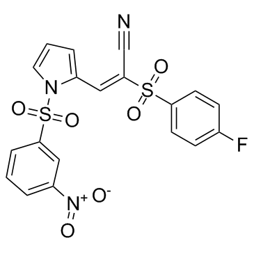
| Chemical Name | (E)-2-(4-fluorophenyl)sulfonyl-3-[1-(3-nitrophenyl)sulfonylpyrrol-2-yl]prop-2-enenitrile |
| Synonyms | AMZ-30; AMZ 30; AMZ30 |
| HS Tariff Code | 2934.99.9001 |
| Storage |
Powder-20°C 3 years 4°C 2 years In solvent -80°C 6 months -20°C 1 month |
| Shipping Condition | Room temperature (This product is stable at ambient temperature for a few days during ordinary shipping and time spent in Customs) |
Biological Activity
| Targets |
PME-1 (IC50 = 0.60 μM) Protein phosphatase methylesterase-1 (PME-1, a serine hydrolase) (IC50 = 600 nM in vitro; IC50 = 3.5 µM in situ) |
| ln Vitro |
1. Compound AMZ30 selectively inactivates PME-1 with an IC50 of 600 nM in human cell lysates (HEK 293T soluble proteome) as determined by gel-based competitive activity-based protein profiling (ABPP) using a fluorophosphonate-rhodamine (FP-Rh) probe. It showed more than 100-fold selectivity over other serine hydrolases in the proteome. [1] 2. Inhibition of PME-1 by AMZ30 is likely covalent and irreversible, as enzyme activity did not recover after gel filtration chromatography. [1] 3. In HEK 293T cell soluble proteomes, AMZ30 (at concentrations up to 100 µM) did not show cross-reactivity with other serine hydrolases in gel-based competitive ABPP assays. [1] 4. AMZ30 (20 µM) significantly reduced the levels of demethylated protein phosphatase 2A (PP2A) and increased the levels of methylated PP2A in HEK 293T cells stably overexpressing PME-1, as determined by Western blot analysis. The reduction in demethylated PP2A was similar in magnitude to that caused by the structurally unrelated inhibitor 1 (ABL127). [1] 5. In untransfected HEK 293T cells expressing basal levels of PME-1, treatment with AMZ30 (20 µM) also decreased the demethylated form of PP2A to a degree similar to treatment with inhibitor 1 (500 nM). [1] 6. AMZ30 (at 100 µM) did not alter the labeling intensity of any proteins by chloroacetamide-rhodamine (CA-Rh) or sulfonate ester-rhodamine (SE-Rh) activity-based probes in soluble proteomes from treated HEK 293T cells, indicating it is not a generally thiol-reactive compound. [1] |
| Enzyme Assay |
1. Competitive ABPP Assay for Serine Hydrolase Activity: Soluble proteomes (e.g., from HEK 293T or MDA-MB-231 cells) are diluted to a protein concentration of 1 mg/mL in phosphate-buffered saline (PBS, pH 7.5). The proteome samples (25 µL total reaction volume) are first incubated with the test compound (dissolved in DMSO) or DMSO vehicle control for 30 minutes at 25°C. Subsequently, the FP-rhodamine activity-based probe is added to a final concentration of 2 µM, and the labeling reaction is allowed to proceed for 45 minutes at 25°C. The reactions are then quenched by adding 2× SDS-PAGE loading buffer (reducing). The samples are separated by SDS-PAGE using 10% acrylamide gels. The labeled serine hydrolases are visualized in-gel using a flatbed fluorescence scanner. Inhibition is evidenced by a reduction in the fluorescence intensity of the PME-1 band compared to the DMSO control. [1] 2. IC50 Determination (In Vitro): For IC50 determination, the soluble proteome (1 mg/mL) is incubated with a range of concentrations of the test compound (performed in triplicate) for 45 minutes at 37°C. The samples are then labeled with FP-rhodamine (2 µM) for 45 minutes, processed for SDS-PAGE, and visualized by in-gel fluorescence scanning. The percentage of PME-1 activity remaining is quantified by measuring the integrated optical intensity of the PME-1 band using image analysis software. The IC50 value is calculated by fitting the dose-response data with appropriate software. [1] 3. Reversibility of Inhibition Assay: Purified recombinant PME-1 protein (500 nM in PBS) is incubated with either DMSO or inhibitor AMZ30 (50 µM) at 25°C for 30 minutes. An aliquot is removed for direct labeling with FP-rhodamine (Fraction A). The remainder of the reaction mixture is passed through a size-exclusion chromatography column (e.g., Sephadex G-25) to remove unbound inhibitor. The eluted protein (Fraction B) is then labeled with FP-rhodamine under the same conditions. Both fractions are analyzed by SDS-PAGE and in-gel fluorescence scanning. Lack of recovery of PME-1 activity in Fraction B compared to Fraction A indicates irreversible inhibition. [1] |
| Cell Assay |
1. In Situ (Cellular) Inhibition and IC50 Determination: HEK 293T cells are cultured in standard media. The test compound (AMZ30) is directly added to the cell culture media at various concentrations, and the cells are incubated at 37°C for a specified time (e.g., 1 hour). Cells are then washed with ice-cold PBS, harvested by scraping, and pelleted by centrifugation. The cell pellet is resuspended in PBS, sonicated, and centrifuged to obtain the soluble proteome fraction. This soluble proteome is diluted to 1 mg/mL protein concentration and then subjected to the standard competitive ABPP labeling protocol with FP-rhodamine as described in the "Enzyme Assay" section. The in situ IC50 value is determined from the dose-response curve generated from the fluorescence intensity of the PME-1 band in proteomes derived from compound-treated cells. [1] 2. Assessment of Cellular Target Selectivity (SILAC-ABPP): HEK 293T cells are grown in "light" or "heavy" stable isotope labeling by amino acids in cell culture (SILAC) media. "Light" cells are treated with compound AMZ30 (20 µM), while "heavy" cells are treated with DMSO vehicle, both for 1 hour at 37°C. After treatment, cells are harvested, and soluble proteomes are prepared. Equal amounts (by protein weight) of "light" and "heavy" proteomes are combined and labeled with a biotinylated fluorophosphonate (FP-biotin) activity-based probe. The labeled proteins are enriched using streptavidin beads, followed by on-bead tryptic digestion. The resulting peptides are analyzed by multidimensional liquid chromatography coupled with tandem mass spectrometry (MudPIT). The relative abundance of peptides from serine hydrolases in the "light" (inhibitor-treated) versus "heavy" (control) samples is quantified to assess the selectivity of the inhibitor across the serine hydrolase family in a cellular context. [1] 3. Analysis of PP2A Methylation State by Western Blot: HEK 293T cells (either wild-type or stably overexpressing PME-1) are treated with compounds (e.g., AMZ30 at 20 µM) or DMSO for a specified time (e.g., 1 hour). Cells are harvested, and total cell lysates are prepared by sonication in PBS. The lysates are denatured in SDS-PAGE loading buffer, separated by SDS-PAGE (10% acrylamide), and transferred to a membrane for Western blotting. The membrane is probed with specific antibodies that recognize the demethylated form of the PP2A catalytic subunit, the methylated form of PP2A, or total PP2A. An antibody against a loading control protein (e.g., α-tubulin) is also used. Band intensities are visualized and quantified using an imaging system to assess changes in PP2A methylation status upon inhibitor treatment. [1] 4. Assessment of General Protein Reactivity in Cells: Soluble proteomes prepared from HEK 293T cells treated with AMZ30 or DMSO are incubated with broad-spectrum activity-based probes that target reactive cysteine residues (CA-rhodamine) or other nucleophilic residues (SE-rhodamine). After labeling, samples are analyzed by SDS-PAGE and in-gel fluorescence scanning. The lack of changes in the labeling profiles of these probes in inhibitor-treated samples compared to DMSO controls indicates the compound does not promiscuously modify reactive residues on proteins. [1] |
| Toxicity/Toxicokinetics |
1. Compound AMZ30 (at 100 µM) did not show general reactivity towards protein nucleophilic residues (cysteines and others) in HEK 293T cell proteomes, as assessed by lack of effect on labeling by CA-rhodamine and SE-rhodamine probes, suggesting it is not a broadly toxic, thiol-reactive electrophile under the tested conditions. [1] |
| References |
[1]. Discovery and optimization of sulfonyl acrylonitriles as selective, covalent inhibitors of protein phosphatase methylesterase-1. J Med Chem. 2011 Jul 28;54(14):5229-36. |
| Additional Infomation |
2-(4-fluorophenyl)sulfonyl-3-[1-(3-nitrophenyl)sulfonyl-2-pyrrolyl]-2-propenenitrile is a sulfonamide. 1. Background and Significance: Protein phosphatase methylesterase-1 (PME-1) regulates the methylation state of protein phosphatase 2A (PP2A), a key enzyme involved in numerous cellular processes. Altered PP2A methylation has been implicated in cancer and Alzheimer's disease, making PME-1 a potential therapeutic target. [1] 2. Compound Class and Probe Utility: AMZ30 (NIH Probe ML136) belongs to the sulfonyl acrylonitrile class of covalent inhibitors. It is structurally unrelated to the previously reported aza-β-lactam (ABL) class of PME-1 inhibitors (e.g., compound 1). This structural diversity makes AMZ30 and the ABL inhibitors valuable as a paired set of pharmacological probes to confidently attribute observed cellular effects to PME-1 inhibition and to study the role of PP2A methylation. [1] 3. Mechanism Speculation: Based on structural analogs, it is speculated that AMZ30 inhibits PME-1 via a 1,4-Michael addition mechanism, where the activated olefin serves as the electrophile for covalent attachment to the active-site serine nucleophile of PME-1. However, direct MS evidence of the adduct was not obtained, possibly due to lability during sample preparation. [1] 4. Scaffold Potential: Several analogs of the lead sulfonyl acrylonitrile scaffold showed activity against other serine hydrolases (e.g., acyl-peptide hydrolase (APEH), prolyl endopeptidase (PREP)), suggesting that further optimization of this chemical phenotype could yield selective inhibitors for other members of this large enzyme family. [1] |
Solubility Data
| Solubility (In Vitro) | DMSO: 92~100 mg/mL (199.4~216.7 mM) |
| Solubility (In Vivo) |
Note: Listed below are some common formulations that may be used to formulate products with low water solubility (e.g. < 1 mg/mL), you may test these formulations using a minute amount of products to avoid loss of samples. Injection Formulations (e.g. IP/IV/IM/SC) Injection Formulation 1: DMSO : Tween 80: Saline = 10 : 5 : 85 (i.e. 100 μL DMSO stock solution → 50 μL Tween 80 → 850 μL Saline) *Preparation of saline: Dissolve 0.9 g of sodium chloride in 100 mL ddH ₂ O to obtain a clear solution. Injection Formulation 2: DMSO : PEG300 :Tween 80 : Saline = 10 : 40 : 5 : 45 (i.e. 100 μL DMSO → 400 μLPEG300 → 50 μL Tween 80 → 450 μL Saline) Injection Formulation 3: DMSO : Corn oil = 10 : 90 (i.e. 100 μL DMSO → 900 μL Corn oil) Example: Take the Injection Formulation 3 (DMSO : Corn oil = 10 : 90) as an example, if 1 mL of 2.5 mg/mL working solution is to be prepared, you can take 100 μL 25 mg/mL DMSO stock solution and add to 900 μL corn oil, mix well to obtain a clear or suspension solution (2.5 mg/mL, ready for use in animals). Injection Formulation 4: DMSO : 20% SBE-β-CD in saline = 10 : 90 [i.e. 100 μL DMSO → 900 μL (20% SBE-β-CD in saline)] *Preparation of 20% SBE-β-CD in Saline (4°C,1 week): Dissolve 2 g SBE-β-CD in 10 mL saline to obtain a clear solution. Injection Formulation 5: 2-Hydroxypropyl-β-cyclodextrin : Saline = 50 : 50 (i.e. 500 μL 2-Hydroxypropyl-β-cyclodextrin → 500 μL Saline) Injection Formulation 6: DMSO : PEG300 : castor oil : Saline = 5 : 10 : 20 : 65 (i.e. 50 μL DMSO → 100 μLPEG300 → 200 μL castor oil → 650 μL Saline) Injection Formulation 7: Ethanol : Cremophor : Saline = 10: 10 : 80 (i.e. 100 μL Ethanol → 100 μL Cremophor → 800 μL Saline) Injection Formulation 8: Dissolve in Cremophor/Ethanol (50 : 50), then diluted by Saline Injection Formulation 9: EtOH : Corn oil = 10 : 90 (i.e. 100 μL EtOH → 900 μL Corn oil) Injection Formulation 10: EtOH : PEG300:Tween 80 : Saline = 10 : 40 : 5 : 45 (i.e. 100 μL EtOH → 400 μLPEG300 → 50 μL Tween 80 → 450 μL Saline) Oral Formulations Oral Formulation 1: Suspend in 0.5% CMC Na (carboxymethylcellulose sodium) Oral Formulation 2: Suspend in 0.5% Carboxymethyl cellulose Example: Take the Oral Formulation 1 (Suspend in 0.5% CMC Na) as an example, if 100 mL of 2.5 mg/mL working solution is to be prepared, you can first prepare 0.5% CMC Na solution by measuring 0.5 g CMC Na and dissolve it in 100 mL ddH2O to obtain a clear solution; then add 250 mg of the product to 100 mL 0.5% CMC Na solution, to make the suspension solution (2.5 mg/mL, ready for use in animals). Oral Formulation 3: Dissolved in PEG400 Oral Formulation 4: Suspend in 0.2% Carboxymethyl cellulose Oral Formulation 5: Dissolve in 0.25% Tween 80 and 0.5% Carboxymethyl cellulose Oral Formulation 6: Mixing with food powders Note: Please be aware that the above formulations are for reference only. InvivoChem strongly recommends customers to read literature methods/protocols carefully before determining which formulation you should use for in vivo studies, as different compounds have different solubility properties and have to be formulated differently. (Please use freshly prepared in vivo formulations for optimal results.) |
| Preparing Stock Solutions | 1 mg | 5 mg | 10 mg | |
| 1 mM | 2.1671 mL | 10.8356 mL | 21.6713 mL | |
| 5 mM | 0.4334 mL | 2.1671 mL | 4.3343 mL | |
| 10 mM | 0.2167 mL | 1.0836 mL | 2.1671 mL |