AEBSF HCl (known also as AEBSF Hydrochloride) is a novel, broad spectrum, covalently/irreversibly inhibitor of serine proteases, such as chymotrypsin, plasmin, thrombin, and trypsin. Different cell lines were found to produce less Aβ when exposed to AEBSF. AEBSF demonstrated a dose-dependent decrease of Aβ with an IC50 value of roughly 1 mM in K293 cells transfected with APP695 (K695sw). AEBSF exhibited an inhibitory effect with an IC50 value of approximately 300 μM in HS695 and SKN695 cells transfected with wild-type APP695.
Physicochemical Properties
| Molecular Formula | C8H11CLFNO2S | |
| Molecular Weight | 239.69 | |
| Exact Mass | 239.018 | |
| Elemental Analysis | C, 40.09; H, 4.63; Cl, 14.79; F, 7.93; N, 5.84; O, 13.35; S, 13.38 | |
| CAS # | 30827-99-7 | |
| Related CAS # | 34284-75-8;30827-99-7 (HCl); | |
| PubChem CID | 186136 | |
| Appearance | White to off-white solid powder | |
| Boiling Point | 292.5ºC at 760 mmHg | |
| Melting Point | 175-177 °C | |
| Flash Point | 130.7ºC | |
| LogP | 3.429 | |
| Hydrogen Bond Donor Count | 2 | |
| Hydrogen Bond Acceptor Count | 4 | |
| Rotatable Bond Count | 3 | |
| Heavy Atom Count | 14 | |
| Complexity | 239 | |
| Defined Atom Stereocenter Count | 0 | |
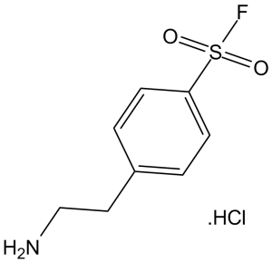
| SMILES | O=S(C1=CC=C(CCN)C=C1)(F)=O.[H]Cl |
|
| InChi Key | WRDABNWSWOHGMS-UHFFFAOYSA-N | |
| InChi Code | InChI=1S/C8H10FNO2S.ClH/c9-13(11,12)8-3-1-7(2-4-8)5-6-10;/h1-4H,5-6,10H2;1H | |
| Chemical Name | 4-(2-aminoethyl)benzenesulfonyl fluoride;hydrochloride | |
| Synonyms |
|
|
| HS Tariff Code | 2934.99.9001 | |
| Storage |
Powder-20°C 3 years 4°C 2 years In solvent -80°C 6 months -20°C 1 month Note: Please store this product in a sealed and protected environment, avoid exposure to moisture. |
|
| Shipping Condition | Room temperature (This product is stable at ambient temperature for a few days during ordinary shipping and time spent in Customs) |
Biological Activity
| Targets |
serine protease AEBSF HCl (4-(2-aminoethyl)-benzenesulfonyl fluoride hydrochloride) inhibits serine proteases involved in amyloid beta-protein (Aβ) production (presumed to be β-secretase-related serine proteases) [1] - AEBSF HCl targets serine proteases secreted by human macrophages (e.g., cathepsin G, elastase) [2] |
| ln Vitro |
AEBSF directly inhibits β-secretase in five distinct human cell lines, both neural and nonneural, to prevent the constitutive synthesis of Aβ[1]. AEBSF, as a serine protease inhibitor that prevents human macrophages from lysing leukemic cells, but it does not stop macrophages from secreting TNF-α and IL-1β[2]. AEBSF also modifies the pattern of protein secretion, which prevents HeLa cells from adhering to HUVECs and interferes with the growth of blastocysts on endometrial cells[4]. In rat pheochromocytoma PC12 cells and primary rat cortical neurons, treatment with 100 μM AEBSF HCl for 48 hours reduced Aβ40 production by ~50% and Aβ42 production by ~45% (detected via sandwich ELISA); Western blot analysis showed decreased levels of the β-secretase-cleaved APP fragment (C99) without affecting total APP expression [1] - In co-cultures of human peripheral blood-derived macrophages and human chronic myelogenous leukemia K562 cells, 100 μM AEBSF HCl inhibited macrophage-mediated K562 cell lysis by ~70% (measured via 51Cr release assay) and reduced macrophage phagocytic activity by ~40% (assessed via latex bead ingestion) [2] - In Vero cell cultures infected with Toxoplasma gondii tachyzoites (MOI = 1), treatment with 200 μM AEBSF HCl for 24 hours inhibited parasite invasion into host cells by ~55% (counted via Giemsa staining) and reduced parasite replication by ~40% (qRT-PCR for T. gondii 18S rRNA) [3] - In primary cultures of rat endometrial epithelial cells, 150 μM AEBSF HCl reduced cell adhesion to fibronectin by ~60% (measured via crystal violet staining of adherent cells) and decreased the expression of integrin αvβ3 (a key adhesion molecule) by ~35% (flow cytometry) [4] |
| ln Vivo |
AEBSF (76.8 mg/kg daily, i.p.) results in prolongation of the survival of mice that have been given a lethal T. gondii infection[3]. AEBSF also lowers the cockroach allergen-induced murine model's airway response and underlying inflammation[4]. In a murine model of acute Toxoplasma gondii infection (BALB/c mice, intraperitoneal inoculation with 1×10³ tachyzoites), intraperitoneal injection of AEBSF HCl at 20 mg/kg once daily for 5 days (starting 1 day post-infection) increased mouse survival rate from ~30% (control) to ~60% and reduced T. gondii burden in brain tissue by ~50% (quantitative PCR) [3] - In a rat model of embryo implantation (Sprague-Dawley rats, mated to fertile males), subcutaneous injection of AEBSF HCl at 10 mg/kg once daily from day 3 to day 5 post-coitus reduced embryo implantation rate from ~80% (control) to ~30% (counted via uterine dissection on day 7 post-coitus) [4] |
| Enzyme Assay |
Serine protease activity assay for Aβ production (from [1] abstract description): Recombinant β-secretase-like serine protease was mixed with a fluorescent peptide substrate (Mca-SEVNLDAEFK(DNP)-RR) in reaction buffer (50 mM Tris-HCl pH 6.5, 0.1% CHAPS). AEBSF HCl was added at concentrations ranging from 25 μM to 200 μM, and the mixture was incubated at 37°C for 1 hour. Fluorescence intensity was measured at excitation 320 nm/emission 405 nm, and enzyme inhibition rate was calculated relative to the vehicle control [1] - Macrophage serine protease assay (from [2] abstract description): Human macrophage-conditioned medium was mixed with chromogenic substrates for cathepsin G (S-2390) or elastase (S-2484) in 50 mM Tris-HCl pH 7.5. AEBSF HCl was added at 10 μM to 150 μM, and the mixture was incubated at 37°C for 30 minutes. Absorbance at 405 nm was measured to quantify protease activity, and inhibition was determined [2] |
| Cell Assay |
HeLa cells suspended in RPMI-1640 medium with 10% FCS are plated into a 96-well microplate, with 5×10³ cells/200 μL allocated to each well. The cells are treated with varying doses of AEBSF (0, 25, 50, and 100 μg/mL) for 48 hours following a 24-hour incubation period at 37°C. Subsequently, each well receives 20 μL of fresh 3-(4,5)-dimethylthiahiazo (-z-y1)-3,5-diphenytetrazoliumromide (MTT) reagent (5 μg/μL), and the cells are cultured for an additional 4 hours at 37°C in 5% CO2. After carefully discarding the media, add 150 μL of DMSO. On a microplate reader, absorbance is measured at two different wavelengths: 540 and 620 nm. PC12 cell Aβ production assay (from [1] abstract description): Rat PC12 cells were cultured in DMEM supplemented with 10% horse serum and 5% fetal bovine serum until 70% confluence. Cells were treated with AEBSF HCl (25 μM, 50 μM, 100 μM, 200 μM) for 48 hours. Culture supernatants were collected to measure Aβ40/Aβ42 levels via sandwich ELISA. Cells were lysed in RIPA buffer, and proteins were separated by SDS-PAGE; Western blot was performed with antibodies against APP, C99 (β-secretase cleavage product), and GAPDH (internal control) [1] - Macrophage-K562 cell co-culture assay (from [2] abstract description): Human macrophages were isolated from peripheral blood via density gradient centrifugation and cultured in RPMI 1640 with 10% fetal bovine serum. K562 cells were labeled with 51Cr (sodium chromate) for 1 hour, then co-cultured with macrophages at a 1:10 (macrophage:K562) ratio. AEBSF HCl (25 μM to 150 μM) was added to the co-culture, which was incubated for 4 hours. 51Cr release into the supernatant was measured via gamma counting to calculate K562 cell lysis rate [2] - T. gondii invasion assay (from [3] abstract description): Vero cells were cultured in MEM with 10% fetal bovine serum until 80% confluence. T. gondii tachyzoites were pre-incubated with AEBSF HCl (50 μM to 250 μM) for 30 minutes, then added to Vero cells (MOI = 1). After 24 hours of incubation, cells were fixed with methanol, stained with Giemsa, and the number of infected cells (with intracellular tachyzoites) was counted under a microscope [3] - Endometrial cell adhesion assay (from [4] abstract description): Primary rat endometrial epithelial cells were isolated from uteri of day 4 post-coitus rats and cultured in DMEM/F12 with 10% fetal bovine serum. Fibronectin-coated 96-well plates were seeded with 5×10⁴ cells/well, and AEBSF HCl (50 μM to 200 μM) was added. After 2 hours of incubation at 37°C, non-adherent cells were washed away, and adherent cells were stained with crystal violet. Absorbance at 570 nm was measured to quantify adhesion [4] |
| Animal Protocol |
Mice that receive a 2.5×10³ parasite injection are randomly allocated to one of the following treatment groups: vehicle alone (control group), pyrimethamine alone at various doses, LY311727 alone at various doses, AEBSF alone at various doses, or AEBSF 76.8 mg/kg plus pyrimethamine 10 mg/kg. Each treatment group has ten animals in it. After the parasite inoculation, treatment is started 24 hours later and lasts for seven days in a row. In live mice, mouse survival is tracked every day for 15 days after infection. Every experiment is run three times, and the results displayed are the total of those runs. Murine T. gondii infection model (from [3] abstract description): Female BALB/c mice (6-8 weeks old) were intraperitoneally inoculated with 1×10³ T. gondii RH strain tachyzoites. One day post-infection, AEBSF HCl was dissolved in sterile physiological saline (0.9% NaCl) and administered via intraperitoneal injection at 20 mg/kg. Injections were repeated once daily for 5 consecutive days. Vehicle control mice received an equal volume of physiological saline. Mouse survival was monitored for 14 days; surviving mice were euthanized on day 14, and brain tissues were collected to quantify T. gondii burden via qRT-PCR [3] - Rat embryo implantation model (from [4] abstract description): Female Sprague-Dawley rats (8-10 weeks old) were mated to fertile males (presence of vaginal plug = day 0 post-coitus). On day 3 post-coitus, AEBSF HCl was dissolved in a mixture of 5% DMSO and 95% physiological saline, then administered via subcutaneous injection at 10 mg/kg. Injections were repeated once daily on day 4 and day 5 post-coitus. Vehicle control rats received 5% DMSO/physiological saline. On day 7 post-coitus, rats were euthanized, uteri were dissected, and the number of implanted embryos (visible as small swellings) was counted [4] |
| Toxicity/Toxicokinetics |
In primary rat cortical neurons, treatment with AEBSF HCl at concentrations up to 200 μM for 48 hours did not affect cell viability (MTT assay, viability >90% compared to control) [1] - In human macrophages, 150 μM AEBSF HCl treatment for 4 hours did not induce significant cytotoxicity (trypan blue exclusion assay, viability >85%) [2] - In BALB/c mice treated with 20 mg/kg AEBSF HCl (intraperitoneal, 5 days), no significant changes in body weight or serum ALT/AST levels (liver toxicity markers) were observed [3] - In Sprague-Dawley rats treated with 10 mg/kg AEBSF HCl (subcutaneous, 3 days), no abnormal histopathological changes were found in the uterus, liver, or kidney [4] |
| References |
[1]. Inhibition of amyloid beta-protein production in neural cells by the serine protease inhibitor AEBSF. Neuron. 1996 Jul;17(1):171-9. [2]. Lysis of leukemic cells by human macrophages: inhibition by 4-(2-aminoethyl)-benzenesulfonyl fluoride (AEBSF), a serine protease inhibitor. J Leukoc Biol. 1996 Sep;60(3):328-36. [3]. Evaluation of two inhibitors of invasion: LY311727 [3-(3-acetamide-1-benzyl-2-ethyl-indolyl-5-oxy)propane phosphonic acid] and AEBSF [4-(2-aminoethyl)-benzenesulphonyl fluoride] in acute murine toxoplasmosis. J Antimicrob Chemother. [4]. Serine protease inhibitor 4-(2-aminoethyl)benzenesulfonyl fluoride hydrochloride (AEBSF) inhibits the rat embryo implantation in vivo and interferes with cell adhesion in vitro. Contraception. 2011 Dec;84(6):642-8. |
| Additional Infomation |
AEBSF HCl is a synthetic, cell-permeable serine protease inhibitor widely used as a research tool to study the role of serine proteases in biological processes (e.g., Aβ production, immune cell function, parasite invasion) [1,2,3] - The mechanism by which AEBSF HCl reduces Aβ production involves inhibition of serine proteases that mediate the rate-limiting cleavage of amyloid precursor protein (APP) into Aβ, making it a potential tool for Alzheimer's disease research [1] - In immunology, AEBSF HCl is used to dissect the role of macrophage serine proteases in cytotoxicity and phagocytosis, as these proteases are critical for macrophage-mediated killing of tumor cells [2] - AEBSF HCl exhibits anti-parasitic activity against T. gondii by inhibiting parasite serine proteases required for host cell invasion, suggesting potential applications in anti-infective research [3] - By interfering with endometrial cell adhesion (a key step in embryo implantation), AEBSF HCl provides a theoretical basis for the development of novel contraceptive strategies targeting serine protease-dependent adhesion pathways [4] |
Solubility Data
| Solubility (In Vitro) |
|
|||
| Solubility (In Vivo) |
Solubility in Formulation 1: ≥ 2.08 mg/mL (8.68 mM) (saturation unknown) in 10% DMSO + 40% PEG300 + 5% Tween80 + 45% Saline (add these co-solvents sequentially from left to right, and one by one), clear solution. For example, if 1 mL of working solution is to be prepared, you can add 100 μL of 20.8 mg/mL clear DMSO stock solution to 400 μL PEG300 and mix evenly; then add 50 μL Tween-80 to the above solution and mix evenly; then add 450 μL normal saline to adjust the volume to 1 mL. Preparation of saline: Dissolve 0.9 g of sodium chloride in 100 mL ddH₂ O to obtain a clear solution. Solubility in Formulation 2: ≥ 2.08 mg/mL (8.68 mM) (saturation unknown) in 10% DMSO + 90% (20% SBE-β-CD in Saline) (add these co-solvents sequentially from left to right, and one by one), clear solution. For example, if 1 mL of working solution is to be prepared, you can add 100 μL of 20.8 mg/mL clear DMSO stock solution to 900 μL of 20% SBE-β-CD physiological saline solution and mix evenly. Preparation of 20% SBE-β-CD in Saline (4°C,1 week): Dissolve 2 g SBE-β-CD in 10 mL saline to obtain a clear solution. Solubility in Formulation 3: ≥ 2.08 mg/mL (8.68 mM) (saturation unknown) in 10% DMSO + 90% Corn Oil (add these co-solvents sequentially from left to right, and one by one), clear solution. For example, if 1 mL of working solution is to be prepared, you can add 100 μL of 20.8 mg/mL clear DMSO stock solution to 900 μL of corn oil and mix evenly. Solubility in Formulation 4: 100 mg/mL (417.21 mM) in PBS (add these co-solvents sequentially from left to right, and one by one), clear solution; with ultrasonication. Solubility in Formulation 5: 100 mg/mL (417.21 mM) in Saline (add these co-solvents sequentially from left to right, and one by one), clear solution; with ultrasonication. Preparation of saline: Dissolve 0.9 g of sodium chloride in 100 mL ddH₂ O to obtain a clear solution. (Please use freshly prepared in vivo formulations for optimal results.) |
| Preparing Stock Solutions | 1 mg | 5 mg | 10 mg | |
| 1 mM | 4.1721 mL | 20.8603 mL | 41.7206 mL | |
| 5 mM | 0.8344 mL | 4.1721 mL | 8.3441 mL | |
| 10 mM | 0.4172 mL | 2.0860 mL | 4.1721 mL |