AC-73 (AC73) is a CD147 inhibitor with anticancer activity. Acting by disrupting CD147 dimerization and thereby mainly suppressing the CD147/ERK1/2/STAT3/MMP-2 pathways, which are crucial for cancer progression.
Physicochemical Properties
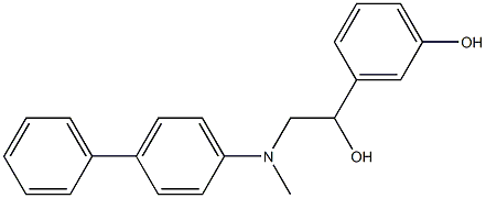
| Molecular Formula | C21H21NO2 |
| Molecular Weight | 319.396945714951 |
| Exact Mass | 319.157 |
| Elemental Analysis | C, 78.97; H, 6.63; N, 4.39; O, 10.02 |
| CAS # | 775294-71-8 |
| Related CAS # | 775294-71-8; |
| PubChem CID | 2989791 |
| Appearance | White to off-white solid powder |
| LogP | 2.8 |
| Hydrogen Bond Donor Count | 3 |
| Hydrogen Bond Acceptor Count | 3 |
| Rotatable Bond Count | 6 |
| Heavy Atom Count | 24 |
| Complexity | 348 |
| Defined Atom Stereocenter Count | 0 |
| InChi Key | UECKKYNEEBRMIL-UHFFFAOYSA-N |
| InChi Code | InChI=1S/C21H21NO2/c23-20-8-4-7-19(13-20)21(24)15-22-14-16-9-11-18(12-10-16)17-5-2-1-3-6-17/h1-13,21-24H,14-15H2 |
| Chemical Name | 3-{2-[(Biphenyl-4-ylmethyl)-amino]-1-hydroxy-ethyl}-phenol |
| Synonyms | AC-73; AC 73; AC73 |
| HS Tariff Code | 2934.99.9001 |
| Storage |
Powder-20°C 3 years 4°C 2 years In solvent -80°C 6 months -20°C 1 month |
| Shipping Condition | Room temperature (This product is stable at ambient temperature for a few days during ordinary shipping and time spent in Customs) |
Biological Activity
| Targets | CD147 |
| ln Vitro | Treatment with AC-73 (5-10 μM; 24 hours) dramatically decreased the capacity of SMMC-7721 and Huh-7 cells to migrate in a dose-dependent manner, as well as the invasion of both HCC cells. The effects were seen within 24 hours. Treatment with AC-73 decreases HCC metastases. The treatment of two HCC cells with AC-73 at a maximum dosage of 20 μM did not significantly affect the viability of the cells. Glu64 and Glu73 in the N-terminal IgC2 domain, which are situated in the dimer interface of CD147, are potential binding sites for AC-73 on CD147 [1]. At a dose of 10 μM, AC-73 (5-10 μM; 24 hours; SMMC-7721 cells) treatment can considerably reduce MMP-2 and MMP-9 mRNA expression, with MMP-2 being the most affected, but not MMP-2. Impact. 1. MMP-11, MMP-13, MMP-7, or MMP-8. Gelatin zymography tests and RT-qPCR analysis demonstrated that AC-73 can dose-dependently lower MMP-2 mRNA expression and protein secretion [1]. The phosphorylation of ERK1/2 and STAT3 is dose-dependently inhibited by AC-73 (5–20 μM; 6 hours; SMMC-7721 cells) treatment [1]. |
| ln Vivo | The incidence of metastases in nude mice was dramatically decreased by AC-73 therapy (25–50 mg/kg; 4 weeks; male BALB/c nu/nu mice). In a dose-dependent manner, AC-73 suppresses the phosphorylation of STAT3 and ERK1/2. Moreover, AC-73 lowers MMP-2. In vivo tumor cell growth cannot be inhibited by AC-73 [1]. |
| Enzyme Assay |
Surface plasmon resonance (SPR) assay[1] SPR measurements were performed using the ProteOn XPR36 system with ProteOn GLH sensor chips. ProteOn PBS/Tween running buffer (phosphate-buffered saline, pH 7.4, with 0.005% Tween 20) containing 0.1% DMSO was used as a running buffer throughout, and all experiments were performed at 25°C. Purified CD147wt or CD147mt was immobilized on a GLH chip. Each compound was used at the same concentration (100 μM) and simultaneously injected in the horizontal direction, with running buffer injected as a control. Dissociation was monitored for 15 min. The data were analyzed using ProteOn Manager Software, version 2.0. Non-denaturing SDS-PAGE assay[1] To evaluate whether AC-73 could inhibit the dimerization of CD147 in a Prokaryotic expression system, non-denaturing SDS-PAGE followed by a Western blot assay was used as previously described. Both CD147wt and CD147mts were purified, and 5 μg of each was added to various concentrations of AC-73 (0, 0.1, 0.25, or 0.5 μM) and mixed with 5× Laemmli sample buffer lacking SDS, followed by resolution by 10% SDS-PAGE without boiling and immunoblotting with anti-His6 antibody. |
| Cell Assay |
RT-PCR[1] Cell Types: SMMC-7721 Cell Tested Concentrations: 5 μM or 10 μM Incubation Duration: 24 hrs (hours) Experimental Results: MMP-2 and MMP-9 mRNA expression was Dramatically inhibited at 10 μM concentration. MMP-2 expression at the mRNA level and secretion at the protein level were dose-dependently diminished using RT-qPCR analysis and gelatin zymography experiments. Western Blot Analysis [1] Cell Types: SMMC-7721 Cell Tested Concentrations: 5 μM, 10 μM or 20 μM Incubation Duration: 6 hrs (hours) Experimental Results: The phosphorylation of ERK1/2 and STAT3 was dose-dependently inhibited. |
| Animal Protocol |
Animal/Disease Models: Male BALB/c nu/nu (nude) mice (4-6 weeks) with SMMC-7721 cells [1] Doses: 25 mg/kg, 50 mg/kg Route of Administration: injection; daily; 3 consecutive Weekly Experimental Results: Dramatically diminished the incidence of metastasis in nude mice. Inhibits the phosphorylation of ERK1/2 and STAT3 in a dose-dependent manner. MMP-2 is also diminished. For in vivo experiments, AC-73 was dissolved in Cremophor EL/ethanol (50:50; Cremophor EL, 95% ethyl alcohol) at 4-fold of the highest dose and stored at room temperature.[1] Establishment of the orthotopic transplant nude mouse model of HCC metastasis[1] Male BALB/c nu/nu mice, 4 to 6 weeks of age, were provided by the Laboratory Animal Research Center of FMMU, and the animal study was reviewed and approved by the FMMU Animal Care and Use Committee. The mice were housed in a standard animal laboratory under constant environmental conditions, including a 12-h light and dark cycle, with free access to water and food. A mixture of SMMC-7721 cells (1 × 10⁶) in 0.1 ml culture medium and the same volume of diluted Matrigel was injected into the left liver lobe of the nude mice. The treatment was started 1 week after implantation. The mice were divided into three groups: vehicle control (Cremophor EL/ethanol), AC-73 (25 mg/kg/day) and AC-73 (50 mg/kg/day). The mice were sacrificed 4 weeks after implantation. The number of intrahepatic metastases was calculated and statistically analyzed. Tumor tissues were then fixed, embedded in paraffin, and serially sectioned at a thickness of 4 mm. IHC staining was performed, and the sections were examined by a pathologist to verify the presence of tumors. Toxicity test[1] 6-week-old male nude mice were divided into 4 groups randomly (n = 5). Mice were injected with normal saline (control), Cremophor EL/ethanol (vehicle), 25 mg/kg/day and 50 mg/kg/day of AC-73. The body weight of each mouse was recorded daily. After 20 more days, mice were killed and selected tissues were fixed in4% paraformaldehyde. Serial histologic sections of their removed hearts, lungs, testis, spleens, kidneys and livers were stained by Hematoxylin and eosin (H&E). The concentration of the serum GPT/ALT and GOT/AST were measured by extracting the eyeball blood using a commercial AST or ALT assay kit. Liver tissue apoptosis were detected by tunel stain (Tunel stain kit). |
| References |
[1]. A novel small-molecule compound targeting CD147 inhibits the motility and invasion of hepatocellular carcinoma cells. Oncotarget. 2016 Feb 23;7(8):9429-47. [2]. The small-molecule compound AC-73 targeting CD147 inhibits leukemic cell proliferation, induces autophagy and increases the chemotherapeutic sensitivity of acute myeloid leukemia cells. Haematologica. 2019 May;104(5):973-985. |
Solubility Data
| Solubility (In Vitro) | DMSO : ≥ 250 mg/mL (~782.72 mM) |
| Solubility (In Vivo) |
Solubility in Formulation 1: ≥ 2.08 mg/mL (6.51 mM) (saturation unknown) in 10% DMSO + 40% PEG300 + 5% Tween80 + 45% Saline (add these co-solvents sequentially from left to right, and one by one), clear solution. For example, if 1 mL of working solution is to be prepared, you can add 100 μL of 20.8 mg/mL clear DMSO stock solution to 400 μL PEG300 and mix evenly; then add 50 μL Tween-80 to the above solution and mix evenly; then add 450 μL normal saline to adjust the volume to 1 mL. Preparation of saline: Dissolve 0.9 g of sodium chloride in 100 mL ddH₂ O to obtain a clear solution. Solubility in Formulation 2: ≥ 2.08 mg/mL (6.51 mM) (saturation unknown) in 10% DMSO + 90% (20% SBE-β-CD in Saline) (add these co-solvents sequentially from left to right, and one by one), clear solution. For example, if 1 mL of working solution is to be prepared, you can add 100 μL of 20.8 mg/mL clear DMSO stock solution to 900 μL of 20% SBE-β-CD physiological saline solution and mix evenly. Preparation of 20% SBE-β-CD in Saline (4°C,1 week): Dissolve 2 g SBE-β-CD in 10 mL saline to obtain a clear solution. Solubility in Formulation 3: ≥ 2.08 mg/mL (6.51 mM) (saturation unknown) in 10% DMSO + 90% Corn Oil (add these co-solvents sequentially from left to right, and one by one), clear solution. For example, if 1 mL of working solution is to be prepared, you can add 100 μL of 20.8 mg/mL clear DMSO stock solution to 900 μL of corn oil and mix evenly. (Please use freshly prepared in vivo formulations for optimal results.) |
| Preparing Stock Solutions | 1 mg | 5 mg | 10 mg | |
| 1 mM | 3.1309 mL | 15.6544 mL | 31.3087 mL | |
| 5 mM | 0.6262 mL | 3.1309 mL | 6.2617 mL | |
| 10 mM | 0.3131 mL | 1.5654 mL | 3.1309 mL |