ABX-1431 is a highly potent, selective, covalent, irreversible, and orally bioavailable, CNS-penetrant MGLL (serine hydrolase monoacylglycerol lipase) inhibitor that has potential use in the treatment of neurological disorders. Monoacylglycerol lipase (MGLL or MAGL) is a critical point of regulation of both endocannabinoid and eicosanoid signaling pathways in the brain, thereby providing novel therapeutic opportunities for neurological and neurodegenerative diseases. Activity-based protein profiling experiments verify the exquisite selectivity of ABX-1431 for MGLL versus other members of the serine hydrolase class. In vivo, ABX-1431 inhibits MGLL activity in rodent brain (ED50 = 0.5–1.4 mg/kg), increases brain 2-AG concentrations, and suppresses pain behavior in the rat formalin pain model. ABX-1431 is currently under evaluation in human clinical trials.
Physicochemical Properties
| Molecular Formula | C20H22F9N3O2 | |
| Molecular Weight | 507.39 | |
| Exact Mass | 507.156 | |
| CAS # | 1446817-84-0 | |
| Related CAS # |
|
|
| PubChem CID | 71657619 | |
| Appearance | White to light yellow solid powder | |
| LogP | 5.2 | |
| Hydrogen Bond Donor Count | 0 | |
| Hydrogen Bond Acceptor Count | 13 | |
| Rotatable Bond Count | 5 | |
| Heavy Atom Count | 34 | |
| Complexity | 670 | |
| Defined Atom Stereocenter Count | 0 | |
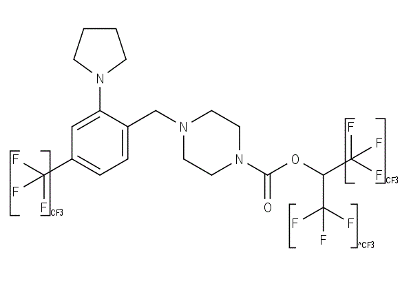
| SMILES | N1(C2C=C(C(F)(F)F)C=CC=2CN2CCN(C(OC(C(F)(F)F)C(F)(F)F)=O)CC2)CCCC1 |
|
| InChi Key | SQZJGTOZFRNWCX-UHFFFAOYSA-N | |
| InChi Code | InChI=1S/C20H22F9N3O2/c21-18(22,23)14-4-3-13(15(11-14)31-5-1-2-6-31)12-30-7-9-32(10-8-30)17(33)34-16(19(24,25)26)20(27,28)29/h3-4,11,16H,1-2,5-10,12H2 | |
| Chemical Name | 1,1,1,3,3,3-hexafluoropropan-2-yl 4-[[2-pyrrolidin-1-yl-4-(trifluoromethyl)phenyl]methyl]piperazine-1-carboxylate | |
| Synonyms |
|
|
| HS Tariff Code | 2934.99.9001 | |
| Storage |
Powder-20°C 3 years 4°C 2 years In solvent -80°C 6 months -20°C 1 month |
|
| Shipping Condition | Room temperature (This product is stable at ambient temperature for a few days during ordinary shipping and time spent in Customs) |
Biological Activity
| Targets |
Monoacylglycerol Lipase (MAGL) (IC50 for recombinant human MAGL: 3.2 nM; Ki for recombinant human MAGL: 1.8 nM) [1] |
||
| ln Vitro |
Elcubragistat, also known as ABX-1431, is a highly effective human MGLL inhibitor (IC50=0.014 µM) that exhibits selectivity levels of >200 against PLA2G7 and >100 against ABHD6. Elcubragistat has an IC50 of 0.0022 µM for inhibiting human PC3 cells. >100-fold selectivity for MGLL over PLA2G7 (IC50=494 µM) and ABHD6 (IC50=0.253 µM) is maintained in the cell-based assay[1]. 1. Potent and selective MAGL inhibition: ABX-1431 reversibly inhibited recombinant human MAGL enzyme activity in a dose-dependent manner, with an IC50 of 3.2 nM and a Ki of 1.8 nM (fluorescent substrate assay). It exhibited high selectivity for MAGL over other lipid-processing enzymes, including diacylglycerol lipases (DAGLα, DAGLβ; IC50 > 10 μM), fatty acid amide hydrolase (FAAH; IC50 > 10 μM), and serine hydrolases (e.g., carboxylesterase 1, CES1; IC50 > 20 μM) [1] 2. Elevation of endocannabinoid 2-arachidonoylglycerol (2-AG) levels in cells: In primary rat cortical neurons and human astrocytes, ABX-1431 (1-100 nM) dose-dependently increased intracellular and extracellular 2-AG concentrations (LC-MS/MS detection). At 10 nM, 2-AG levels were elevated by ~3.5-fold in rat cortical neurons and ~2.8-fold in human astrocytes after 2-hour treatment. No significant change in anandamide (AEA) levels was observed, confirming MAGL-specific activity [1] 3. Inhibition of proinflammatory mediator production: In LPS-stimulated human astrocytes, ABX-1431 (5-50 nM) dose-dependently reduced the secretion of proinflammatory cytokines TNF-α (by ~42% at 50 nM) and IL-6 (by ~38% at 50 nM), and chemokine CXCL10 (by ~35% at 50 nM) (ELISA detection). It also downregulated LPS-induced mRNA expression of iNOS (by ~40% at 50 nM) and COX-2 (by ~32% at 50 nM) (qPCR analysis) [1] 4. Lack of cytotoxicity: ABX-1431 showed no significant cytotoxicity to primary rat cortical neurons, human astrocytes, or HEK293 cells at concentrations up to 10 μM (MTT assay), with cell viability > 90% compared to vehicle controls [1] |
||
| ln Vivo |
Elcubragistat (ABX-1431) raises brain 2-AG concentrations, reduces pain behavior in the rat formalin pain paradigm, and inhibits MGLL activity in rodent brains (ED50=0.5-1.4 mg/kg)[1]. 1. MAGL inhibition and 2-AG elevation in rodent brains: C57BL/6 mice were orally administered ABX-1431 at doses of 3 mg/kg, 10 mg/kg, and 30 mg/kg. At 1 hour post-administration, brain MAGL activity was inhibited by ~55% (3 mg/kg), ~75% (10 mg/kg), and ~88% (30 mg/kg) compared to vehicle controls. Corresponding brain 2-AG levels were elevated by ~2.2-fold, ~4.0-fold, and ~6.5-fold, respectively (LC-MS/MS detection). The effect persisted for ~6 hours at the 10 mg/kg dose (MAGL inhibition ~50%, 2-AG elevation ~2.8-fold) [1] 2. Amelioration of experimental autoimmune encephalomyelitis (EAE) in mice: Female C57BL/6 mice were immunized to induce EAE (a model of multiple sclerosis). ABX-1431 was administered orally at 10 mg/kg/day starting from day 7 post-immunization (onset of symptoms). Compared to vehicle controls, ABX-1431 significantly reduced the mean clinical EAE score (from 3.2 to 1.5 at day 21) and decreased spinal cord inflammation, as evidenced by ~45% fewer CD4+ T cells and ~50% fewer macrophages/microglia infiltrating the spinal cord (immunohistochemical analysis). It also reduced spinal cord levels of proinflammatory cytokines TNF-α (by ~52%) and IL-1β (by ~48%) [1] 3. Attenuation of neuropathic pain in rats: Sprague-Dawley rats subjected to chronic constriction injury (CCI) of the sciatic nerve were treated with ABX-1431 (10 mg/kg, p.o., once daily for 7 days starting 7 days post-CCI). ABX-1431 significantly reduced mechanical allodynia (paw withdrawal threshold increased by ~60%) and thermal hyperalgesia (paw withdrawal latency increased by ~55%) compared to vehicle controls. Spinal cord 2-AG levels were elevated by ~3.8-fold, and TNF-α mRNA expression was downregulated by ~40% [1] |
||
| Enzyme Assay |
1. MAGL enzyme activity inhibition assay (fluorescent substrate-based): - Recombinant human MAGL protein was expressed and purified, then diluted in assay buffer (50 mM Tris-HCl, pH 7.4, 150 mM NaCl, 1 mM EDTA) to a final concentration of 1 nM [1] - Serial dilutions of ABX-1431 (0.1 nM-100 nM) were mixed with the diluted MAGL enzyme and incubated at room temperature for 15 minutes to allow binding [1] - The fluorescent substrate 4-methylumbelliferyl oleate (final concentration 20 μM) was added to initiate the reaction, and the mixture was incubated at 37°C for 30 minutes [1] - Fluorescence intensity (excitation: 360 nm, emission: 460 nm) was measured using a microplate reader. The percentage inhibition of MAGL activity was calculated relative to vehicle control, and IC50 values were derived from nonlinear regression analysis of dose-response curves. Ki values were determined using the Cheng-Prusoff equation [1] 2. Selectivity assay against other lipid-processing enzymes: - Recombinant enzymes (DAGLα, DAGLβ, FAAH, CES1) were prepared in assay buffer at optimal concentrations for their respective substrates [1] - ABX-1431 (0.1 nM-10 μM) was incubated with each enzyme, followed by addition of enzyme-specific fluorescent or radiolabeled substrates (e.g., DAGLα substrate: 4-methylumbelliferyl diacylglycerol; FAAH substrate: [³H]-anandamide) [1] - Enzyme activity was measured as described for MAGL, and IC50 values were calculated. Selectivity indices (IC50 for off-target enzyme / IC50 for MAGL) were determined to assess specificity [1] 3. Surface Plasmon Resonance (SPR) binding assay: - Purified human MAGL was immobilized on a CM5 sensor chip via amine coupling to a surface density of ~1000 resonance units (RU) [1] - Serial dilutions of ABX-1431 (0.5 nM-50 nM) were injected over the MAGL-coated chip at a constant flow rate of 30 μL/min in running buffer (50 mM Tris-HCl, pH 7.4, 150 mM NaCl, 0.05% Tween 20) [1] - Sensorgrams were recorded, and dissociation constants (Ki) were calculated using a 1:1 binding model with reference subtraction. Association (ka) and dissociation (kd) rate constants were derived from global fitting of the sensorgrams [1] |
||
| Cell Assay |
1. Intracellular 2-AG elevation assay in primary neurons: - Primary rat cortical neurons were isolated from embryonic day 18 rats, seeded into 24-well plates at 5×10⁵ cells/well, and cultured for 7 days in neurobasal medium supplemented with B27 [1] - ABX-1431 (1 nM-100 nM) was added to the cultures, and cells were incubated for 2 hours at 37°C with 5% CO₂ [1] - Cells were lysed with ice-cold methanol, and lipids were extracted by liquid-liquid extraction with chloroform. 2-AG concentrations in the extracts were quantified using LC-MS/MS with a calibration curve of synthetic 2-AG [1] 2. LPS-induced inflammation modulation assay in astrocytes: - Human astrocytes were seeded into 24-well plates at 2×10⁵ cells/well and cultured until 80% confluency [1] - Cells were pretreated with ABX-1431 (5 nM-50 nM) for 1 hour, then stimulated with LPS (1 μg/mL) for 24 hours [1] - Culture supernatant was collected for TNF-α, IL-6, and CXCL10 detection by ELISA. Total RNA was extracted from cells, reverse-transcribed into cDNA, and qPCR was performed to measure iNOS and COX-2 mRNA expression (normalized to GAPDH) [1] 3. Cell viability assay (MTT): - Primary rat cortical neurons, human astrocytes, and HEK293 cells were seeded into 96-well plates at 1×10⁴ cells/well (neurons) or 5×10³ cells/well (astrocytes/HEK293) and cultured overnight [1] - Serial dilutions of ABX-1431 (0.1 nM-10 μM) were added, and cells were incubated for 48 hours [1] - MTT reagent was added to each well, incubated for 4 hours at 37°C, and formazan crystals were dissolved in DMSO. Absorbance was measured at 570 nm, and cell viability was calculated relative to vehicle control [1] |
||
| Animal Protocol |
|
||
| ADME/Pharmacokinetics |
1. Absorption: In rats, oral administration of ABX-1431 (10 mg/kg) resulted in a peak plasma concentration (Cmax) of 87 ng/mL at 1 hour (Tmax), with an oral bioavailability of ~45% [1] 2. Distribution: ABX-1431 distributed widely into tissues, with a steady-state volume of distribution (Vss) of ~1.8 L/kg in rats. Brain-to-plasma concentration ratio was ~0.8 at 1 hour post-oral administration (10 mg/kg), indicating good blood-brain barrier penetration [1] 3. Metabolism: ABX-1431 was primarily metabolized in the liver via cytochrome P450 (CYP) enzymes, with CYP3A4 as the major isoform involved. The main metabolites were inactive hydroxylated derivatives, which were further glucuronidated for excretion [1] 4. Excretion: In rats, ~65% of the administered dose was excreted in feces and ~25% in urine within 72 hours, primarily as metabolites. The elimination half-life (t1/2) was ~3.2 hours in rats and ~4.1 hours in mice [1] 5. Plasma protein binding: In human plasma, ABX-1431 showed high protein binding (~92%), with no concentration-dependent binding at 10 nM-10 μM [1] |
||
| Toxicity/Toxicokinetics |
1. In vitro toxicity: ABX-1431 had no significant cytotoxicity to primary neurons, astrocytes, or HEK293 cells at concentrations up to 10 μM (cell viability > 90% vs. control). It did not induce apoptosis in neurons (Annexin V/PI staining) at concentrations up to 5 μM [1] 2. Acute in vivo toxicity: Single oral administration of ABX-1431 at doses up to 300 mg/kg in rats and 500 mg/kg in mice did not cause mortality or acute toxicity signs (e.g., lethargy, convulsions, abnormal behavior). Body weight and food intake remained unchanged for 14 days post-administration [1] 3. Subchronic in vivo toxicity: Rats were administered ABX-1431 (10 mg/kg, 30 mg/kg, 100 mg/kg) orally once daily for 28 days. No significant changes in body weight, organ weights (liver, kidney, brain, spleen), or clinical chemistry parameters (ALT, AST, creatinine, BUN) were observed. Histological analysis of major organs revealed no drug-related abnormalities [1] 4. Drug-drug interaction potential: ABX-1431 did not inhibit or induce major CYP450 isoforms (CYP1A2, CYP2C9, CYP2C19, CYP2D6, CYP3A4) at concentrations up to 10 μM, indicating low potential for drug-drug interactions [1] |
||
| References |
[1]. Identification of ABX-1431, a Selective Inhibitor of Monoacylglycerol Lipase and Clinical Candidate for Treatment of Neurological Disorders. J Med Chem. 2018 Oct 25;61(20):9062-9084. |
||
| Additional Infomation |
ABX-1431 is under investigation in clinical trial NCT03625453 (Study of ABX-1431 in Adult Patients With Tourette Syndrome or Chronic Motor Tic Disorder). Elcubragistat is an orally bioavailable inhibitor of the serine hydrolase monoacylglycerol lipase (MGLL), with potential use for the treatment of various central nervous system (CNS) diseases and with potential analgesic and anti-neuroinflammatory activities. Upon oral administration, elcubragistat targets and binds to MGLL, thereby inhibiting MGLL activity and preventing the breakdown and inactivation of endogenous 2-arachidonoylglycerol (2-AG). Increased 2-AG levels results in enhanced activation of the cannabinoid receptor 1 (CB1) in the CNS and enhanced CB1 endocannabinoid signaling in active neural circuits. Activation of CB1 helps modulate the endocannabinoid system and reduce neurotransmitter release, thereby decreasing overactive neural signaling. This induces analgesic, anti-inflammatory and various neurological effects that are caused by dysregulation of the endocannabinoid system and overactive neural signaling, including anxiolytic effects, reduced spasticity and decreased neurodegenerative effects. In addition, MGLL inhibition by elcubragistat depletes the supply of the inflammatory signaling molecule arachidonic acid, thereby further alleviating pain and inflammation. CB1 plays a key role in the regulation of neurotransmission; increased CB1 activation decreases overactive neural signaling. MGLL, an enzyme that catalyzes the breakdown of 2-AG, regulates the activation of CB1 and CB2 to modulate neurotransmission and inflammatory signaling, respectively. 1. ABX-1431 is a selective, reversible small-molecule inhibitor of monoacylglycerol lipase (MAGL), a key enzyme involved in the catabolism of the endocannabinoid 2-arachidonoylglycerol (2-AG) [1] 2. Its core mechanism of action involves inhibiting MAGL-mediated hydrolysis of 2-AG, leading to elevated endogenous 2-AG levels in tissues (particularly the central nervous system). 2-AG acts on cannabinoid receptors (CB1, CB2) to exert anti-inflammatory, analgesic, and neuroprotective effects [1] 3. ABX-1431 is a clinical candidate for the treatment of neurological disorders associated with neuroinflammation and pain, including multiple sclerosis (MS) and neuropathic pain, based on preclinical efficacy in EAE and CCI models [1] 4. The compound exhibits favorable pharmacokinetic properties, including good oral bioavailability, blood-brain barrier penetration, and a manageable half-life, supporting once-daily oral dosing [1] 5. High selectivity for MAGL minimizes off-target effects on other lipid-processing enzymes, reducing the risk of adverse effects related to disruption of other endocannabinoid or lipid signaling pathways [1] |
Solubility Data
| Solubility (In Vitro) |
|
|||
| Solubility (In Vivo) |
Solubility in Formulation 1: ≥ 2.08 mg/mL (4.10 mM) (saturation unknown) in 10% DMSO + 40% PEG300 + 5% Tween80 + 45% Saline (add these co-solvents sequentially from left to right, and one by one), clear solution. For example, if 1 mL of working solution is to be prepared, you can add 100 μL of 20.8 mg/mL clear DMSO stock solution to 400 μL PEG300 and mix evenly; then add 50 μL Tween-80 to the above solution and mix evenly; then add 450 μL normal saline to adjust the volume to 1 mL. Preparation of saline: Dissolve 0.9 g of sodium chloride in 100 mL ddH₂ O to obtain a clear solution. Solubility in Formulation 2: 2.08 mg/mL (4.10 mM) in 10% DMSO + 90% (20% SBE-β-CD in Saline) (add these co-solvents sequentially from left to right, and one by one), suspension solution; with ultrasonication. For example, if 1 mL of working solution is to be prepared, you can add 100 μL of 20.8 mg/mL clear DMSO stock solution to 900 μL of 20% SBE-β-CD physiological saline solution and mix evenly. Preparation of 20% SBE-β-CD in Saline (4°C,1 week): Dissolve 2 g SBE-β-CD in 10 mL saline to obtain a clear solution. Solubility in Formulation 3: ≥ 2.08 mg/mL (4.10 mM) (saturation unknown) in 10% DMSO + 90% Corn Oil (add these co-solvents sequentially from left to right, and one by one), clear solution. For example, if 1 mL of working solution is to be prepared, you can add 100 μL of 20.8 mg/mL clear DMSO stock solution to 900 μL of corn oil and mix evenly. (Please use freshly prepared in vivo formulations for optimal results.) |
| Preparing Stock Solutions | 1 mg | 5 mg | 10 mg | |
| 1 mM | 1.9709 mL | 9.8544 mL | 19.7087 mL | |
| 5 mM | 0.3942 mL | 1.9709 mL | 3.9417 mL | |
| 10 mM | 0.1971 mL | 0.9854 mL | 1.9709 mL |