ABT 491 Hydrochloride is a novel and potent platelet-activating factor receptor (PAF-R) antagonist
Physicochemical Properties
| Molecular Formula | C28H22N5O2F.HCL |
| Molecular Weight | 515.97 |
| Exact Mass | 515.152 |
| CAS # | 189689-94-9 |
| Related CAS # | 170499-15-7 |
| PubChem CID | 154086 |
| Appearance | Typically exists as solid at room temperature |
| Boiling Point | 725.2ºC at 760mmHg |
| Melting Point | 170-178ºC(lit.) |
| Flash Point | 392.4ºC |
| Vapour Pressure | 6.9E-21mmHg at 25°C |
| LogP | 5.425 |
| Hydrogen Bond Donor Count | 1 |
| Hydrogen Bond Acceptor Count | 5 |
| Rotatable Bond Count | 5 |
| Heavy Atom Count | 37 |
| Complexity | 888 |
| Defined Atom Stereocenter Count | 0 |
| SMILES | Cl.CN(C(N1C=C(C(C2C=CC(CN3C(C)=NC4C=NC=CC3=4)=C(F)C=2)=O)C2=C(C=CC=C12)C#C)=O)C |
| InChi Key | AWRGBOKANQBIBM-UHFFFAOYSA-N |
| InChi Code | InChI=1S/C28H22FN5O2.ClH/c1-5-18-7-6-8-25-26(18)21(16-34(25)28(36)32(3)4)27(35)19-9-10-20(22(29)13-19)15-33-17(2)31-23-14-30-12-11-24(23)33;/h1,6-14,16H,15H2,2-4H3;1H |
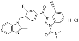
| Chemical Name | 1H-Indole-1-carboxamide, 4-ethynyl-3-(3-fluoro-4-((2-methyl-1H-imidazo(4,5-c)pyridin-1-yl)methyl)benzoyl)-N,N-dimethyl-, monohydrochloride |
| Synonyms | ABT 491 hydrochloride; ABT-491; ABT491; ABT-491 hydrochloride; 189689-94-9; ABT 491 hydrochloride; ABT-491; 4-Ethynyl-N,N-dimethyl-3-[3-fluoro-4-[(2-methyl-1H-imidazo-[4,5-c]pyridin-1-yl)methyl]benzoyl]-1H-indole-1-carboxamide hydrochloride; N7IG7Z867J; ABT 491 |
| HS Tariff Code | 2934.99.9001 |
| Storage |
Powder-20°C 3 years 4°C 2 years In solvent -80°C 6 months -20°C 1 month |
| Shipping Condition | Room temperature (This product is stable at ambient temperature for a few days during ordinary shipping and time spent in Customs) |
Biological Activity
| Targets | PAF-R/platelet-activating factor receptor |
| ln Vitro |
ABT-491 (4-ethynyl-N, N-dimethyl-3-[3-fluoro-4-[(2-methyl-1H-imidazo-[4,5-c]pyridin-1-yl)methy l]benzoyl]-1H- indole-1-carboxamide hydrochloride) is a novel PAF (platelet-activating factor) receptor antagonist with a K(i) for inhibiting PAF binding to human platelets of 0.6 nM. Binding kinetics of ABT-491 to the PAF receptor is consistent with a relatively slow off-rate of the antagonist when compared to PAF. Inhibition of PAF binding is selective and is correlated with functional antagonism of PAF-mediated cellular responses (Ca2+ mobilization, priming, and degranulation) [1].
Inhibition of PAF binding [1] The intrinsic PAF binding activity of ABT-491 has been evaluated by measuring the inhibition of [3H]C18-PAF binding to the PAF receptor of isolated rabbit platelet membranes and intact human platelets. Results, shown in Fig. 2A, illustrate the potency of ABT-491 in these assays. The inhibitory binding constants (Ki) computed from these results for ABT-491 are 1.8 and 0.57 nM, respectively and are within 3-fold the potency of PAF itself (Kd, 0.6 nM). The Hill coefficients obtained from the binding results (1.1 and 0.8) approach unity, indicating, as has previously been shown for PAF (Hwang and Lam, 1986), that ABT-491 interacts with a single class of binding sites. The nature of inhibition of PAF binding exhibited by ABT-491 was examined using rabbit platelet membranes. In a competition binding experiment, membrane preparations were incubated (1 h) with increasing concentrations of [3H]PAF in the absence and presence of ABT-491 (0.5–1.9 nM). Scatchard analysis of these results revealed that the maximal number of receptor sites (Bmax, x-intercept) was not affected whereas the apparent dissociation constant (Kd, −1/slope) increased with respect to increasing concentrations of ABT-491 (Fig. 2B). These results strongly indicate that ABT-491 is a competitive inhibitor of PAF binding to the high-affinity receptor. However, although ABT-491 acts as a competitive receptor antagonist under equilibrium conditions, there is a time-dependent component to its inhibitory action. This is apparent from the Scatchard analysis of results from a binding assay in which membranes were preincubated for 1 h with ABT-491 prior to a 30 min incubation with [3H]PAF. Under these experimental conditions, the Bmax was altered with little effect on the apparent Kd, indicative of non-competitive inhibition (Fig. 2C). These results suggest that ABT-491, in comparison to PAF, may have a relatively slow off-rate on the PAF receptor. To assess the specificity of inhibition for PAF binding, ABT-491 was evaluated in a wide variety of receptor, ion channel, and membrane binding assays. ABT-491 did not exhibit significant interaction at test concentrations up to 10 μM in any of these 45 different binding assays (data not shown). Antagonism of PAF-induced platelet responses [1] Platelet degranulation and release of granular constituents is a consequence of PAF-mediated platelet activation (McManus et al., 1981). The ability of ABT-491 to inhibit platelet degranulation was demonstrated using PAF-induced [14C]serotonin release from rabbit platelets. In the presence of increasing concentrations of ABT-491 (1–3 nM 5 min prior to PAF), a rightward and parallel shift in the PAF dose–response curves for [14C]serotonin release was observed (Fig. 3A). From Schild analysis of the rightward shift, the A2 value for ABT-491 for functional inhibition of PAF binding was calculated as 0.9 nM. Similar results (A2=1.3 nM) were obtained in an additional experiment, yielding a mean A2 value of 1.1 nM (not shown). At concentrations of ABT-491 greater than 3 nM the maximal response to PAF was not restored with agonist concentrations as high as 10 μM. These results, suggestive of non-competitive antagonism, are consistent with the kinetics observed in the binding studies with platelet membranes preincubated with ABT-491. In other experiments, ABT-491 (10 μM) did not cause release of [14C]serotonin and did not inhibit release in response to thrombin (0.2 U/ml) or Ca2+ ionophore (0.3 μM). Taken together these results indicate that ABT-491 selectively and potently inhibits functional activity coupled to the platelet PAF receptor. Antagonism of PAF-induced neutrophil responses [1] Three PAF-mediated neutrophil responses were examined in this study: intracellular Ca2+ mobilization, degranulation, and priming (Korchak et al., 1988; Dewald and Baggiolini, 1987; Baggiolini and Dewald, 1986; Gay, 1993). The Ca2+ response induced by PAF is rapid and transient, reaching a maximum in 30–45 s then returning to near-basal levels within 120 s (Fig. 4A). The response was inhibited in a concentration-dependent manner by preincubation with ABT-491 that resulted in a rightward shift in the PAF dose–response curve, suggestive of competitive antagonism (Fig. 3C). Schild analysis of the data yielded an A2 value of 25 nM. The antagonist appears to be selective since ABT-491, at a concentration (1 μM) that completely inhibited the PAF-induced response, did not inhibit the Ca2+ response to leukotriene B4 and C5a (not shown). |
| ln Vivo |
Administration of ABT-491 in vivo leads to potent inhibition of PAF-induced inflammatory responses (increased vascular permeability, hypotension, and edema) and PAF-induced lethality. Oral potency (ED50) was between 0.03 and 0.4 mg/kg in rat, mouse, and guinea-pig. When administered intravenously in these species, ABT-491 exhibited ED50 values between 0.005 and 0.016 mg/kg. An oral dose of 0.5 mg/kg in rat provided > 50% protection for 8 h against cutaneous PAF challenge. ABT-491 administered orally was also effective in inhibiting lipopolysaccharide-induced hypotension (ED50 = 0.04 mg/kg), gastrointestinal damage (0.05 mg/kg, 79% inhibition), and lethality (1 mg/kg, 85% vs. 57% survival). The potency of this novel antagonist suggests that ABT-491 will be useful in the treatment of PAF-mediated diseases.[1] In vivo models of PAF-induced inflammation [1] PAF induces localized cutaneous vascular permeability and edema characteristic of acute inflammation (Hwang et al., 1985b; Qu et al., 1990). ABT-491 possessed potent activity as an antagonist of these responses. In rat, an orally administrated dose of 1 mg/kg of ABT-491 resulted in 90% inhibition of the PAF-induced permeability response (Fig. 5A). The inhibition was dose related and regression analysis yielded an ED50 value of 0.094 mg/kg. In addition, this compound exhibited potent activity when administered intravenously (ED50=0.003 mg/kg). In guinea-pig, ABT-491 was several fold less potent. Nevertheless, when administered intravenously, a dose of 50 μg/kg produced near-maximal antagonism (75%) of the permeability response (Fig. 5B). As in the rat, the inhibition in guinea-pig was dose related for both intravenous and oral administration (ED50=0.016 and 0.29 mg/kg, respectively). Administration of ABT-491 to rats at a dose (1 mg/kg i.v.) sufficient to provide maximal (87%) inhibition of the PAF-induced response had no significant effect (<5%) on the permeability responses induced by serotonin and histamine (not shown).[1] The effect of ABT-491 on PAF-induced edema was assessed in mice. Pre-administration of ABT-491 to mice subjected to subcutaneous injections of PAF in the paw inhibited the resulting edema formation in a dose-related manner (Fig. 5C). The potency (ED50) of ABT-491 for blocking edema was 0.069 mg/kg when administered intravenously and 0.38 mg/kg when administered orally. These values are comparable to those for inhibiting PAF-induced responses in the guinea pig and rat, and further illustrate the effectiveness of ABT-491 in blocking PAF-mediated inflammatory responses. PAF- and LPS-induced shock [1] Following systemic administration, PAF causes an acute drop in systemic arterial pressure (Blank et al., 1979). At higher doses of PAF, death results from acute cardio-pulmonary failure and complement activation (Carlson et al., 1987; Sun and Hsueh, 1991). The hypotensive response induced in guinea pig by intraarterial administration of PAF was antagonized in a dose-dependent fashion by pretreatment with ABT-491 (Fig. 7A). The ED50 values derived from regression analysis for intraarterial and oral administration of ABT-491 were 0.005 and 0.026 mg/kg (Fig. 7A, insert).[1] LPS given intraarterially to rats also results in hypotension. The initial response is transient but is followed by a sustained decrease in systemic blood pressure (Fig. 7B). ABT-491, when administered intraarterially (0.1 mg/kg) or orally (1 mg/kg) 1 h prior to LPS, blocked the second-phase response. Inhibition was dose-dependent and analysis of the dose–response relationship (Fig. 7B, insert) yielded ED50 values for intraarterial and oral administration of 0.004 and 0.036 mg/kg respectively. In addition to being effective when given prior to LPS, ABT-491 was also capable of reversing hypotension when administered post LPS. As shown in Fig. 7B, administration of ABT-491 restored blood pressure as late as 1 h after LPS challenge.[1] Other hallmarks of septic shock, including gastrointestinal injury and lethality, are evident following the administration of LPS. Marked hyperemia and overt luminal bleeding are prominent in the small bowel of the rat within 30 min post LPS. Microscopically, congestion of mucosal microvessels, single cell necrosis and isolated loss of villi are evident (Wallace et al., 1987). In the current study the extent of damage was followed by scoring the gross appearance of the small intestine and by measuring intraluminal hemoglobin. ABT-491 (0.050 mg/kg), given orally 60 min prior to LPS challenge, inhibited intestinal bleeding by 79% (P<0.05). A 10-fold higher dose resulted in complete protection and a normal appearing intestine.[1] Studies on the lethal effect of PAF were conducted with mice. Intravenous administration of PAF (3–1000 μg/kg) resulted in an increase in mortality that was dose-dependent (LD100 approximately 30 μg/kg, Fig. 8). Pre-treatment with ABT-491 (1, 10 and 100 μg/kg, p.o.) resulted in a rightward shift in the dose–response curve, indicative of competitive antagonism. Administration of 1 mg/kg ABT-491 provided total protection against lethality induced by a PAF challenge as high as 1 mg/kg, greater than 30-fold the PAF LD100. These observations are consistent with potent antagonism of the cardio-pulmonary responses to PAF and provide additional evidence that in vivo administration of ABT-491 results in antagonism of systemic PAF responses.[1] The effect of ABT-491 on long-term survival was also evaluated in the rat endotoxemia model. LPS (8.5 mg/kg, i.v.) resulted in 43% mortality within 24 h. Pretreatment with ABT-491 (1 mg/kg, p.o.) produced a marked reduction in mortality (15% vs. 43%, P<0.05). Hypoxic ischemic brain injury (HIBI) is a common cause of neonatal mortality and morbidity. To date, no study has investigated the role of platelet-activating factor (PAF) antagonists on neuronal apoptosis in neonatal rat model of HIBI. In the present study, we evaluated the effect of a highly potent and selective PAF antagonist (ABT-491) on neuronal apoptosis in neonatal rat model of HIBI. Seven-day-old Wistar rat pups were subjected to right common carotid artery ligation and hypoxia (92% nitrogen and 8% oxygen) for 2 h. They were treated with ABT-491 or saline either immediately before or after hypoxia. In sham group animals, neither ligation, nor hypoxia was performed. Neuronal apoptosis was evaluated by the terminal-transferase mediated dUTP biotin nick-end-labeling (TUNEL) and caspase-3 staining methods. Administration of ABT-491 either before or after hypoxia resulted in significant reduction of the numbers of apoptotic cells in both hemispheres, when compared to saline treatment group. The numbers of apoptotic cells in right hemispheres in all groups were significantly higher than that in the left hemispheres. These results suggested that ABT-491, a highly potent and selective PAF antagonist, administration either before or after hypoxia reduces apoptosis and we propose that ABT-491 may be a novel approach in the treatment of HIBI [2]. |
| Enzyme Assay |
Binding assays [1] The PAF receptor binding assays with rabbit platelet membranes and washed human platelets were based upon the procedures described by Hwang et al. (1983), Hwang et al. (1985a)and Hwang and Lam (1986), and detailed elsewhere (Albert et al., 1996a). In brief, the standard membrane binding assay, conducted in Millititre-GV microtiter filter plates, contained 10 μg platelet membrane protein, 0.6 nM of [3H]C18-PAF and test compound in buffer containing 0.25% bovine serum albumin in a final volume of 100 μl. The human platelet assay, conducted in 96-well glass fiber filtration plates, contained 30×106 washed platelets, 0.6 nM [3H]C18-PAF and test compound in a total volume 250 μl assay buffer. Test compounds were dissolved in dimethyl sulfoxide and diluted into buffer vehicle (final concentration <0.1%). Both assays were conducted at ambient temperature for 60 min. After the incubation period, the filter plates were subjected to vacuum filtration, washed with 1 ml of ice-cold assay buffer, and assayed for radioactivity with a microtiter scintillation counter. Specific binding was defined as the difference between total binding of 0.6 nM [3H]C18-PAF (3H radioactivity in the absence of added PAF) and nonspecific binding (3H radioactivity in the presence of 1 μM PAF). Compound binding is 3H radioactivity in the presence of the test compound. Percent inhibition is calculated as [(Total binding−Compound binding)/Specific binding]×100%. |
| Cell Assay |
PAF-induced platelet responses [1] For the rabbit platelet release assay, washed rabbit platelets labeled with [14C]serotonin were prepared as described previously (Ostermann et al., 1983; Albert et al., 1996a). Aliquots of the platelet suspension were incubated at 37°C with various concentrations of test compounds or vehicle (Tyrode buffer with 1.3 mM CaCl2) for 5 min. Various concentrations of PAF or vehicle buffer were then added and the reaction mixture was incubated for an additional 6 min. To assess specificity of inhibition, thrombin (0.05–0.2 U/ml) or Ca2+ ionophore A23187 (0.3 μM) was substituted for PAF. The release reaction was terminated by cooling on ice and by the addition of 3 mM EDTA (final concentration) in saline. The platelet suspension was then centrifuged (1500×g, 15 min, 4°C) and the supernatant was collected for measurement of released 14C radioactivity by liquid scintillation spectrometry. To assay β-thromboglobulin release in blood peripheral human blood was obtained from healthy human volunteers and treated with heparin (20 U/ml) and incubated with 0.2% bovine serum albumin in normal saline (vehicle) or ABT-491 at the indicated concentration for 5 min at room temperature. Aliquots of the blood were then incubated with PAF or vehicle for an additional 5 min at 37°C and placed in ice-water. The blood samples were centrifuged (1000×g for 15 min) and the plasma was removed and stored frozen until analyzed. Plasma levels of β-thromboglobulin were measured using commercially available immunoassay kits following the manufacturer's recommendations. PAF-induced neutrophil responses [1] The assays for PAF-induced elastase release, reactive oxygen species production and intracellular Ca2+ were described in detail elsewhere (Albert et al., 1996a). In brief, for the elastase release assay, neutrophils (1 million/ml in Tyrode buffer, pH 7.4) were pretreated with drug for 6–8 min at room temperature and then exposed to cytochalasin B (1 μg/ml) for 15 min at 37°C (Dewald and Baggiolini, 1987). Various concentrations of PAF in Tyrode buffer containing 0.2% bovine serum albumin were then added and the incubation was continued for an additional 10 min until terminated by rapid cooling. The cell suspensions were then centrifuged at 5°C (700×g for 15 min). Elastase activity in the supernatant was then measured with a fluorescence plate reader using the synthetic substrate N-succinyl-l-alanyl-l-alanyl-l-prolyl-l-valine-amino-4-methylcoumarin (0.25 mM) at excitation and emission wavelengths of 370 and 460 nm, respectively. Relative activity was computed as the ratio of the rate of fluorescence increase obtained with stimulated and non-stimulated cells. For the reactive oxygen species assay, cells were resuspended (5×10⁶ cells/ml) in Dulbecco's phosphate-buffered saline containing 0.25% bovine serum albumin and 20 μM luminol (Allen, 1986). The cells (70 μl) were preincubated in microtiter trays at room temperature with 10 μl drug or vehicle (1% dimethyl sulfoxide in Hanks' balanced salt solution). After 5 min, 10 μl PAF or vehicle was added and the preincubation continued for an additional 2 min at room temperature. At the end of the preincubation, 10 μl formyl-methionyl-leucinyl-phenylalanine (fMLP) or vehicle was added. The resulting light output was recorded in a microtiter scintillation counter at 20-s intervals for 5 min. Response to PAF and fMLP challenge was measured by calculating the area under the curve (using the trapezoidal rule) for the response to the challenge over the 5-min period. To measure intracellular Ca2+, neutrophils suspended in Hanks' balanced salt solution containing 0.035% NaHCO3, pH 7.0, were incubated with 5 μM indo-1 AM for 30 min at room temperature, washed, and resuspended at a concentration of (1–2)×106 cells/ml. The cells were pretreated with receptor antagonist for 60 s followed by the addition of agonist at the indicated concentrations. Fluorescence was measured at an excitation wavelength of 350 nm and emission wavelengths of 485 nm (low Ca2+) and 410 nm (high Ca2+) using an SLM 8000 spectrofluoromete. Intracellular Ca2+ concentration was calculated from the ratio of fluorescence at the two emission wavelengths (Grynkiewicz et al., 1985). Values are expressed as percent inhibition relative to control response to PAF in the absence of antagonist. |
| Animal Protocol |
Animal preparation and surgical procedure [2] Rat pups were anaesthetized by halothane inhalation and duration of anaesthesia was less than 5 min. Hypoxic ischemia was induced according to the Levine–Rice model (Rice et al., 1981). A median incision was made in the neck. Under the microscopic magnification, the right common carotid artery was dissected and ligated with a 6-zero silk suture. After the wound was sutured, the animals were allowed to have 3 h recovery and feeding period. Except for the sham group, rats were then placed in a plastic chamber and exposed to a continuous flow of 8% oxygen–92% nitrogen for 2 h. After hypoxic period, the rats allowed to have a 2 h recovery period in an open chamber without any supplemental oxygen. The animals in the sham group were placed in an open chamber for the same intervals. The chambers were partially submerged in a water bath at 37 °C to maintain a constant thermal environment. After these procedures, all the pups were euthanized by decapitation. The brains were removed and paraffin-embedded for pathological evaluation. Saline treatment group: 0.5 mL saline was injected intraperitoneally either immediately before (n = 20) or after (n = 20) hypoxia. Fourteen rats (6 before hypoxia, 8 after hypoxia) in this group were died during the procedure. ABT-491treatment group: The rat pups were administered intraperitoneally 0.4 mg/kg ABT-491 which was dissolved in saline, either immediately before (n = 20) or after (n = 20) hypoxia. Five rats (4 before hypoxia, 1 after hypoxia) in this group were died during the procedure. PAF and endotoxin-induced shock [1] To measure PAF or endotoxin-induced hypotension, the carotid artery of guinea pigs and rats maintained under light to moderate anesthesia by inhalation of Penthrane (Abbott Laboratories, North Chicago, IL, USA) was cannulated with PE-50 tubing. The tubing was tunneled subcutaneously to the posterior neck, exteriorized and connected to a pressure transducer and polygraph (Model MI2, Modular Instruments, Southeastern, PA, USA) for monitoring arterial pressure. After cannulation, the animals were allowed to recover for at least 1 h. After the recovery period a baseline pressure for each animal was established by monitoring arterial pressure over a 15 min period. There was no significant difference between baseline values among control and experimental groups. For pre-treatment studies antagonist or vehicle (0.9% saline) was given 15 min (intraarterial) or 1 h (p.o.) prior to agonist challenge. Following pretreatment, guinea pigs and rats were challenged with PAF (0.6 μg/kg) or endotoxin (LPS, 25 mg/kg in PBS, pH 7.4), administered as an intraarterial bolus. For studies of the ability of ABT-491 to reverse ongoing hypotension, drug was administered 60 min following LPS challenge. Measurements of arterial pressure were taken at 1 min intervals until the conclusion of the experiment. To correct for the small degree of animal to animal variation (<10%), pressure values were expressed as percent initial baseline. Response to PAF or LPS challenge and drug was determined by calculating the area under the curve of percent baseline vs. time (using the trapezoidal rule) for the response to the challenge over the experimental period. From these areas, percent inhibition of the agonist-induced response was calculated as (drug−agonist)/(vehicle−agonist). Intestinal bleeding induced by LPS treatment was determined by measuring hemoglobin which leaked into the gastrointestinal lumen. ABT-491 was given p.o. to conscious rats (6–7 per group) followed 60 min later by LPS (25 mg/kg). Rats were killed 30 min after LPS challenge and a 15 cm segment of intestine, starting from the duodenum, was collected from each rat. Luminal content was collected by rinsing the intestine segment. Hemoglobin concentration in the rinse was measured with a hemoglobinometer (Coulter Electronic, Hialeah, FL, USA) by comparison to a calibration curve obtained with standard hemoglobin solutions. Lethality studies were conducted with PAF (3–1000 μg/kg) and LPS (8.5 mg/kg) in mice and rats, respectively. In both cases ABT-491 was administered orally 30 min prior to PAF or LPS challenge. The lethal effects of PAF, which generally occurred within 30 min following PAF challenge, were monitored over an 8 h period. LPS-induced mortality generally occurred 6–10 h following LPS challenge. Rats were closely monitored over a period of 2 weeks following LPS to determine the total number of deaths. |
| ADME/Pharmacokinetics |
Duration of action [1] The model for PAF-induced cutaneous permeability was used to assess the duration of activity of ABT-491. The compound was administered orally at a dose expected to produce 70–80% inhibition 1 h after administration. The level of inhibition of PAF-induced permeability was then assessed at various times after compound dosing. As shown in Fig. 6, ABT-491 (0.5 mg/kg p.o.) provided protection (>50%) against cutaneous PAF challenge in the rat for greater than 8 h. |
| References |
[1]. Pharmacology of ABT-491, a highly potent platelet-activating factor receptor antagonist. Eur J Pharmacol. 1997 Apr 23;325(1):69-80. [2]. Platelet-activating factor antagonist (ABT-491) decreases neuronal apoptosis in neonatal rat model of hypoxic ischemic brain injury. Brain Res. 2007 Apr 27:1143:193-8. |
| Additional Infomation |
ABT-491 is a recently described highly potent and selective PAF antagonist (Albert et al., 1997). It is a potent antagonist of responses linked to the PAF receptor at the cellular level, especially platelets and neutrophils. Albert et al. (1997) found that ABT-491 was also effective in blocking platelet activation in blood, indicating that the presence of high concentrations of protein and other serum factors slightly alters the ability of ABT-491 to interact with PAF receptor. In the present study, we used 0.4 mg/kg ABT-491 intraperitoneally. The dose of ABT-491 in the treatment of HIBI has not been reported previously. It has been demonstrated that ABT-491 was effective in inhibiting lipopolysaccharide-induced hypotension (ED50 = 0.04 mg/kg) and gastrointestinal damage (0.05 mg/kg), it was found to be lethal at a dose of 1 mg/kg (Albert et al., 1997). The exact mechanism of the apoptotic function of PAF is still unclear. However, it has been demonstrated that PAF elicits apoptosis in enterocytes via a mechanism that involves Bax translocation to mitochondria, collapse of mitochondrial membrane potential, and caspase activation (Lu et al., 2004). There is strong evidence that PAF was involved in cellular apoptosis with Fas–Fas ligand (FasL) and cytochrome-C release in several cell types. Wu et al. (2003) demonstrated that PAF promotes mucosal apoptosis via FasL-mediating caspase-9 active pathway in rat small intestine after ischemia–reperfusion. On the other hand, recent studies have emphasized the anti-apoptotic effect of PAF antagonists and the protective effect of PAF antagonists against apoptotic changes in several tissues and cells (Murohisa et al., 2002, Loucks et al., 2003, Grypioti et al., 2006). Our study is the first report in the literature to show that the neuronal apoptotic changes were reduced by ABT-491, a highly potent and selective PAF antagonist, in neonatal rat model of HIBI. In light of these data, we propose that inhibition of apoptosis in hypoxic ischemia represents an important protective effect provided by PAF antagonist. Therapy using PAF antagonists may be a new approach in the treatment of HIBI. Although there has been relatively little research about the role of PAF antagonists in neonatal HIBI, no study has investigated the role of PAF antagonists on neuronal apoptosis in this model. The current study shows that ABT-491, a highly potent and selective PAF antagonist, administration either before or after hypoxia reduces apoptosis and we propose that ABT-491 may be a novel approach for the therapy of HIBI. Although the present study demonstrated the immediate neuroprotective effect of ABT-491 in HIBI, the long term effects of this drug should be investigated and further studies are required to elucidate the mechanism of HIBI that involves PAF activation and the possible role of platelets in HIBI. [2] PAF has also been previously implicated as a mediator of endotoxin-induced gastrointestinal damage and lethality in the rat (Albert et al., 1996b; Torley et al., 1992; Wallace et al., 1987; Terashita et al., 1985). In the present study 0.050 mg/kg ABT-491 given orally 60 min prior to LPS challenge was effective in blocking by 79% LPS-induced damage. Thus the oral potency for blocking gastrointestinal damage correlates well with activity for the hypotensive response and places ABT-491 as one of the most potent PAF receptor antagonists evaluated in this model. ABT-491 also was effective in increasing the long-term survival rate of rats administered LPS; however, the oral dose (1 mg/kg) necessary to achieve maximum effect was higher than that required for protection against gastrointestinal damage. Furthermore, the effect was somewhat less than that achieved in the PAF-induced lethality model (85% vs. 100% survival). The higher dose and less efficacy in the LPS lethality model may be a reflection of the longer coverage time required for protecting against LPS-induced mortality (most deaths occur 6–8 h post LPS) compared to the acute hypotensive and organ injury responses (30–60 min). They may also reflect the role of other mediators in the response to LPS. In conclusion ABT-491 is a novel antagonist that is active at nanomolar and sub-nanomolar concentrations in selectively inhibiting PAF binding and PAF-mediated cellular responses. The receptor antagonist is orally active at sub-mg/kg doses and exhibits a bioduration of >8 h. Its oral activity coupled with its aqueous solubility provide a variety of dosing options and make ABT-491 an attractive candidate for clinical studies. Thus ABT-491, along with several other structurally distinct antagonists (e.g., BBT-882 and SR-27,417), may prove useful in defining the role of PAF in human diseases such as asthma, allergic rhinitis and sepsis.[1] |
Solubility Data
| Solubility (In Vitro) | May dissolve in DMSO (in most cases), if not, try other solvents such as H2O, Ethanol, or DMF with a minute amount of products to avoid loss of samples |
| Solubility (In Vivo) |
Note: Listed below are some common formulations that may be used to formulate products with low water solubility (e.g. < 1 mg/mL), you may test these formulations using a minute amount of products to avoid loss of samples. Injection Formulations (e.g. IP/IV/IM/SC) Injection Formulation 1: DMSO : Tween 80: Saline = 10 : 5 : 85 (i.e. 100 μL DMSO stock solution → 50 μL Tween 80 → 850 μL Saline) *Preparation of saline: Dissolve 0.9 g of sodium chloride in 100 mL ddH ₂ O to obtain a clear solution. Injection Formulation 2: DMSO : PEG300 :Tween 80 : Saline = 10 : 40 : 5 : 45 (i.e. 100 μL DMSO → 400 μLPEG300 → 50 μL Tween 80 → 450 μL Saline) Injection Formulation 3: DMSO : Corn oil = 10 : 90 (i.e. 100 μL DMSO → 900 μL Corn oil) Example: Take the Injection Formulation 3 (DMSO : Corn oil = 10 : 90) as an example, if 1 mL of 2.5 mg/mL working solution is to be prepared, you can take 100 μL 25 mg/mL DMSO stock solution and add to 900 μL corn oil, mix well to obtain a clear or suspension solution (2.5 mg/mL, ready for use in animals). Injection Formulation 4: DMSO : 20% SBE-β-CD in saline = 10 : 90 [i.e. 100 μL DMSO → 900 μL (20% SBE-β-CD in saline)] *Preparation of 20% SBE-β-CD in Saline (4°C,1 week): Dissolve 2 g SBE-β-CD in 10 mL saline to obtain a clear solution. Injection Formulation 5: 2-Hydroxypropyl-β-cyclodextrin : Saline = 50 : 50 (i.e. 500 μL 2-Hydroxypropyl-β-cyclodextrin → 500 μL Saline) Injection Formulation 6: DMSO : PEG300 : castor oil : Saline = 5 : 10 : 20 : 65 (i.e. 50 μL DMSO → 100 μLPEG300 → 200 μL castor oil → 650 μL Saline) Injection Formulation 7: Ethanol : Cremophor : Saline = 10: 10 : 80 (i.e. 100 μL Ethanol → 100 μL Cremophor → 800 μL Saline) Injection Formulation 8: Dissolve in Cremophor/Ethanol (50 : 50), then diluted by Saline Injection Formulation 9: EtOH : Corn oil = 10 : 90 (i.e. 100 μL EtOH → 900 μL Corn oil) Injection Formulation 10: EtOH : PEG300:Tween 80 : Saline = 10 : 40 : 5 : 45 (i.e. 100 μL EtOH → 400 μLPEG300 → 50 μL Tween 80 → 450 μL Saline) Oral Formulations Oral Formulation 1: Suspend in 0.5% CMC Na (carboxymethylcellulose sodium) Oral Formulation 2: Suspend in 0.5% Carboxymethyl cellulose Example: Take the Oral Formulation 1 (Suspend in 0.5% CMC Na) as an example, if 100 mL of 2.5 mg/mL working solution is to be prepared, you can first prepare 0.5% CMC Na solution by measuring 0.5 g CMC Na and dissolve it in 100 mL ddH2O to obtain a clear solution; then add 250 mg of the product to 100 mL 0.5% CMC Na solution, to make the suspension solution (2.5 mg/mL, ready for use in animals). Oral Formulation 3: Dissolved in PEG400 Oral Formulation 4: Suspend in 0.2% Carboxymethyl cellulose Oral Formulation 5: Dissolve in 0.25% Tween 80 and 0.5% Carboxymethyl cellulose Oral Formulation 6: Mixing with food powders Note: Please be aware that the above formulations are for reference only. InvivoChem strongly recommends customers to read literature methods/protocols carefully before determining which formulation you should use for in vivo studies, as different compounds have different solubility properties and have to be formulated differently. (Please use freshly prepared in vivo formulations for optimal results.) |
| Preparing Stock Solutions | 1 mg | 5 mg | 10 mg | |
| 1 mM | 1.9381 mL | 9.6905 mL | 19.3810 mL | |
| 5 mM | 0.3876 mL | 1.9381 mL | 3.8762 mL | |
| 10 mM | 0.1938 mL | 0.9690 mL | 1.9381 mL |